Show PDB file:   
         Plain Text   HTML   (compressed file size)
QuickSearch:   
by PDB,NDB,UniProt,PROSITE Code or Search Term(s)  
(-)Asymmetric Unit
(-)Biological Unit 1
(-)Biological Unit 2
collapse expand < >
Image Asymmetric Unit
Asymmetric Unit  (Jmol Viewer)
Image Biological Unit 1
Biological Unit 1  (Jmol Viewer)
Image Biological Unit 2
Biological Unit 2  (Jmol Viewer)

(-) Description

Title :  STRUCTURE OF THE MERS-COV FUSION CORE
 
Authors :  J. Gao, G. Lu, J. Qi, Y. Li, Y. Wu, Y. Deng, H. Geng, H. Xiao, W. Tan, J. Yan, G
Date :  12 Sep 13  (Deposition) - 09 Oct 13  (Release) - 28 Jun 17  (Revision)
Method :  X-RAY DIFFRACTION
Resolution :  1.90
Chains :  Asym. Unit :  A,B
Biol. Unit 1:  A  (3x)
Biol. Unit 2:  B  (3x)
Keywords :  Mers-Cov, Viral Envelope Proteins, Spike, Trimer Of Hairpins, 6-Helix Bundle, Membrane Fusion, Viral Protein (Keyword Search: [Gene Ontology, PubMed, Web (Google))
 
Reference :  J. Gao, G. Lu, J. Qi, Y. Li, Y. Wu, Y. Deng, H. Geng, H. Li, Q. Wang, H. Xiao, W. Tan, J. Yan, G. F. Gao
Structure Of The Fusion Core And Inhibition Of Fusion By A Heptad-Repeat Peptide Derived From The S Protein Of Mers-Co
J. Virol. 2013
PubMed-ID: 24067982  |  Reference-DOI: 10.1128/JVI.02433-13

(-) Compounds

Molecule 1 - HR1 OF S PROTEIN, LINKER, HR2 OF S PROTEIN
    ChainsA, B
    EngineeredYES
    Expression SystemESCHERICHIA COLI
    Expression System PlasmidPET-21A
    Expression System StrainBL21(DE3)
    Expression System Taxid562
    Expression System Vector TypePLASMID
    GeneSPIKE, S
    Organism CommonMERS-COV
    Organism ScientificHUMAN BETACORONAVIRUS 2C EMC/2012, SYNTHETIC CONSTRUCT
    Organism Taxid1235996, 32630
    Other DetailsAMINO ACIDS 992-1054 (HR1) AND AMINO ACIDS 1252-1286 (HR2) LINKED BY A 22-RESIDUE LINKER (LVPRGSGGSGGSGGLEVLFQGP)
    StrainHCOV-EMC

 Structural Features

(-) Chains, Units

  12
Asymmetric Unit AB
Biological Unit 1 (3x)A 
Biological Unit 2 (3x) B

Summary Information (see also Sequences/Alignments below)

(-) Ligands, Modified Residues, Ions  (0, 0)

(no "Ligand,Modified Residues,Ions" information available for 4MOD)

(-) Sites  (0, 0)

(no "Site" information available for 4MOD)

(-) SS Bonds  (0, 0)

(no "SS Bond" information available for 4MOD)

(-) Cis Peptide Bonds  (1, 1)
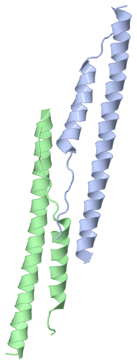
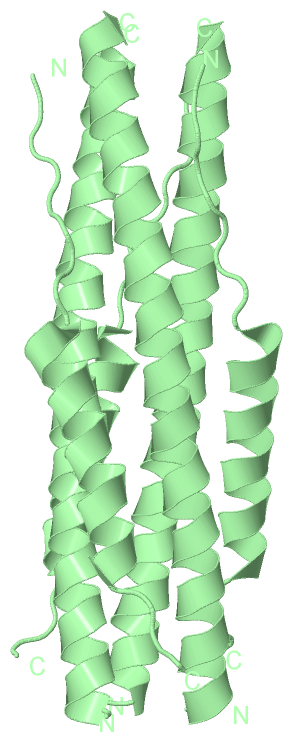
Asymmetric Unit
No.Residues
1Gln A:1254 -Ile A:1255

 Sequence-Structure Mapping

(-) SAPs(SNPs)/Variants  (0, 0)

(no "SAP(SNP)/Variant" information available for 4MOD)

(-) PROSITE Motifs  (0, 0)

(no "PROSITE Motif" information available for 4MOD)

(-) Exons   (0, 0)

(no "Exon" information available for 4MOD)

(-) Sequences/Alignments

Asymmetric Unit
   Reformat: Number of residues per line =  ('0' or empty: single-line sequence representation)
  Number of residues per labelling interval =   
  UniProt sequence: complete  aligned part    
   Show mapping: SCOP domains CATH domains Pfam domains Secondary structure (by author)
SAPs(SNPs) PROSITE motifs Exons
(details for a mapped element are shown in a popup box when the mouse pointer rests over it)
Chain A from PDB  Type:PROTEIN  Length:76
                                                                                                             
               SCOP domains ---------------------------------------------------------------------------- SCOP domains
               CATH domains ---------------------------------------------------------------------------- CATH domains
               Pfam domains ---------------------------------------------------------------------------- Pfam domains
         Sec.struct. author .hhhhhhhhhhhhhhhhhhhhhhhhhhhhhhhhhhhhhhhhhhhhhh........hhhhhhhhhhhhhhhhh.... Sec.struct. author
                 SAPs(SNPs) ---------------------------------------------------------------------------- SAPs(SNPs)
                    PROSITE ---------------------------------------------------------------------------- PROSITE
                 Transcript ---------------------------------------------------------------------------- Transcript
                4mod A  993 NQKLIANKFNQALGAMQTGFTTTNEAFQKVQDAVNNNAQALSKLASEQINTTLLDLTYEMLSLQQVVKALNESYID 1282
                                  1002      1012      1022      1032      1256      1266      1276      
                                                                       1039|                            
                                                                        1254                            

Chain B from PDB  Type:PROTEIN  Length:76
                                                                                                             
               SCOP domains ---------------------------------------------------------------------------- SCOP domains
               CATH domains ---------------------------------------------------------------------------- CATH domains
               Pfam domains ---------------------------------------------------------------------------- Pfam domains
         Sec.struct. author .hhhhhhhhhhhhhhhhhhhhhhhhhhhhhhhhhhhhhhhhhhhhhh........hhhhhhhhhhhhhhhhh.... Sec.struct. author
                 SAPs(SNPs) ---------------------------------------------------------------------------- SAPs(SNPs)
                    PROSITE ---------------------------------------------------------------------------- PROSITE
                 Transcript ---------------------------------------------------------------------------- Transcript
                4mod B  993 NQKLIANKFNQALGAMQTGFTTTNEAFQKVQDAVNNNAQALSKLASEQINTTLLDLTYEMLSLQQVVKALNESYID 1282
                                  1002      1012      1022      1032      1256      1266      1276      
                                                                       1039|                            
                                                                        1254                            

   Legend:   → Mismatch (orange background)
  - → Gap (green background, '-', border residues have a numbering label)
    → Modified Residue (blue background, lower-case, 'x' indicates undefined single-letter code, labelled with number + name)
  x → Chemical Group (purple background, 'x', labelled with number + name, e.g. ACE or NH2)
  extra numbering lines below/above indicate numbering irregularities and modified residue names etc., number ends below/above '|'

 Classification and Annotation

(-) SCOP Domains  (0, 0)

(no "SCOP Domain" information available for 4MOD)

(-) CATH Domains  (0, 0)

(no "CATH Domain" information available for 4MOD)

(-) Pfam Domains  (0, 0)

(no "Pfam Domain" information available for 4MOD)

(-) Gene Ontology  (5, 5)

Asymmetric Unit(hide GO term definitions)

 Visualization

(-) Interactive Views

Asymmetric Unit
  Complete Structure
    Jena3D(integrated viewing of ligand, site, SAP, PROSITE, SCOP information)
    WebMol | AstexViewer[tm]@PDBe
(Java Applets, require no local installation except for Java; loading may be slow)
    STRAP
(Java WebStart application, automatic local installation, requires Java; full application with system access!)
    RasMol
(require local installation)
    Molscript (VRML)
(requires installation of a VRML viewer; select preferred view via VRML and generate a mono or stereo PDF format file)
 
  Ligands, Modified Residues, Ions
(no "Ligands, Modified Residues, Ions" information available for 4mod)
 
  Sites
(no "Sites" information available for 4mod)
 
  Cis Peptide Bonds
    Gln A:1254 - Ile A:1255   [ RasMol ]  
 
Biological Units
  Complete Structure
    Biological Unit 1  [ Jena3D ]
    Biological Unit 2  [ Jena3D ]

(-) Still Images

Jmol
  protein: cartoon or spacefill or dots and stick; nucleic acid: cartoon and stick; ligands: spacefill; active site: stick
Molscript
  protein, nucleic acid: cartoon; ligands: spacefill; active site: ball and stick

 Databases and Analysis Tools

(-) Databases

Access by PDB/NDB ID
  4mod
    Family and Domain InformationProDom | SYSTERS
    General Structural InformationGlycoscienceDB | MMDB | NDB | OCA | PDB | PDBe | PDBj | PDBsum | PDBWiki | PQS | PROTEOPEDIA
    Orientation in MembranesOPM
    Protein SurfaceSURFACE
    Secondary StructureDSSP (structure derived) | HSSP (homology derived)
    Structural GenomicsGeneCensus
    Structural NeighboursCE | VAST
    Structure ClassificationCATH | Dali | SCOP
    Validation and Original DataBMRB Data View | BMRB Restraints Grid | EDS | PROCHECK | RECOORD | WHAT_CHECK
 
Access by UniProt ID/Accession number
  K0BRG7_9BETC | K0BRG7
    Comparative Protein Structure ModelsModBase
    Genomic InformationEnsembl
    Protein-protein InteractionDIP
    Sequence, Family and Domain InformationInterPro | Pfam | SMART | UniProtKB/TrEMBL
 
Access by Enzyme Classificator   (EC Number)
  (no 'Enzyme Classificator' available)
    General Enzyme InformationBRENDA | EC-PDB | Enzyme | IntEnz
    PathwayKEGG | MetaCyc
 
Access by Disease Identifier   (MIM ID)
  (no 'MIM ID' available)
    Disease InformationOMIM
 
Access by GenAge ID
  (no 'GenAge ID' available)
    Age Related InformationGenAge

(-) Analysis Tools

Access by PDB/NDB ID
    Domain InformationXDom
    Interatomic Contacts of Structural UnitsCSU
    Ligand-protein ContactsLPC
    Protein CavitiescastP
    Sequence and Secondary StructurePDBCartoon
    Structure AlignmentSTRAP(Java WebStart application, automatic local installation, requires Java; full application with system access!)
    Structure and Sequence BrowserSTING
 
Access by UniProt ID/Accession number
  K0BRG7_9BETC | K0BRG7
    Protein Disorder PredictionDisEMBL | FoldIndex | GLOBPLOT (for more information see DisProt)

 Related Entries

(-) Entries Sharing at Least One Protein Chain (UniProt ID)

UniProtKB/TrEMBL
        K0BRG7_9BETC | K0BRG74kqz 4kr0 5do2

(-) Related Entries Specified in the PDB File

1wdf STRUCTURE OF THE MHV FUSION CORE
1wnc STRUCTURE OF THE SARS-COV FUSION CORE