Show PDB file:   
         Plain Text   HTML   (compressed file size)
QuickSearch:   
by PDB,NDB,UniProt,PROSITE Code or Search Term(s)  
(-)Asym./Biol. Unit
(-)Asym./Biol. Unit - sites
collapse expand < >
Image Asym./Biol. Unit
Asym./Biol. Unit  (Jmol Viewer)
Image Asym./Biol. Unit - sites
Asym./Biol. Unit - sites  (Jmol Viewer)

(-) Description

Title :  CRYSTAL STRUCTURE OF GH18 CHITINASE (G77W/E119Q MUTANT) FROM CYCAS REVOLUTA IN COMPLEX WITH (GLCNAC)4
 
Authors :  N. Umemoto, T. Numata, T. Ohnuma, T. Osawa, T. Taira, T. Fukamizo
Date :  11 Sep 13  (Deposition) - 17 Sep 14  (Release) - 17 Sep 14  (Revision)
Method :  X-RAY DIFFRACTION
Resolution :  1.80
Chains :  Asym./Biol. Unit :  A
Keywords :  Chitinase, Hydrolase, Carbohydrate (Keyword Search: [Gene Ontology, PubMed, Web (Google))
 
Reference :  N. Umemoto, T. Numata, T. Ohnuma, T. Osawa, T. Taira, T. Fukamizo
Crystal Structure Of Gh18 Chitinase From Cycad, Cycas Revoluta
To Be Published
PubMed: search

(-) Compounds

Molecule 1 - CHITINASE A
    ChainsA
    EC Number3.2.1.14
    EngineeredYES
    Expression SystemESCHERICHIA COLI
    Expression System PlasmidPET-22B
    Expression System StrainBL21(DE3)
    Expression System Taxid562
    Expression System Vector TypePLASMID
    FragmentUNP RESIDUES 24-370
    GeneCRCHIA
    MutationYES
    Organism CommonSAGO PALM
    Organism ScientificCYCAS REVOLUTA
    Organism Taxid3396

 Structural Features

(-) Chains, Units

  1
Asymmetric/Biological Unit A

Summary Information (see also Sequences/Alignments below)
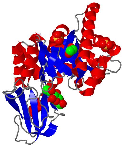
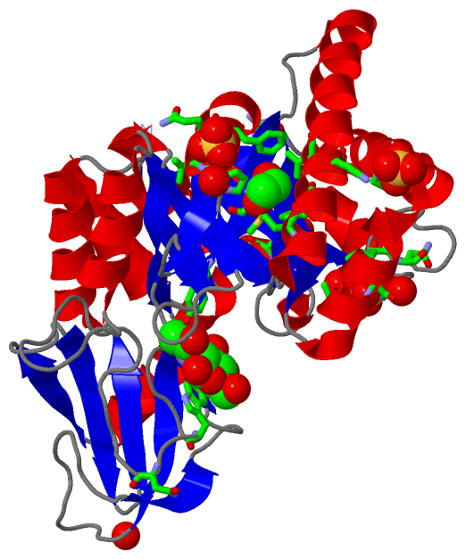
(-) Ligands, Modified Residues, Ions  (3, 6)

Asymmetric/Biological Unit (3, 6)
No.NameCountTypeFull Name
1EDO1Ligand/Ion1,2-ETHANEDIOL
2NAG1Ligand/IonN-ACETYL-D-GLUCOSAMINE
3SO44Ligand/IonSULFATE ION

(-) Sites  (6, 6)

Asymmetric Unit (6, 6)
No.NameEvidenceResiduesDescription
1AC1SOFTWAREMET A:127 , PHE A:130 , VAL A:134 , GLU A:157 , PHE A:178BINDING SITE FOR RESIDUE EDO A 401
2AC2SOFTWARETYR A:160 , ASP A:192 , TRP A:197BINDING SITE FOR RESIDUE NAG A 402
3AC3SOFTWAREPRO A:18 , PRO A:19 , SER A:20 , ASN A:199 , HOH A:570BINDING SITE FOR RESIDUE SO4 A 403
4AC4SOFTWAREGLU A:36 , LEU A:37 , ASN A:38 , VAL A:81 , ASN A:84 , HOH A:606 , HOH A:687BINDING SITE FOR RESIDUE SO4 A 404
5AC5SOFTWAREARG A:62 , HIS A:112 , PRO A:175 , VAL A:176 , GLN A:177 , PHE A:178 , HOH A:615BINDING SITE FOR RESIDUE SO4 A 405
6AC6SOFTWAREARG A:95 , LYS A:132 , HOH A:604BINDING SITE FOR RESIDUE SO4 A 406

(-) SS Bonds  (0, 0)

(no "SS Bond" information available for 4MNM)

(-) Cis Peptide Bonds  (7, 7)

Asymmetric/Biological Unit
No.Residues
1Ala A:33 -Phe A:34
2Ala A:39 -Pro A:40
3Gln A:119 -Tyr A:120
4Glu A:122 -Pro A:123
5Leu A:165 -Phe A:166
6Pro A:263 -Val A:264
7Trp A:327 -Ser A:328

 Sequence-Structure Mapping

(-) SAPs(SNPs)/Variants  (0, 0)

(no "SAP(SNP)/Variant" information available for 4MNM)

(-) PROSITE Motifs  (0, 0)

(no "PROSITE Motif" information available for 4MNM)

(-) Exons   (0, 0)

(no "Exon" information available for 4MNM)

(-) Sequences/Alignments

Asymmetric/Biological Unit
   Reformat: Number of residues per line =  ('0' or empty: single-line sequence representation)
  Number of residues per labelling interval =   
  UniProt sequence: complete  aligned part    
   Show mapping: SCOP domains CATH domains Pfam domains Secondary structure (by author)
SAPs(SNPs) PROSITE motifs Exons
(details for a mapped element are shown in a popup box when the mouse pointer rests over it)
Chain A from PDB  Type:PROTEIN  Length:344
                                                                                                                                                                                                                                                                                                                                                                                        
               SCOP domains -------------------------------------------------------------------------------------------------------------------------------------------------------------------------------------------------------------------------------------------------------------------------------------------------------------------------------------------------------- SCOP domains
               CATH domains -------------------------------------------------------------------------------------------------------------------------------------------------------------------------------------------------------------------------------------------------------------------------------------------------------------------------------------------------------- CATH domains
               Pfam domains -------------------------------------------------------------------------------------------------------------------------------------------------------------------------------------------------------------------------------------------------------------------------------------------------------------------------------------------------------- Pfam domains
         Sec.struct. author .eeeeeee.hhh...hhhhhhhhhh.eeeeeee..............hhhhhhhhhhhhhhhh...eeeeeeee.hhhhhhhhhhhhhhhhhhhhhhhhhhhhhhhhh..eeeeee....hhhhhhhhhhhhhhhhhhhhhhhhhhh....eeeeeee...ee......ee.hhhhhhhhh.eeeee.........................hhhhhhhhhhhh..hhh.eeeeee.eeeeeee...........eeee.....eeehhhhhhhhhh..eeeee....eeeeee..eeeee.hhhhhhhhhhhhhhh...eeeee.hhhh..hhhhhhhhhhhh Sec.struct. author
                 SAPs(SNPs) -------------------------------------------------------------------------------------------------------------------------------------------------------------------------------------------------------------------------------------------------------------------------------------------------------------------------------------------------------- SAPs(SNPs)
                    PROSITE -------------------------------------------------------------------------------------------------------------------------------------------------------------------------------------------------------------------------------------------------------------------------------------------------------------------------------------------------------- PROSITE
                 Transcript -------------------------------------------------------------------------------------------------------------------------------------------------------------------------------------------------------------------------------------------------------------------------------------------------------------------------------------------------------- Transcript
                 4mnm A   3 ALKVGFWPAYSVSEFPPSKINSRLFTHLYYAFAELNAPTFEVRVPPGSEKTAEDFTPTVRRLNPSVKTLISIGGWGSEVRDNFAKLNSDASARQRFVKSSIALARRYGFHGLDLDYQYPEPQLEMENFVKLVSELTAAIREEARTSGKPRLLLTEAVYFHQKLFPWEVVTEYPVQFIAAGLDWVNVMAYDFHGSWENFTGAPAALRDPNSKFTASVGIESFLAAGMPPEKLVLGIPLFGRSWLLKNNNEVGIGAPAVGAGPVDGALSFSEIQNFIRGGAREVFDTTTVSAYAYKDNVWVGYDNQQSVALKVQYAKEKRLGGYFFWSVNQDIDAILPKIASDTWG 346
                                    12        22        32        42        52        62        72        82        92       102       112       122       132       142       152       162       172       182       192       202       212       222       232       242       252       262       272       282       292       302       312       322       332       342    

   Legend:   → Mismatch (orange background)
  - → Gap (green background, '-', border residues have a numbering label)
    → Modified Residue (blue background, lower-case, 'x' indicates undefined single-letter code, labelled with number + name)
  x → Chemical Group (purple background, 'x', labelled with number + name, e.g. ACE or NH2)
  extra numbering lines below/above indicate numbering irregularities and modified residue names etc., number ends below/above '|'

 Classification and Annotation

(-) SCOP Domains  (0, 0)

(no "SCOP Domain" information available for 4MNM)

(-) CATH Domains  (0, 0)

(no "CATH Domain" information available for 4MNM)

(-) Pfam Domains  (0, 0)

(no "Pfam Domain" information available for 4MNM)

(-) Gene Ontology  (4, 4)

Asymmetric/Biological Unit(hide GO term definitions)

 Visualization

(-) Interactive Views

Asymmetric/Biological Unit
  Complete Structure
    Jena3D(integrated viewing of ligand, site, SAP, PROSITE, SCOP information)
    WebMol | AstexViewer[tm]@PDBe
(Java Applets, require no local installation except for Java; loading may be slow)
    STRAP
(Java WebStart application, automatic local installation, requires Java; full application with system access!)
    RasMol
(require local installation)
    Molscript (VRML)
(requires installation of a VRML viewer; select preferred view via VRML and generate a mono or stereo PDF format file)
 
  Ligands, Modified Residues, Ions
    EDO  [ RasMol | Jena3D ]  +environment [ RasMol | Jena3D ]
    NAG  [ RasMol | Jena3D ]  +environment [ RasMol | Jena3D ]
    SO4  [ RasMol | Jena3D ]  +environment [ RasMol | Jena3D ]
 
  Sites
    AC1  [ RasMol ]  +environment [ RasMol ]
    AC2  [ RasMol ]  +environment [ RasMol ]
    AC3  [ RasMol ]  +environment [ RasMol ]
    AC4  [ RasMol ]  +environment [ RasMol ]
    AC5  [ RasMol ]  +environment [ RasMol ]
    AC6  [ RasMol ]  +environment [ RasMol ]
 
  Cis Peptide Bonds
    Ala A:33 - Phe A:34   [ RasMol ]  
    Ala A:39 - Pro A:40   [ RasMol ]  
    Gln A:119 - Tyr A:120   [ RasMol ]  
    Glu A:122 - Pro A:123   [ RasMol ]  
    Leu A:165 - Phe A:166   [ RasMol ]  
    Pro A:263 - Val A:264   [ RasMol ]  
    Trp A:327 - Ser A:328   [ RasMol ]  
 

(-) Still Images

Jmol
  protein: cartoon or spacefill or dots and stick; nucleic acid: cartoon and stick; ligands: spacefill; active site: stick
Molscript
  protein, nucleic acid: cartoon; ligands: spacefill; active site: ball and stick

 Databases and Analysis Tools

(-) Databases

Access by PDB/NDB ID
  4mnm
    Family and Domain InformationProDom | SYSTERS
    General Structural InformationGlycoscienceDB | MMDB | NDB | OCA | PDB | PDBe | PDBj | PDBsum | PDBWiki | PQS | PROTEOPEDIA
    Orientation in MembranesOPM
    Protein SurfaceSURFACE
    Secondary StructureDSSP (structure derived) | HSSP (homology derived)
    Structural GenomicsGeneCensus
    Structural NeighboursCE | VAST
    Structure ClassificationCATH | Dali | SCOP
    Validation and Original DataBMRB Data View | BMRB Restraints Grid | EDS | PROCHECK | RECOORD | WHAT_CHECK
 
Access by UniProt ID/Accession number
  Q4W6L6_CYCRE | Q4W6L6
    Comparative Protein Structure ModelsModBase
    Genomic InformationEnsembl
    Protein-protein InteractionDIP
    Sequence, Family and Domain InformationInterPro | Pfam | SMART | UniProtKB/TrEMBL
 
Access by Enzyme Classificator   (EC Number)
  3.2.1.14
    General Enzyme InformationBRENDA | EC-PDB | Enzyme | IntEnz
    PathwayKEGG | MetaCyc
 
Access by Disease Identifier   (MIM ID)
  (no 'MIM ID' available)
    Disease InformationOMIM
 
Access by GenAge ID
  (no 'GenAge ID' available)
    Age Related InformationGenAge

(-) Analysis Tools

Access by PDB/NDB ID
    Domain InformationXDom
    Interatomic Contacts of Structural UnitsCSU
    Ligand-protein ContactsLPC
    Protein CavitiescastP
    Sequence and Secondary StructurePDBCartoon
    Structure AlignmentSTRAP(Java WebStart application, automatic local installation, requires Java; full application with system access!)
    Structure and Sequence BrowserSTING
 
Access by UniProt ID/Accession number
  Q4W6L6_CYCRE | Q4W6L6
    Protein Disorder PredictionDisEMBL | FoldIndex | GLOBPLOT (for more information see DisProt)

 Related Entries

(-) Entries Sharing at Least One Protein Chain (UniProt ID)

UniProtKB/TrEMBL
        Q4W6L6_CYCRE | Q4W6L63wij 4mnj 4mnk 4mnl 4r5e

(-) Related Entries Specified in the PDB File

4mnj 4mnk 4mnl