Show PDB file:   
         Plain Text   HTML   (compressed file size)
QuickSearch:   
by PDB,NDB,UniProt,PROSITE Code or Search Term(s)  
(-)Asym./Biol. Unit
(-)Asym./Biol. Unit - sites
collapse expand < >
Image Asym./Biol. Unit
Asym./Biol. Unit  (Jmol Viewer)
Image Asym./Biol. Unit - sites
Asym./Biol. Unit - sites  (Jmol Viewer)

(-) Description

Title :  RECOMBINANT KRINGLE IV-10/W72R MUTANT OF HUMAN APOLIPOPROTEIN(A)
 
Authors :  I. Mochalkin, A. Tulinsky, A. Scanu
Date :  26 Aug 98  (Deposition) - 18 May 99  (Release) - 24 Feb 09  (Revision)
Method :  X-RAY DIFFRACTION
Resolution :  2.20
Chains :  Asym./Biol. Unit :  A
Keywords :  Lysine Binding Site, Site Mutation (Keyword Search: [Gene Ontology, PubMed, Web (Google))
 
Reference :  I. Mochalkin, B. Cheng, O. Klezovitch, A. M. Scanu, A. Tulinsky
Recombinant Kringle Iv-10 Modules Of Human Apolipoprotein(A): Structure, Ligand Binding Modes, And Biological Relevance.
Biochemistry V. 38 1990 1999
PubMed-ID: 10026282  |  Reference-DOI: 10.1021/BI9820558
(for further references see the PDB file header)

(-) Compounds

Molecule 1 - APOLIPOPROTEIN
    ChainsA
    EngineeredYES
    Expression SystemESCHERICHIA COLI
    Expression System Taxid562
    FragmentKRINGLE IV-10
    MutationYES
    Organism CommonHUMAN
    Organism ScientificHOMO SAPIENS
    Organism Taxid9606
    Other DetailsKIV-10/W72R MUTANT OF HUMAN APOLIPOPROTEIN(A)

 Structural Features

(-) Chains, Units

  1
Asymmetric/Biological Unit A

Summary Information (see also Sequences/Alignments below)

(-) Ligands, Modified Residues, Ions  (0, 0)

(no "Ligand,Modified Residues,Ions" information available for 4KIV)
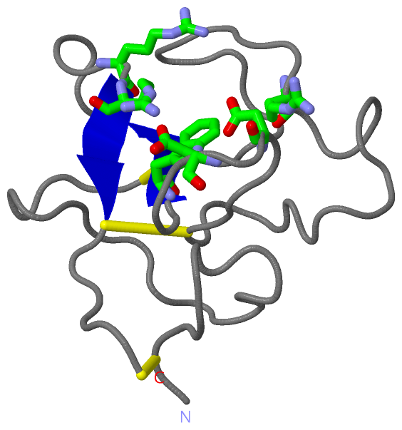
(-) Sites  (1, 1)

Asymmetric Unit (1, 1)
No.NameEvidenceResiduesDescription
1LBSUNKNOWNARG A:35 , ASP A:55 , ASP A:57 , TRP A:62 , ARG A:71 , ARG A:72W72R MUTATION OF THE LBS RESULTS IN LOSS OF THE BINDING CAPACITY OF KIV-10.

(-) SS Bonds  (3, 3)

Asymmetric/Biological Unit
No.Residues
1A:1 -A:80
2A:22 -A:63
3A:51 -A:75

(-) Cis Peptide Bonds  (1, 1)

Asymmetric/Biological Unit
No.Residues
1Thr A:29 -Pro A:30

 Sequence-Structure Mapping

(-) SAPs(SNPs)/Variants  (0, 0)

(no "SAP(SNP)/Variant" information available for 4KIV)

(-) PROSITE Motifs  (0, 0)

(no "PROSITE Motif" information available for 4KIV)

(-) Exons   (0, 0)

(no "Exon" information available for 4KIV)

(-) Sequences/Alignments

Asymmetric/Biological Unit
   Reformat: Number of residues per line =  ('0' or empty: single-line sequence representation)
  Number of residues per labelling interval =   
  UniProt sequence: complete  aligned part    
   Show mapping: SCOP domains CATH domains Pfam domains Secondary structure (by author)
SAPs(SNPs) PROSITE motifs Exons
(details for a mapped element are shown in a popup box when the mouse pointer rests over it)
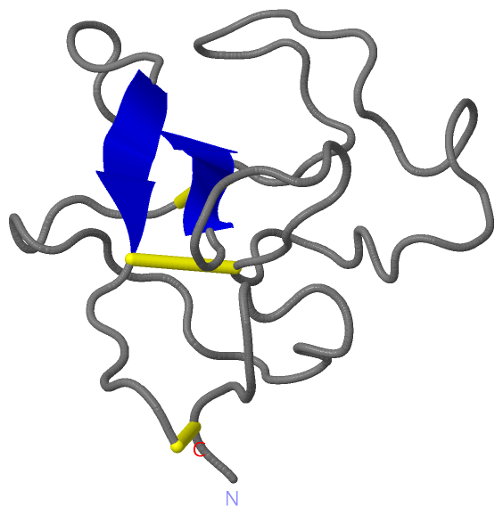
Chain A from PDB  Type:PROTEIN  Length:79
                                                                                                              
               SCOP domains d4kiva_ A: Apolipoprotein A                                                     SCOP domains
               CATH domains 4kivA00 A:0-80 Plasminogen Kringle 4                                            CATH domains
               Pfam domains ------------------------------------------------------------------------------- Pfam domains
         Sec.struct. author ............................................................eee.......eee...... Sec.struct. author
                 SAPs(SNPs) ------------------------------------------------------------------------------- SAPs(SNPs)
                    PROSITE ------------------------------------------------------------------------------- PROSITE
                 Transcript ------------------------------------------------------------------------------- Transcript
                  4kiv A  0 QCYHGNGQSYRGTFSTTVTGRTCQSWSSMTPHRHQRTPENYPNDGLTMNYCRNPDADTGPWCFTMDPSIRREYCNLTRC 80
                                     9        19        29     || 40        50       |61        71         
                                                              35|                   58|                    
                                                               37                    60                    

   Legend:   → Mismatch (orange background)
  - → Gap (green background, '-', border residues have a numbering label)
    → Modified Residue (blue background, lower-case, 'x' indicates undefined single-letter code, labelled with number + name)
  x → Chemical Group (purple background, 'x', labelled with number + name, e.g. ACE or NH2)
  extra numbering lines below/above indicate numbering irregularities and modified residue names etc., number ends below/above '|'

 Classification and Annotation

(-) SCOP Domains  (1, 1)

Asymmetric/Biological Unit

(-) CATH Domains  (1, 1)

Asymmetric/Biological Unit
(-)
Class: Mainly Beta (13760)

(-) Pfam Domains  (0, 0)

(no "Pfam Domain" information available for 4KIV)

(-) Gene Ontology  (18, 18)

Asymmetric/Biological Unit(hide GO term definitions)

 Visualization

(-) Interactive Views

Asymmetric/Biological Unit
  Complete Structure
    Jena3D(integrated viewing of ligand, site, SAP, PROSITE, SCOP information)
    WebMol | AstexViewer[tm]@PDBe
(Java Applets, require no local installation except for Java; loading may be slow)
    STRAP
(Java WebStart application, automatic local installation, requires Java; full application with system access!)
    RasMol
(require local installation)
    Molscript (VRML)
(requires installation of a VRML viewer; select preferred view via VRML and generate a mono or stereo PDF format file)
 
  Ligands, Modified Residues, Ions
(no "Ligands, Modified Residues, Ions" information available for 4kiv)
 
  Sites
    LBS  [ RasMol ]  +environment [ RasMol ]
 
  Cis Peptide Bonds
    Thr A:29 - Pro A:30   [ RasMol ]  
 

(-) Still Images

Jmol
  protein: cartoon or spacefill or dots and stick; nucleic acid: cartoon and stick; ligands: spacefill; active site: stick
Molscript
  protein, nucleic acid: cartoon; ligands: spacefill; active site: ball and stick

 Databases and Analysis Tools

(-) Databases

Access by PDB/NDB ID
  4kiv
    Family and Domain InformationProDom | SYSTERS
    General Structural InformationGlycoscienceDB | MMDB | NDB | OCA | PDB | PDBe | PDBj | PDBsum | PDBWiki | PQS | PROTEOPEDIA
    Orientation in MembranesOPM
    Protein SurfaceSURFACE
    Secondary StructureDSSP (structure derived) | HSSP (homology derived)
    Structural GenomicsGeneCensus
    Structural NeighboursCE | VAST
    Structure ClassificationCATH | Dali | SCOP
    Validation and Original DataBMRB Data View | BMRB Restraints Grid | EDS | PROCHECK | RECOORD | WHAT_CHECK
 
Access by UniProt ID/Accession number
  APOA_HUMAN | P08519
    Comparative Protein Structure ModelsModBase
    Genomic InformationEnsembl
    Protein-protein InteractionDIP
    Sequence, Family and Domain InformationInterPro | Pfam | SMART | UniProtKB/SwissProt
 
Access by Enzyme Classificator   (EC Number)
  (no 'Enzyme Classificator' available)
    General Enzyme InformationBRENDA | EC-PDB | Enzyme | IntEnz
    PathwayKEGG | MetaCyc
 
Access by Disease Identifier   (MIM ID)
  (no 'MIM ID' available)
    Disease InformationOMIM
 
Access by GenAge ID
  (no 'GenAge ID' available)
    Age Related InformationGenAge

(-) Analysis Tools

Access by PDB/NDB ID
    Domain InformationXDom
    Interatomic Contacts of Structural UnitsCSU
    Ligand-protein ContactsLPC
    Protein CavitiescastP
    Sequence and Secondary StructurePDBCartoon
    Structure AlignmentSTRAP(Java WebStart application, automatic local installation, requires Java; full application with system access!)
    Structure and Sequence BrowserSTING
 
Access by UniProt ID/Accession number
  APOA_HUMAN | P08519
    Protein Disorder PredictionDisEMBL | FoldIndex | GLOBPLOT (for more information see DisProt)

 Related Entries

(-) Entries Sharing at Least One Protein Chain (UniProt ID)

UniProtKB/Swiss-Prot
        APOA_HUMAN | P085191i71 1jfn 1kiv 2feb 3kiv 4bv5 4bv7 4bvc 4bvd 4bvv 4bvw

(-) Related Entries Specified in the PDB File

(no "Related Entries Specified in the PDB File" available for 4KIV)