Show PDB file:   
         Plain Text   HTML   (compressed file size)
QuickSearch:   
by PDB,NDB,UniProt,PROSITE Code or Search Term(s)  
(-)Asymmetric Unit
(-)Asym. Unit - sites
(-)Biological Unit 1
(-)Biol. Unit 1 - sites
(-)Biological Unit 2
collapse expand < >
Image Asymmetric Unit
Asymmetric Unit  (Jmol Viewer)
Image Asym. Unit - sites
Asym. Unit - sites  (Jmol Viewer)
Image Biological Unit 1
Biological Unit 1  (Jmol Viewer)
Image Biol. Unit 1 - sites
Biol. Unit 1 - sites  (Jmol Viewer)
Image Biological Unit 2
Biological Unit 2  (Jmol Viewer)

(-) Description

Title :  STRUCTURE OF STAPHYLOCOCCUS AUREUS MNTC
 
Authors :  K. D. Parris, L. Mosyak
Date :  11 Apr 13  (Deposition) - 17 Jul 13  (Release) - 11 Sep 13  (Revision)
Method :  X-RAY DIFFRACTION
Resolution :  2.20
Chains :  Asym. Unit :  A,B
Biol. Unit 1:  A  (1x)
Biol. Unit 2:  B  (1x)
Keywords :  Mn2+ Specific Mntabc Transporter, Transport Protein (Keyword Search: [Gene Ontology, PubMed, Web (Google))
 
Reference :  A. Gribenko, L. Mosyak, S. Ghosh, K. Parris, K. Svenson, J. Moran, L. Chu, S. Li, T. Liu, V. L. Woods, K. U. Jansen, B. A. Green, A. S. Anderson, Y. V. Matsuka
Three-Dimensional Structure And Biophysical Characterizatio Of Staphylococcus Aureus Cell Surface Antigen-Manganese Transporter Mntc.
J. Mol. Biol. V. 425 3429 2013
PubMed-ID: 23827136  |  Reference-DOI: 10.1016/J.JMB.2013.06.033

(-) Compounds

Molecule 1 - ABC SUPERFAMILY ATP BINDING CASSETTE TRANSPORTER, BINDING PROTEIN
    ChainsA, B
    EngineeredYES
    Expression SystemESCHERICHIA COLI
    Expression System Taxid562
    GeneMNTC, SAA6159_00586
    Organism ScientificSTAPHYLOCOCCUS AUREUS
    Organism Taxid869816
    StrainJKD6159

 Structural Features

(-) Chains, Units

  12
Asymmetric Unit AB
Biological Unit 1 (1x)A 
Biological Unit 2 (1x) B

Summary Information (see also Sequences/Alignments below)

(-) Ligands, Modified Residues, Ions  (1, 2)

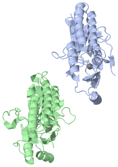
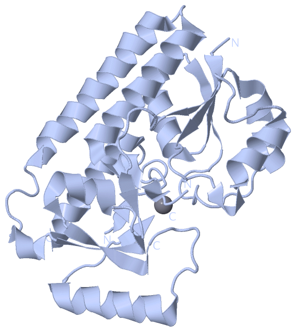
Asymmetric Unit (1, 2)
No.NameCountTypeFull Name
1MN2Ligand/IonMANGANESE (II) ION
Biological Unit 1 (0, 0)
No.NameCountTypeFull Name
1MN-1Ligand/IonMANGANESE (II) ION
Biological Unit 2 (0, 0)
No.NameCountTypeFull Name
1MN-1Ligand/IonMANGANESE (II) ION

(-) Sites  (2, 2)

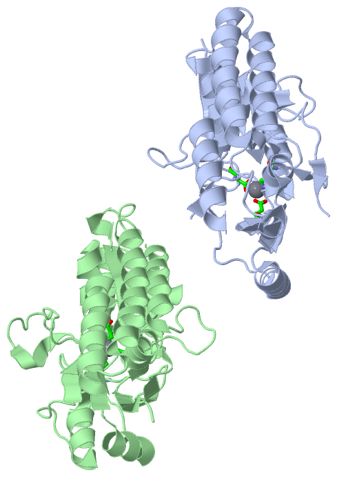
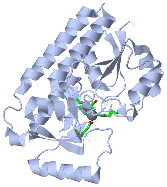
Asymmetric Unit (2, 2)
No.NameEvidenceResiduesDescription
1AC1SOFTWAREHIS A:50 , HIS A:123 , GLU A:189 , ASP A:264BINDING SITE FOR RESIDUE MN A 301
2AC2SOFTWAREHIS B:50 , HIS B:123 , GLU B:189 , ASP B:264BINDING SITE FOR RESIDUE MN B 301

(-) SS Bonds  (0, 0)

(no "SS Bond" information available for 4K3V)

(-) Cis Peptide Bonds  (0, 0)

(no "Cis Peptide Bond" information available for 4K3V)

 Sequence-Structure Mapping

(-) SAPs(SNPs)/Variants  (0, 0)

(no "SAP(SNP)/Variant" information available for 4K3V)

(-) PROSITE Motifs  (0, 0)

(no "PROSITE Motif" information available for 4K3V)

(-) Exons   (0, 0)

(no "Exon" information available for 4K3V)

(-) Sequences/Alignments

Asymmetric Unit
   Reformat: Number of residues per line =  ('0' or empty: single-line sequence representation)
  Number of residues per labelling interval =   
  UniProt sequence: complete  aligned part    
   Show mapping: SCOP domains CATH domains Pfam domains Secondary structure (by author)
SAPs(SNPs) PROSITE motifs Exons
(details for a mapped element are shown in a popup box when the mouse pointer rests over it)
Chain A from PDB  Type:PROTEIN  Length:253
                                                                                                                                                                                                                                                                                             
               SCOP domains d4k3va_ A: automated matches                                                                                                                                                                                                                                  SCOP domains
               CATH domains ------------------------------------------------------------------------------------------------------------------------------------------------------------------------------------------------------------------------------------------------------------- CATH domains
               Pfam domains ------------------------------------------------------------------------------------------------------------------------------------------------------------------------------------------------------------------------------------------------------------- Pfam domains
         Sec.struct. author ..eeeee.hhhhhhhhhhhhh..eeeee.............hhhhhhhhhhh.eeee......hhhhhhhhhhhhh........eee.......ee....ee.hhhhhhhhhhhhhhhhhhhhhhhh..hhhhhhhhhhhhhhhhhhhhhhhh......hhhh.eeeeee..hhhhhhhhh.eeeeee........hhhhhhhhhhhhhhh......ee..ee.............hhhhhhhhhhhhhhhhh Sec.struct. author
                 SAPs(SNPs) ------------------------------------------------------------------------------------------------------------------------------------------------------------------------------------------------------------------------------------------------------------- SAPs(SNPs)
                    PROSITE ------------------------------------------------------------------------------------------------------------------------------------------------------------------------------------------------------------------------------------------------------------- PROSITE
                 Transcript ------------------------------------------------------------------------------------------------------------------------------------------------------------------------------------------------------------------------------------------------------------- Transcript
                 4k3v A  14 GKLKVVTTNSILYDMAKNVGGDNVDIHSIVPVGQDPHEYEVKPKDIKKLTDADVILYNGLNLETGNGWFEKALEQAGKSLKDKKVIAVSKDVKPIYLNDKQDPHAWLSLDNGIKYVKTIQQTFIDNDKKHKADYEKQGNKYIAQLEKLNNDSKDKFNDIPKEQRAMITSEGAFKYFSKQYGITPGYIWEINTEKQGTPEQMRQAIEFVKKHKLKHLLVETGEVYTDSIGKEGTKGDSYYKMMKSNIETVHGSM 291
                                    23        33        43        53        63        73        83        93       103       119       129       139       149       159       169       179       189       199       209       219       229       239|      268       278       288   
                                                                                                                           111|                                                                                                                      239|                                
                                                                                                                            118                                                                                                                       259                                

Chain B from PDB  Type:PROTEIN  Length:278
                                                                                                                                                                                                                                                                                                                      
               SCOP domains d4k3vb_ B: automated matches                                                                                                                                                                                                                                                           SCOP domains
               CATH domains -------------------------------------------------------------------------------------------------------------------------------------------------------------------------------------------------------------------------------------------------------------------------------------- CATH domains
               Pfam domains -------------------------------------------------------------------------------------------------------------------------------------------------------------------------------------------------------------------------------------------------------------------------------------- Pfam domains
         Sec.struct. author ..eeeee.hhhhhhhhhhhhh..eeeee.............hhhhhhhhhhh.eeee......hhhhhhhhhhhhh........eee...............hhhhh..hhhhhhhhhhhhhhhhhhhhhhhh..hhhhhhhhhhhhhhhhhhhhhhhh......hhhh.eeeeee..hhhhhhhhh.eeeeee........hhhhhhhhhhhhhhh...eeeee....hhhhhhhhhhhh.eeeeee.............hhhhhhhhhhhhhhhhh Sec.struct. author
                 SAPs(SNPs) -------------------------------------------------------------------------------------------------------------------------------------------------------------------------------------------------------------------------------------------------------------------------------------- SAPs(SNPs)
                    PROSITE -------------------------------------------------------------------------------------------------------------------------------------------------------------------------------------------------------------------------------------------------------------------------------------- PROSITE
                 Transcript -------------------------------------------------------------------------------------------------------------------------------------------------------------------------------------------------------------------------------------------------------------------------------------- Transcript
                 4k3v B  14 GKLKVVTTNSILYDMAKNVGGDNVDIHSIVPVGQDPHEYEVKPKDIKKLTDADVILYNGLNLETGNGWFEKALEQAGKSLKDKKVIAVSKDVKPIYLNGEEGNKDKQDPHAWLSLDNGIKYVKTIQQTFIDNDKKHKADYEKQGNKYIAQLEKLNNDSKDKFNDIPKEQRAMITSEGAFKYFSKQYGITPGYIWEINTEKQGTPEQMRQAIEFVKKHKLKHLLVETSVDKKAMESLSEETKKDIFGEVYTDSIGKEGTKGDSYYKMMKSNIETVHGSM 291
                                    23        33        43        53        63        73        83        93       103       113       123       133       143       153       163       173       183       193       203       213       223       233       243       253       263       273       283        

   Legend:   → Mismatch (orange background)
  - → Gap (green background, '-', border residues have a numbering label)
    → Modified Residue (blue background, lower-case, 'x' indicates undefined single-letter code, labelled with number + name)
  x → Chemical Group (purple background, 'x', labelled with number + name, e.g. ACE or NH2)
  extra numbering lines below/above indicate numbering irregularities and modified residue names etc., number ends below/above '|'

 Classification and Annotation

(-) SCOP Domains  (1, 2)

Asymmetric Unit

(-) CATH Domains  (0, 0)

(no "CATH Domain" information available for 4K3V)

(-) Pfam Domains  (0, 0)

(no "Pfam Domain" information available for 4K3V)

(-) Gene Ontology  (0, 0)

Asymmetric Unit(hide GO term definitions)
    (no "Gene Ontology" information available for 4K3V)

 Visualization

(-) Interactive Views

Asymmetric Unit
  Complete Structure
    Jena3D(integrated viewing of ligand, site, SAP, PROSITE, SCOP information)
    WebMol | AstexViewer[tm]@PDBe
(Java Applets, require no local installation except for Java; loading may be slow)
    STRAP
(Java WebStart application, automatic local installation, requires Java; full application with system access!)
    RasMol
(require local installation)
    Molscript (VRML)
(requires installation of a VRML viewer; select preferred view via VRML and generate a mono or stereo PDF format file)
 
  Ligands, Modified Residues, Ions
    MN  [ RasMol | Jena3D ]  +environment [ RasMol | Jena3D ]
 
  Sites
    AC1  [ RasMol ]  +environment [ RasMol ]
    AC2  [ RasMol ]  +environment [ RasMol ]
 
  Cis Peptide Bonds
(no "Cis Peptide Bonds" information available for 4k3v)
 
Biological Units
  Complete Structure
    Biological Unit 1  [ Jena3D ]
    Biological Unit 2  [ Jena3D ]

(-) Still Images

Jmol
  protein: cartoon or spacefill or dots and stick; nucleic acid: cartoon and stick; ligands: spacefill; active site: stick
Molscript
  protein, nucleic acid: cartoon; ligands: spacefill; active site: ball and stick

 Databases and Analysis Tools

(-) Databases

Access by PDB/NDB ID
  4k3v
    Family and Domain InformationProDom | SYSTERS
    General Structural InformationGlycoscienceDB | MMDB | NDB | OCA | PDB | PDBe | PDBj | PDBsum | PDBWiki | PQS | PROTEOPEDIA
    Orientation in MembranesOPM
    Protein SurfaceSURFACE
    Secondary StructureDSSP (structure derived) | HSSP (homology derived)
    Structural GenomicsGeneCensus
    Structural NeighboursCE | VAST
    Structure ClassificationCATH | Dali | SCOP
    Validation and Original DataBMRB Data View | BMRB Restraints Grid | EDS | PROCHECK | RECOORD | WHAT_CHECK
 
Access by UniProt ID/Accession number
  (no 'UniProt ID/Accession number' available) |
    Comparative Protein Structure ModelsModBase
    Genomic InformationEnsembl
    Protein-protein InteractionDIP
    Sequence, Family and Domain InformationInterPro | Pfam | SMART | UniProt
 
Access by Enzyme Classificator   (EC Number)
  (no 'Enzyme Classificator' available)
    General Enzyme InformationBRENDA | EC-PDB | Enzyme | IntEnz
    PathwayKEGG | MetaCyc
 
Access by Disease Identifier   (MIM ID)
  (no 'MIM ID' available)
    Disease InformationOMIM
 
Access by GenAge ID
  (no 'GenAge ID' available)
    Age Related InformationGenAge

(-) Analysis Tools

Access by PDB/NDB ID
    Domain InformationXDom
    Interatomic Contacts of Structural UnitsCSU
    Ligand-protein ContactsLPC
    Protein CavitiescastP
    Sequence and Secondary StructurePDBCartoon
    Structure AlignmentSTRAP(Java WebStart application, automatic local installation, requires Java; full application with system access!)
    Structure and Sequence BrowserSTING
 
Access by UniProt ID/Accession number
  (no 'UniProt ID/Accession number' available) |
    Protein Disorder PredictionDisEMBL | FoldIndex | GLOBPLOT

 Related Entries

(-) Entries Sharing at Least One Protein Chain (UniProt ID)

(no "Entries Sharing at Least One Protein Chain" available for 4K3V)

(-) Related Entries Specified in the PDB File

(no "Related Entries Specified in the PDB File" available for 4K3V)