Show PDB file:   
         Plain Text   HTML   (compressed file size)
QuickSearch:   
by PDB,NDB,UniProt,PROSITE Code or Search Term(s)  
(-)Asymmetric Unit
(-)Asym. Unit - sites
(-)Biological Unit 1
(-)Biol. Unit 1 - sites
(-)Biological Unit 2
(-)Biological Unit 3
collapse expand < >
Image Asymmetric Unit
Asymmetric Unit  (Jmol Viewer)
Image Asym. Unit - sites
Asym. Unit - sites  (Jmol Viewer)
Image Biological Unit 1
Biological Unit 1  (Jmol Viewer)
Image Biol. Unit 1 - sites
Biol. Unit 1 - sites  (Jmol Viewer)
Image Biological Unit 2
Biological Unit 2  (Jmol Viewer)
Image Biological Unit 3
Biological Unit 3  (Jmol Viewer)

(-) Description

Title :  CRYSTAL STRUCTURE OF MAMP
 
Authors :  M. Siponen, D. Pignol, P. Arnoux
Date :  07 Mar 13  (Deposition) - 09 Oct 13  (Release) - 13 Nov 13  (Revision)
Method :  X-RAY DIFFRACTION
Resolution :  1.80
Chains :  Asym. Unit :  A,B
Biol. Unit 1:  A  (2x)
Biol. Unit 2:  B  (2x)
Biol. Unit 3:  A,B  (2x)
Keywords :  Pdz, C-Type Heme, Cytochrome, Electron Transport (Keyword Search: [Gene Ontology, PubMed, Web (Google))
 
Reference :  M. I. Siponen, P. Legrand, M. Widdrat, S. R. Jones, W. J. Zhang, M. C. Chang, D. Faivre, P. Arnoux, D. Pignol
Structural Insight Into Magnetochrome-Mediated Magnetite Biomineralization.
Nature V. 502 681 2013
PubMed-ID: 24097349  |  Reference-DOI: 10.1038/NATURE12573

(-) Compounds

Molecule 1 - MAMP
    ChainsA, B
    EngineeredYES
    Expression SystemESCHERICHIA COLI
    Expression System Taxid562
    FragmentUNP RESIDUES 26-268
    Organism ScientificMAGNETOCOCCUS
    Organism Taxid156889

 Structural Features

(-) Chains, Units

  12
Asymmetric Unit AB
Biological Unit 1 (2x)A 
Biological Unit 2 (2x) B
Biological Unit 3 (2x)AB

Summary Information (see also Sequences/Alignments below)
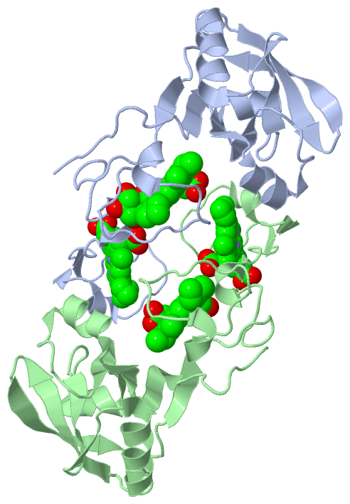
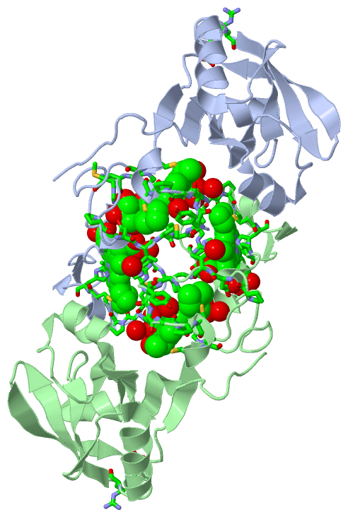
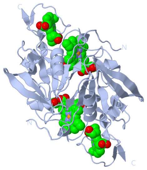
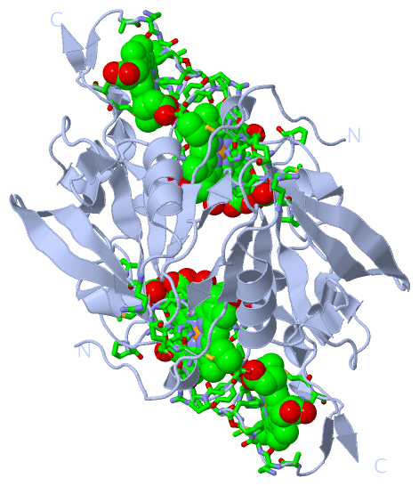
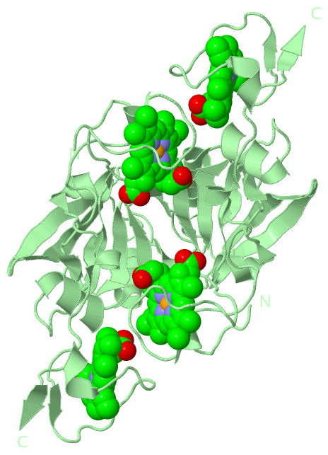
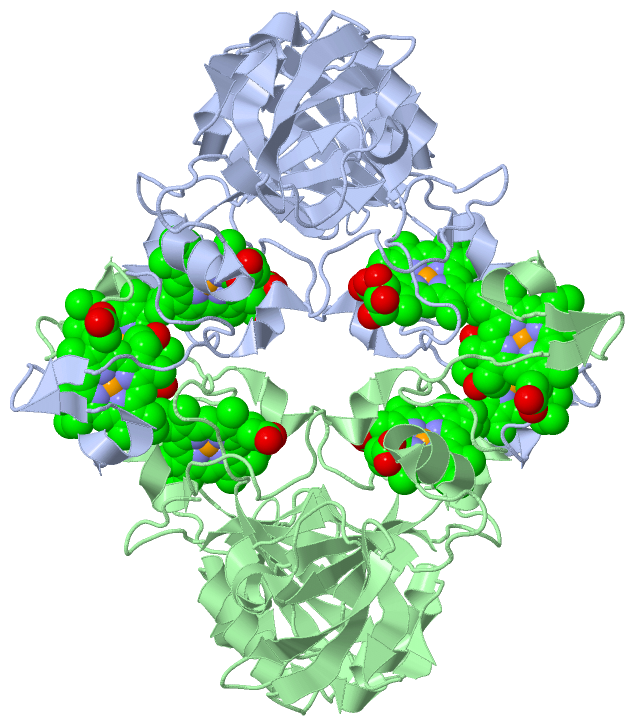
(-) Ligands, Modified Residues, Ions  (2, 5)

Asymmetric Unit (2, 5)
No.NameCountTypeFull Name
1GOL1Ligand/IonGLYCEROL
2HEC4Ligand/IonHEME C
Biological Unit 1 (2, 6)
No.NameCountTypeFull Name
1GOL2Ligand/IonGLYCEROL
2HEC4Ligand/IonHEME C
Biological Unit 2 (1, 4)
No.NameCountTypeFull Name
1GOL-1Ligand/IonGLYCEROL
2HEC4Ligand/IonHEME C
Biological Unit 3 (2, 10)
No.NameCountTypeFull Name
1GOL2Ligand/IonGLYCEROL
2HEC8Ligand/IonHEME C

(-) Sites  (5, 5)

Asymmetric Unit (5, 5)
No.NameEvidenceResiduesDescription
1AC1SOFTWAREARG A:164 , GLU A:184 , TYR A:208 , ALA A:242 , LYS A:243 , MET A:244 , PRO A:245 , HIS A:246 , ARG A:249 , GLY A:250 , PRO A:251 , CYS A:252 , CYS A:255 , HIS A:256 , HOH A:738 , PRO B:200 , HOH B:693BINDING SITE FOR RESIDUE HEC A 501
2AC2SOFTWAREPRO A:196 , ASP A:202 , ILE A:203 , MET A:204 , PRO A:205 , HIS A:206 , ARG A:209 , GLY A:210 , PRO A:211 , CYS A:212 , CYS A:215 , HIS A:216 , GOL A:503 , HOH A:604 , HOH A:658 , HOH A:707 , HOH A:741 , ARG B:249BINDING SITE FOR RESIDUE HEC A 502
3AC3SOFTWAREMET A:197 , ASP A:202 , HIS A:223 , HEC A:502BINDING SITE FOR RESIDUE GOL A 503
4AC4SOFTWAREPRO A:200 , ARG A:247 , HOH A:642 , ARG B:164 , GLU B:184 , TYR B:208 , PRO B:235 , ALA B:242 , LYS B:243 , PRO B:245 , HIS B:246 , ARG B:249 , GLY B:250 , PRO B:251 , CYS B:252 , CYS B:255 , HIS B:256 , HOH B:652BINDING SITE FOR RESIDUE HEC B 501
5AC5SOFTWAREPRO B:196 , ASP B:202 , ILE B:203 , PRO B:205 , HIS B:206 , ARG B:209 , GLY B:210 , PRO B:211 , CYS B:212 , CYS B:215 , HIS B:216 , HOH B:633 , HOH B:643 , HOH B:666 , HOH B:684 , HOH B:734 , HOH B:760 , HOH B:788BINDING SITE FOR RESIDUE HEC B 502

(-) SS Bonds  (0, 0)

(no "SS Bond" information available for 4JJ0)

(-) Cis Peptide Bonds  (2, 2)

Asymmetric Unit
No.Residues
1Thr A:225 -Pro A:226
2Thr B:225 -Pro B:226

 Sequence-Structure Mapping

(-) SAPs(SNPs)/Variants  (0, 0)

(no "SAP(SNP)/Variant" information available for 4JJ0)

(-) PROSITE Motifs  (0, 0)

(no "PROSITE Motif" information available for 4JJ0)

(-) Exons   (0, 0)

(no "Exon" information available for 4JJ0)

(-) Sequences/Alignments

Asymmetric Unit
   Reformat: Number of residues per line =  ('0' or empty: single-line sequence representation)
  Number of residues per labelling interval =   
  UniProt sequence: complete  aligned part    
   Show mapping: SCOP domains CATH domains Pfam domains Secondary structure (by author)
SAPs(SNPs) PROSITE motifs Exons
(details for a mapped element are shown in a popup box when the mouse pointer rests over it)
Chain A from PDB  Type:PROTEIN  Length:181
                                                                                                                                                                                                                     
               SCOP domains ------------------------------------------------------------------------------------------------------------------------------------------------------------------------------------- SCOP domains
               CATH domains ------------------------------------------------------------------------------------------------------------------------------------------------------------------------------------- CATH domains
               Pfam domains ------------------------------------------------------------------------------------------------------------------------------------------------------------------------------------- Pfam domains
         Sec.struct. author ...........eeee..eeeee.hhhhhhhhh.......eeeeeehhhhhhh......eeeee..ee..hhhhhhhhhhhh....eeeeeeee..eeeeeeee........eee.................hhhhh...........hhhhh......ee...........hhhhh..ee. Sec.struct. author
                 SAPs(SNPs) ------------------------------------------------------------------------------------------------------------------------------------------------------------------------------------- SAPs(SNPs)
                    PROSITE ------------------------------------------------------------------------------------------------------------------------------------------------------------------------------------- PROSITE
                 Transcript ------------------------------------------------------------------------------------------------------------------------------------------------------------------------------------- Transcript
                 4jj0 A  80 GGFVAPNVQFSEAHWQGMEALPLSIELKRKLKLPLDLEGLLIDETSLNAAVSGLLAGDVLVAINGRKVKTLKKMQKETRRVQMDRRASLTVYRKGRLLTLTLSEEKNLGLAQVETAPMILPGDIMPHPYRGPCTQCHAIGTTGHITPDPDGIVLPPGPIRAGAKMPHRDRGPCAACHAIIQ 260
                                    89        99       109       119       129       139       149       159       169       179       189       199       209       219       229       239       249       259 

Chain B from PDB  Type:PROTEIN  Length:180
                                                                                                                                                                                                                    
               SCOP domains ------------------------------------------------------------------------------------------------------------------------------------------------------------------------------------ SCOP domains
               CATH domains ------------------------------------------------------------------------------------------------------------------------------------------------------------------------------------ CATH domains
               Pfam domains ------------------------------------------------------------------------------------------------------------------------------------------------------------------------------------ Pfam domains
         Sec.struct. author ........eeeeee..eeeee.hhhhhhhhh.......eeeee.hhhhhhhh.....eeeee..ee..hhhhhhhhhhhh....eeeeeeee..eeeeeeee........eeee................hhhhh...........hhhhh......ee...........hhhhh..ee. Sec.struct. author
                 SAPs(SNPs) ------------------------------------------------------------------------------------------------------------------------------------------------------------------------------------ SAPs(SNPs)
                    PROSITE ------------------------------------------------------------------------------------------------------------------------------------------------------------------------------------ PROSITE
                 Transcript ------------------------------------------------------------------------------------------------------------------------------------------------------------------------------------ Transcript
                 4jj0 B  81 GFVAPNVQFSEAHWQGMEALPLSIELKRKLKLPLDLEGLLIDETSLNAAVSGLLAGDVLVAINGRKVKTLKKMQKETRRVQMDRRASLTVYRKGRLLTLTLSEEKNLGLAQVETAPMILPGDIMPHPYRGPCTQCHAIGTTGHITPDPDGIVLPPGPIRAGAKMPHRDRGPCAACHAIIQ 260
                                    90       100       110       120       130       140       150       160       170       180       190       200       210       220       230       240       250       260

   Legend:   → Mismatch (orange background)
  - → Gap (green background, '-', border residues have a numbering label)
    → Modified Residue (blue background, lower-case, 'x' indicates undefined single-letter code, labelled with number + name)
  x → Chemical Group (purple background, 'x', labelled with number + name, e.g. ACE or NH2)
  extra numbering lines below/above indicate numbering irregularities and modified residue names etc., number ends below/above '|'

 Classification and Annotation

(-) SCOP Domains  (0, 0)

(no "SCOP Domain" information available for 4JJ0)

(-) CATH Domains  (0, 0)

(no "CATH Domain" information available for 4JJ0)

(-) Pfam Domains  (0, 0)

(no "Pfam Domain" information available for 4JJ0)

(-) Gene Ontology  (0, 0)

Asymmetric Unit(hide GO term definitions)
    (no "Gene Ontology" information available for 4JJ0)

 Visualization

(-) Interactive Views

Asymmetric Unit
  Complete Structure
    Jena3D(integrated viewing of ligand, site, SAP, PROSITE, SCOP information)
    WebMol | AstexViewer[tm]@PDBe
(Java Applets, require no local installation except for Java; loading may be slow)
    STRAP
(Java WebStart application, automatic local installation, requires Java; full application with system access!)
    RasMol
(require local installation)
    Molscript (VRML)
(requires installation of a VRML viewer; select preferred view via VRML and generate a mono or stereo PDF format file)
 
  Ligands, Modified Residues, Ions
    GOL  [ RasMol | Jena3D ]  +environment [ RasMol | Jena3D ]
    HEC  [ RasMol | Jena3D ]  +environment [ RasMol | Jena3D ]
 
  Sites
    AC1  [ RasMol ]  +environment [ RasMol ]
    AC2  [ RasMol ]  +environment [ RasMol ]
    AC3  [ RasMol ]  +environment [ RasMol ]
    AC4  [ RasMol ]  +environment [ RasMol ]
    AC5  [ RasMol ]  +environment [ RasMol ]
 
  Cis Peptide Bonds
    Thr A:225 - Pro A:226   [ RasMol ]  
    Thr B:225 - Pro B:226   [ RasMol ]  
 
Biological Units
  Complete Structure
    Biological Unit 1  [ Jena3D ]
    Biological Unit 2  [ Jena3D ]
    Biological Unit 3  [ Jena3D ]

(-) Still Images

Jmol
  protein: cartoon or spacefill or dots and stick; nucleic acid: cartoon and stick; ligands: spacefill; active site: stick
Molscript
  protein, nucleic acid: cartoon; ligands: spacefill; active site: ball and stick

 Databases and Analysis Tools

(-) Databases

Access by PDB/NDB ID
  4jj0
    Family and Domain InformationProDom | SYSTERS
    General Structural InformationGlycoscienceDB | MMDB | NDB | OCA | PDB | PDBe | PDBj | PDBsum | PDBWiki | PQS | PROTEOPEDIA
    Orientation in MembranesOPM
    Protein SurfaceSURFACE
    Secondary StructureDSSP (structure derived) | HSSP (homology derived)
    Structural GenomicsGeneCensus
    Structural NeighboursCE | VAST
    Structure ClassificationCATH | Dali | SCOP
    Validation and Original DataBMRB Data View | BMRB Restraints Grid | EDS | PROCHECK | RECOORD | WHAT_CHECK
 
Access by UniProt ID/Accession number
  A0L9W2_MAGMM | A0L9W2
    Comparative Protein Structure ModelsModBase
    Genomic InformationEnsembl
    Protein-protein InteractionDIP
    Sequence, Family and Domain InformationInterPro | Pfam | SMART | UniProtKB/TrEMBL
 
Access by Enzyme Classificator   (EC Number)
  (no 'Enzyme Classificator' available)
    General Enzyme InformationBRENDA | EC-PDB | Enzyme | IntEnz
    PathwayKEGG | MetaCyc
 
Access by Disease Identifier   (MIM ID)
  (no 'MIM ID' available)
    Disease InformationOMIM
 
Access by GenAge ID
  (no 'GenAge ID' available)
    Age Related InformationGenAge

(-) Analysis Tools

Access by PDB/NDB ID
    Domain InformationXDom
    Interatomic Contacts of Structural UnitsCSU
    Ligand-protein ContactsLPC
    Protein CavitiescastP
    Sequence and Secondary StructurePDBCartoon
    Structure AlignmentSTRAP(Java WebStart application, automatic local installation, requires Java; full application with system access!)
    Structure and Sequence BrowserSTING
 
Access by UniProt ID/Accession number
  A0L9W2_MAGMM | A0L9W2
    Protein Disorder PredictionDisEMBL | FoldIndex | GLOBPLOT (for more information see DisProt)

 Related Entries

(-) Entries Sharing at Least One Protein Chain (UniProt ID)

UniProtKB/TrEMBL
        A0L9W2_MAGMM | A0L9W24jj3

(-) Related Entries Specified in the PDB File

4jj3