Show PDB file:   
         Plain Text   HTML   (compressed file size)
QuickSearch:   
by PDB,NDB,UniProt,PROSITE Code or Search Term(s)  
(-)Asymmetric Unit
(-)Asym. Unit - sites
(-)Biological Unit 1
(-)Biol. Unit 1 - sites
(-)Biological Unit 2
(-)Biological Unit 3
(-)Biological Unit 4
collapse expand < >
Image Asymmetric Unit
Asymmetric Unit  (Jmol Viewer)
Image Asym. Unit - sites
Asym. Unit - sites  (Jmol Viewer)
Image Biological Unit 1
Biological Unit 1  (Jmol Viewer)
Image Biol. Unit 1 - sites
Biol. Unit 1 - sites  (Jmol Viewer)
Image Biological Unit 2
Biological Unit 2  (Jmol Viewer)
Image Biological Unit 3
Biological Unit 3  (Jmol Viewer)
Image Biological Unit 4
Biological Unit 4  (Jmol Viewer)

(-) Description

Title :  CRYSTAL STRUCTURE OF CALCIUM2+-FREE WILD-TYPE CD23 LECTIN DOMAIN (CRYSTAL FORM A)
 
Authors :  B. Dhaliwal, N. Yahya, B. J. Sutton
Date :  11 Feb 13  (Deposition) - 28 Aug 13  (Release) - 28 Aug 13  (Revision)
Method :  X-RAY DIFFRACTION
Resolution :  1.90
Chains :  Asym. Unit :  A,B,C,D
Biol. Unit 1:  A  (1x)
Biol. Unit 2:  B  (1x)
Biol. Unit 3:  C  (1x)
Biol. Unit 4:  D  (1x)
Keywords :  Immunoglobulin Fold Lectin, Antibody Receptor, Immune System (Keyword Search: [Gene Ontology, PubMed, Web (Google))
 
Reference :  B. Dhaliwal, M. O. Pang, D. Yuan, N. Yahya, S. M. Fabiane, J. M. Mcdonnell, H. J. Gould, A. J. Beavil, B. J. Sutton
Conformational Plasticity At The Ige-Binding Site Of The B-Cell Receptor Cd23.
Mol. Immunol. V. 56 693 2013
PubMed-ID: 23933509  |  Reference-DOI: 10.1016/J.MOLIMM.2013.07.005

(-) Compounds

Molecule 1 - LOW AFFINITY IMMUNOGLOBULIN EPSILON FC RECEPTOR
    ChainsA, B, C, D
    EngineeredYES
    Expression SystemESCHERICHIA COLI
    Expression System Taxid562
    FragmentSOLUBLE HEAD DOMAIN OF THE B-CELL RECEPTOR CD23 (UNP RESIDUES 156-298)
    GeneCD23A, CLEC4J, FCE2, FCER2, IGEBF
    Organism CommonHUMAN
    Organism ScientificHOMO SAPIENS
    Organism Taxid9606
    SynonymBLAST-2, C-TYPE LECTIN DOMAIN FAMILY 4 MEMBER J, FC-EPSILON- RII, IMMUNOGLOBULIN E-BINDING FACTOR, LYMPHOCYTE IGE RECEPTOR, LOW AFFINITY IMMUNOGLOBULIN EPSILON FC RECEPTOR MEMBRANE-BOUND FORM, LOW AFFINITY IMMUNOGLOBULIN EPSILON FC RECEPTOR SOLUBLE FORM

 Structural Features

(-) Chains, Units

  1234
Asymmetric Unit ABCD
Biological Unit 1 (1x)A   
Biological Unit 2 (1x) B  
Biological Unit 3 (1x)  C 
Biological Unit 4 (1x)   D

Summary Information (see also Sequences/Alignments below)

(-) Ligands, Modified Residues, Ions  (1, 2)

Asymmetric Unit (1, 2)
No.NameCountTypeFull Name
1SO42Ligand/IonSULFATE ION
Biological Unit 1 (0, 0)
No.NameCountTypeFull Name
1SO4-1Ligand/IonSULFATE ION
Biological Unit 2 (0, 0)
No.NameCountTypeFull Name
1SO4-1Ligand/IonSULFATE ION
Biological Unit 3 (1, 1)
No.NameCountTypeFull Name
1SO41Ligand/IonSULFATE ION
Biological Unit 4 (1, 1)
No.NameCountTypeFull Name
1SO41Ligand/IonSULFATE ION

(-) Sites  (2, 2)

Asymmetric Unit (2, 2)
No.NameEvidenceResiduesDescription
1AC1SOFTWAREARG A:263 , SER A:265 , ARG A:267 , HOH A:307 , ARG C:253 , SER C:254 , GLN C:255 , LYS D:229BINDING SITE FOR RESIDUE SO4 C 301
2AC2SOFTWAREARG B:263 , SER B:265 , ARG B:267 , ARG D:253 , SER D:254 , GLN D:255BINDING SITE FOR RESIDUE SO4 D 301

(-) SS Bonds  (14, 14)

Asymmetric Unit
No.Residues
1A:160 -A:288
2A:163 -A:174
3A:191 -A:282
4A:259 -A:273
5B:160 -B:288
6B:163 -B:174
7B:191 -B:282
8B:259 -B:273
9C:191 -C:282
10C:259 -C:273
11D:160 -D:288
12D:163 -D:174
13D:191 -D:282
14D:259 -D:273

(-) Cis Peptide Bonds  (5, 5)

Asymmetric Unit
No.Residues
1Glu A:249 -Pro A:250
2Glu B:249 -Pro B:250
3Glu C:249 -Pro C:250
4Ser C:254 -Gln C:255
5Glu D:249 -Pro D:250

 Sequence-Structure Mapping

(-) SAPs(SNPs)/Variants  (0, 0)

(no "SAP(SNP)/Variant" information available for 4J6J)

(-) PROSITE Motifs  (0, 0)

(no "PROSITE Motif" information available for 4J6J)

(-) Exons   (0, 0)

(no "Exon" information available for 4J6J)

(-) Sequences/Alignments

Asymmetric Unit
   Reformat: Number of residues per line =  ('0' or empty: single-line sequence representation)
  Number of residues per labelling interval =   
  UniProt sequence: complete  aligned part    
   Show mapping: SCOP domains CATH domains Pfam domains Secondary structure (by author)
SAPs(SNPs) PROSITE motifs Exons
(details for a mapped element are shown in a popup box when the mouse pointer rests over it)
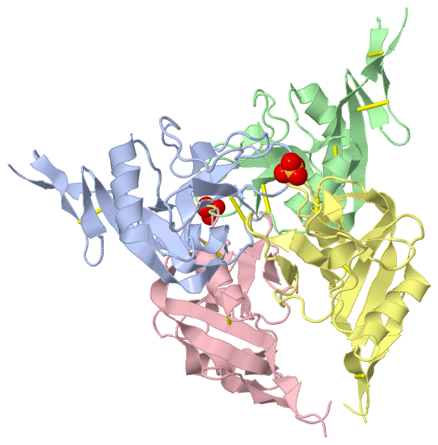
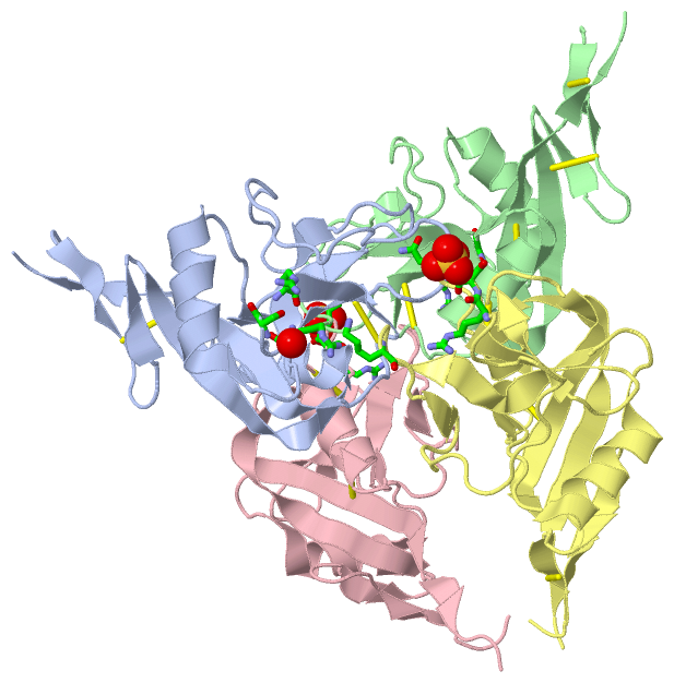
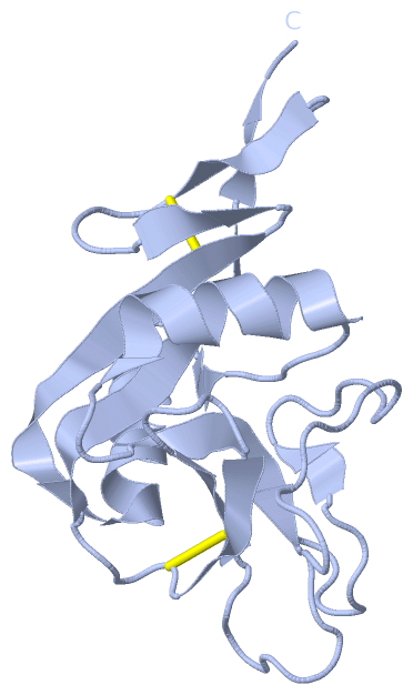
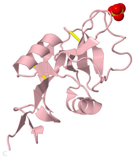
Chain A from PDB  Type:PROTEIN  Length:135
                                                                                                                                                                       
               SCOP domains d4j6ja_ A: Low affinity immunoglobulin epsilon Fc receptor                                                                              SCOP domains
               CATH domains --------------------------------------------------------------------------------------------------------------------------------------- CATH domains
               Pfam domains --------------------------------------------------------------------------------------------------------------------------------------- Pfam domains
         Sec.struct. author ..eeee.....eee..eeeeeeeeeehhhhhhhhhhh...ee....hhhhhhhhhhhh....eeeeee........ee........................eeee.....eeee.....eeeeeeeee.eee.. Sec.struct. author
                 SAPs(SNPs) --------------------------------------------------------------------------------------------------------------------------------------- SAPs(SNPs)
                    PROSITE --------------------------------------------------------------------------------------------------------------------------------------- PROSITE
                 Transcript --------------------------------------------------------------------------------------------------------------------------------------- Transcript
                 4j6j A 157 GFVCNTCPEKWINFQRKCYYFGKGTKQWVHARYACDDMEGQLVSIHSPEEQDFLTKHASHTGSWIGLRNLDLKGEFIWVDGSHVDYSNWAPGEPTSRSQGEDCVMMRGSGRWNDAFCDRKLGAWVCDRLATCTPP 291
                                   166       176       186       196       206       216       226       236       246       256       266       276       286     

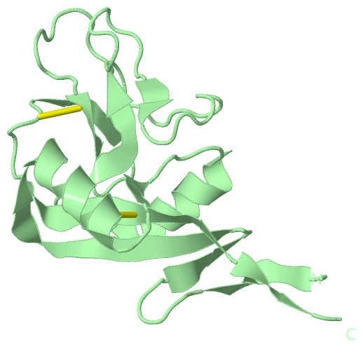
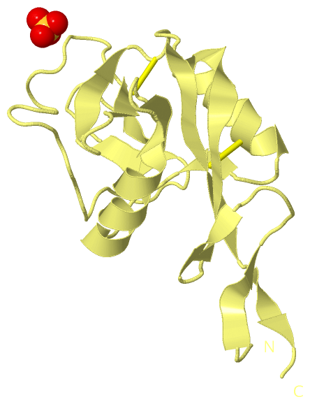
Chain B from PDB  Type:PROTEIN  Length:135
                                                                                                                                                                       
               SCOP domains d4j6jb_ B: Low affinity immunoglobulin epsilon Fc receptor                                                                              SCOP domains
               CATH domains --------------------------------------------------------------------------------------------------------------------------------------- CATH domains
               Pfam domains --------------------------------------------------------------------------------------------------------------------------------------- Pfam domains
         Sec.struct. author ..eeee.....eee..eeeeeeeeeehhhhhhhhhhh...ee....hhhhhhhhhhhh....eeeeee........ee........................eeee.....eeee.....eeeeeeeee.eee.. Sec.struct. author
                 SAPs(SNPs) --------------------------------------------------------------------------------------------------------------------------------------- SAPs(SNPs)
                    PROSITE --------------------------------------------------------------------------------------------------------------------------------------- PROSITE
                 Transcript --------------------------------------------------------------------------------------------------------------------------------------- Transcript
                 4j6j B 157 GFVCNTCPEKWINFQRKCYYFGKGTKQWVHARYACDDMEGQLVSIHSPEEQDFLTKHASHTGSWIGLRNLDLKGEFIWVDGSHVDYSNWAPGEPTSRSQGEDCVMMRGSGRWNDAFCDRKLGAWVCDRLATCTPP 291
                                   166       176       186       196       206       216       226       236       246       256       266       276       286     

Chain C from PDB  Type:PROTEIN  Length:135
                                                                                                                                                                       
               SCOP domains d4j6jc_ C: Low affinity immunoglobulin epsilon Fc receptor                                                                              SCOP domains
               CATH domains --------------------------------------------------------------------------------------------------------------------------------------- CATH domains
               Pfam domains --------------------------------------------------------------------------------------------------------------------------------------- Pfam domains
         Sec.struct. author ...eee.....eee..eeeeeeeeeehhhhhhhhhhh...ee....hhhhhhhhhhhh....eeeeeee......eee........................eeee.....eeee.....eeeeeeeee.ee... Sec.struct. author
                 SAPs(SNPs) --------------------------------------------------------------------------------------------------------------------------------------- SAPs(SNPs)
                    PROSITE --------------------------------------------------------------------------------------------------------------------------------------- PROSITE
                 Transcript --------------------------------------------------------------------------------------------------------------------------------------- Transcript
                 4j6j C 157 GFVCNTCPEKWINFQRKCYYFGKGTKQWVHARYACDDMEGQLVSIHSPEEQDFLTKHASHTGSWIGLRNLDLKGEFIWVDGSHVDYSNWAPGEPTSRSQGEDCVMMRGSGRWNDAFCDRKLGAWVCDRLATCTPP 291
                                   166       176       186       196       206       216       226       236       246       256       266       276       286     

Chain D from PDB  Type:PROTEIN  Length:135
                                                                                                                                                                       
               SCOP domains d4j6jd_ D: Low affinity immunoglobulin epsilon Fc receptor                                                                              SCOP domains
               CATH domains --------------------------------------------------------------------------------------------------------------------------------------- CATH domains
               Pfam domains --------------------------------------------------------------------------------------------------------------------------------------- Pfam domains
         Sec.struct. author ..eeee.....eee..eeeeeeeeeehhhhhhhhhhh...ee....hhhhhhhhhhhh....eeeeeee......eee........................eeee.....eeee.....eeeeeeeee.eee.. Sec.struct. author
                 SAPs(SNPs) --------------------------------------------------------------------------------------------------------------------------------------- SAPs(SNPs)
                    PROSITE --------------------------------------------------------------------------------------------------------------------------------------- PROSITE
                 Transcript --------------------------------------------------------------------------------------------------------------------------------------- Transcript
                 4j6j D 157 GFVCNTCPEKWINFQRKCYYFGKGTKQWVHARYACDDMEGQLVSIHSPEEQDFLTKHASHTGSWIGLRNLDLKGEFIWVDGSHVDYSNWAPGEPTSRSQGEDCVMMRGSGRWNDAFCDRKLGAWVCDRLATCTPP 291
                                   166       176       186       196       206       216       226       236       246       256       266       276       286     

   Legend:   → Mismatch (orange background)
  - → Gap (green background, '-', border residues have a numbering label)
    → Modified Residue (blue background, lower-case, 'x' indicates undefined single-letter code, labelled with number + name)
  x → Chemical Group (purple background, 'x', labelled with number + name, e.g. ACE or NH2)
  extra numbering lines below/above indicate numbering irregularities and modified residue names etc., number ends below/above '|'

 Classification and Annotation

(-) SCOP Domains  (1, 4)

Asymmetric Unit

(-) CATH Domains  (0, 0)

(no "CATH Domain" information available for 4J6J)

(-) Pfam Domains  (0, 0)

(no "Pfam Domain" information available for 4J6J)

(-) Gene Ontology  (17, 17)

Asymmetric Unit(hide GO term definitions)

 Visualization

(-) Interactive Views

Asymmetric Unit
  Complete Structure
    Jena3D(integrated viewing of ligand, site, SAP, PROSITE, SCOP information)
    WebMol | AstexViewer[tm]@PDBe
(Java Applets, require no local installation except for Java; loading may be slow)
    STRAP
(Java WebStart application, automatic local installation, requires Java; full application with system access!)
    RasMol
(require local installation)
    Molscript (VRML)
(requires installation of a VRML viewer; select preferred view via VRML and generate a mono or stereo PDF format file)
 
  Ligands, Modified Residues, Ions
    SO4  [ RasMol | Jena3D ]  +environment [ RasMol | Jena3D ]
 
  Sites
    AC1  [ RasMol ]  +environment [ RasMol ]
    AC2  [ RasMol ]  +environment [ RasMol ]
 
  Cis Peptide Bonds
    Glu A:249 - Pro A:250   [ RasMol ]  
    Glu B:249 - Pro B:250   [ RasMol ]  
    Glu C:249 - Pro C:250   [ RasMol ]  
    Glu D:249 - Pro D:250   [ RasMol ]  
    Ser C:254 - Gln C:255   [ RasMol ]  
 
Biological Units
  Complete Structure
    Biological Unit 1  [ Jena3D ]
    Biological Unit 2  [ Jena3D ]
    Biological Unit 3  [ Jena3D ]
    Biological Unit 4  [ Jena3D ]

(-) Still Images

Jmol
  protein: cartoon or spacefill or dots and stick; nucleic acid: cartoon and stick; ligands: spacefill; active site: stick
Molscript
  protein, nucleic acid: cartoon; ligands: spacefill; active site: ball and stick

 Databases and Analysis Tools

(-) Databases

Access by PDB/NDB ID
  4j6j
    Family and Domain InformationProDom | SYSTERS
    General Structural InformationGlycoscienceDB | MMDB | NDB | OCA | PDB | PDBe | PDBj | PDBsum | PDBWiki | PQS | PROTEOPEDIA
    Orientation in MembranesOPM
    Protein SurfaceSURFACE
    Secondary StructureDSSP (structure derived) | HSSP (homology derived)
    Structural GenomicsGeneCensus
    Structural NeighboursCE | VAST
    Structure ClassificationCATH | Dali | SCOP
    Validation and Original DataBMRB Data View | BMRB Restraints Grid | EDS | PROCHECK | RECOORD | WHAT_CHECK
 
Access by UniProt ID/Accession number
  FCER2_HUMAN | P06734
    Comparative Protein Structure ModelsModBase
    Genomic InformationEnsembl
    Protein-protein InteractionDIP
    Sequence, Family and Domain InformationInterPro | Pfam | SMART | UniProtKB/SwissProt
 
Access by Enzyme Classificator   (EC Number)
  (no 'Enzyme Classificator' available)
    General Enzyme InformationBRENDA | EC-PDB | Enzyme | IntEnz
    PathwayKEGG | MetaCyc
 
Access by Disease Identifier   (MIM ID)
  (no 'MIM ID' available)
    Disease InformationOMIM
 
Access by GenAge ID
  (no 'GenAge ID' available)
    Age Related InformationGenAge

(-) Analysis Tools

Access by PDB/NDB ID
    Domain InformationXDom
    Interatomic Contacts of Structural UnitsCSU
    Ligand-protein ContactsLPC
    Protein CavitiescastP
    Sequence and Secondary StructurePDBCartoon
    Structure AlignmentSTRAP(Java WebStart application, automatic local installation, requires Java; full application with system access!)
    Structure and Sequence BrowserSTING
 
Access by UniProt ID/Accession number
  FCER2_HUMAN | P06734
    Protein Disorder PredictionDisEMBL | FoldIndex | GLOBPLOT (for more information see DisProt)

 Related Entries

(-) Entries Sharing at Least One Protein Chain (UniProt ID)

UniProtKB/Swiss-Prot
        FCER2_HUMAN | P067341hli 1kje 1t8c 1t8d 2h2r 2h2t 4ezm 4g96 4g9a 4gi0 4gj0 4gjx 4gk1 4gko 4j6k 4j6l 4j6m 4j6n 4j6p 4j6q 4ki1 5lgk

(-) Related Entries Specified in the PDB File

4g96