Show PDB file:   
         Plain Text   HTML   (compressed file size)
QuickSearch:   
by PDB,NDB,UniProt,PROSITE Code or Search Term(s)  
(-)Asymmetric Unit
(-)Asym. Unit - sites
(-)Biological Unit 1
(-)Biol. Unit 1 - sites
(-)Biological Unit 2
(-)Biological Unit 3
(-)Biological Unit 4
(-)Biological Unit 5
(-)Biological Unit 6
(-)Biological Unit 7
collapse expand < >
Image Asymmetric Unit
Asymmetric Unit  (Jmol Viewer)
Image Asym. Unit - sites
Asym. Unit - sites  (Jmol Viewer)
Image Biological Unit 1
Biological Unit 1  (Jmol Viewer)
Image Biol. Unit 1 - sites
Biol. Unit 1 - sites  (Jmol Viewer)
Image Biological Unit 2
Biological Unit 2  (Jmol Viewer)
Image Biological Unit 3
Biological Unit 3  (Jmol Viewer)
Image Biological Unit 4
Biological Unit 4  (Jmol Viewer)
Image Biological Unit 5
Biological Unit 5  (Jmol Viewer)
Image Biological Unit 6
Biological Unit 6  (Jmol Viewer)
Image Biological Unit 7
Biological Unit 7  (Jmol Viewer)

(-) Description

Title :  CRYSTAL STRUCTURE OF A POLYADENYLATE-BINDING PROTEIN 3 (PABPC3) FROM HOMO SAPIENS AT 2.30 A RESOLUTION
 
Authors :  Joint Center For Structural Genomics (Jcsg), Partnership For Biology (Tcell)
Date :  22 Jan 13  (Deposition) - 06 Feb 13  (Release) - 06 Feb 13  (Revision)
Method :  X-RAY DIFFRACTION
Resolution :  2.30
Chains :  Asym. Unit :  A,B,C,D
Biol. Unit 1:  A,B,C,D  (1x)
Biol. Unit 2:  A,B  (1x)
Biol. Unit 3:  C,D  (1x)
Biol. Unit 4:  A  (1x)
Biol. Unit 5:  B  (1x)
Biol. Unit 6:  C  (1x)
Biol. Unit 7:  D  (1x)
Keywords :  Pabp Unique Domain, Pf00658 Family, Structural Genomics, Joint Center For Structural Genomics, Jcsg, Protein Structure Initiative, Psi- Biology, Rna-Binding Protein, Rna Binding Protein (Keyword Search: [Gene Ontology, PubMed, Web (Google))
 
Reference :  Joint Center For Structural Genomics (Jcsg), Partnership For T-Cell Biology (Tcell)
Crystal Structure Of A Polyadenylate-Binding Protein 3 (Pabpc3) From Homo Sapiens At 2. 30 A Resolution
To Be Published
PubMed: search

(-) Compounds

Molecule 1 - POLYADENYLATE-BINDING PROTEIN 3
    ChainsA, B, C, D
    EngineeredYES
    Expression SystemESCHERICHIA COLI
    Expression System PlasmidSPEEDET
    Expression System StrainPB1
    Expression System Taxid562
    Expression System Vector TypePLASMID
    FragmentPABC DOMAIN RESIDUES 535-631
    GeneBC027617, PABP3, PABPC3, PABPL3
    Organism CommonHUMAN
    Organism ScientificHOMO SAPIENS
    Organism Taxid9606
    SynonymPABP-3, POLY(A)-BINDING PROTEIN 3, TESTIS-SPECIFIC POLY(A)- BINDING PROTEIN

 Structural Features

(-) Chains, Units

  1234
Asymmetric Unit ABCD
Biological Unit 1 (1x)ABCD
Biological Unit 2 (1x)AB  
Biological Unit 3 (1x)  CD
Biological Unit 4 (1x)A   
Biological Unit 5 (1x) B  
Biological Unit 6 (1x)  C 
Biological Unit 7 (1x)   D

Summary Information (see also Sequences/Alignments below)

(-) Ligands, Modified Residues, Ions  (2, 18)

Asymmetric Unit (2, 18)
No.NameCountTypeFull Name
1CL2Ligand/IonCHLORIDE ION
2MSE16Mod. Amino AcidSELENOMETHIONINE
Biological Unit 1 (1, 16)
No.NameCountTypeFull Name
1CL-1Ligand/IonCHLORIDE ION
2MSE16Mod. Amino AcidSELENOMETHIONINE
Biological Unit 2 (1, 8)
No.NameCountTypeFull Name
1CL-1Ligand/IonCHLORIDE ION
2MSE8Mod. Amino AcidSELENOMETHIONINE
Biological Unit 3 (1, 8)
No.NameCountTypeFull Name
1CL-1Ligand/IonCHLORIDE ION
2MSE8Mod. Amino AcidSELENOMETHIONINE
Biological Unit 4 (1, 4)
No.NameCountTypeFull Name
1CL-1Ligand/IonCHLORIDE ION
2MSE4Mod. Amino AcidSELENOMETHIONINE
Biological Unit 5 (1, 4)
No.NameCountTypeFull Name
1CL-1Ligand/IonCHLORIDE ION
2MSE4Mod. Amino AcidSELENOMETHIONINE
Biological Unit 6 (1, 4)
No.NameCountTypeFull Name
1CL-1Ligand/IonCHLORIDE ION
2MSE4Mod. Amino AcidSELENOMETHIONINE
Biological Unit 7 (1, 4)
No.NameCountTypeFull Name
1CL-1Ligand/IonCHLORIDE ION
2MSE4Mod. Amino AcidSELENOMETHIONINE

(-) Sites  (2, 2)

Asymmetric Unit (2, 2)
No.NameEvidenceResiduesDescription
1AC1SOFTWAREHIS A:569 , THR A:571BINDING SITE FOR RESIDUE CL A 701
2AC2SOFTWAREHIS C:569 , PRO C:570 , THR C:571BINDING SITE FOR RESIDUE CL C 701

(-) SS Bonds  (0, 0)

(no "SS Bond" information available for 4IVE)

(-) Cis Peptide Bonds  (0, 0)

(no "Cis Peptide Bond" information available for 4IVE)

 Sequence-Structure Mapping

(-) SAPs(SNPs)/Variants  (0, 0)

(no "SAP(SNP)/Variant" information available for 4IVE)

(-) PROSITE Motifs  (0, 0)

(no "PROSITE Motif" information available for 4IVE)

(-) Exons   (0, 0)

(no "Exon" information available for 4IVE)

(-) Sequences/Alignments

Asymmetric Unit
   Reformat: Number of residues per line =  ('0' or empty: single-line sequence representation)
  Number of residues per labelling interval =   
  UniProt sequence: complete  aligned part    
   Show mapping: SCOP domains CATH domains Pfam domains Secondary structure (by author)
SAPs(SNPs) PROSITE motifs Exons
(details for a mapped element are shown in a popup box when the mouse pointer rests over it)
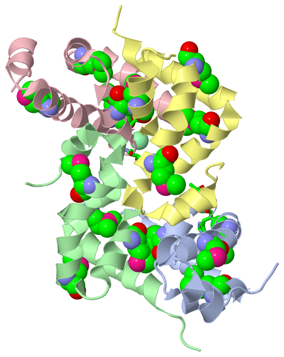
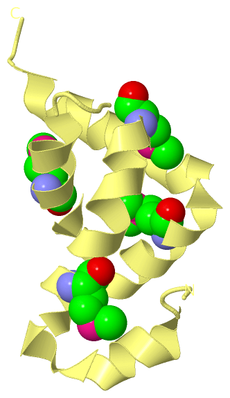
Chain A from PDB  Type:PROTEIN  Length:76
                                                                                                            
               SCOP domains d4ivea_ A: automated matches                                                 SCOP domains
               CATH domains ---------------------------------------------------------------------------- CATH domains
               Pfam domains ---------------------------------------------------------------------------- Pfam domains
         Sec.struct. author ....hhhhhhh.hhhhhhhhhhhhhhhhhhhhh..hhhhhhhhhh..hhhhhhhhhhhhhhhhhhhhhhhhhhh.. Sec.struct. author
                 SAPs(SNPs) ---------------------------------------------------------------------------- SAPs(SNPs)
                    PROSITE ---------------------------------------------------------------------------- PROSITE
                 Transcript ---------------------------------------------------------------------------- Transcript
                 4ive A 537 QETLTASRLASAPPQKQKQmLGERLFPLIQAmHPTLAGKITGmLLEIDNSELLYmLESPESLRSKVDEAVAVLQAH 612
                                   546       556       566 |     576  |    586    |  596       606      
                                             556-MSE     568-MSE    579-MSE     591-MSE                 

Chain B from PDB  Type:PROTEIN  Length:77
                                                                                                             
               SCOP domains d4iveb_ B: automated matches                                                  SCOP domains
               CATH domains ----------------------------------------------------------------------------- CATH domains
               Pfam domains ----------------------------------------------------------------------------- Pfam domains
         Sec.struct. author ...hhhhhhhhhhhhhhhhhhhhhhhhhhhhh..hhhhhhhhhh..hhhhhhhhhhhhhhhhhhhhhhhhhhh.... Sec.struct. author
                 SAPs(SNPs) ----------------------------------------------------------------------------- SAPs(SNPs)
                    PROSITE ----------------------------------------------------------------------------- PROSITE
                 Transcript ----------------------------------------------------------------------------- Transcript
                 4ive B 538 ETLTASRLASAPPQKQKQmLGERLFPLIQAmHPTLAGKITGmLLEIDNSELLYmLESPESLRSKVDEAVAVLQAHQA 614
                                   547       557       567|      577 |     587   |   597       607       
                                            556-MSE     568-MSE    579-MSE     591-MSE                   

Chain C from PDB  Type:PROTEIN  Length:76
                                                                                                            
               SCOP domains d4ivec_ C: automated matches                                                 SCOP domains
               CATH domains ---------------------------------------------------------------------------- CATH domains
               Pfam domains ---------------------------------------------------------------------------- Pfam domains
         Sec.struct. author ...hhhhhhh.hhhhhhhhhhhhhhhhhhhhh..hhhhhhhhhh..hhhhhhhhh.hhhhhhhhhhhhhhhhh... Sec.struct. author
                 SAPs(SNPs) ---------------------------------------------------------------------------- SAPs(SNPs)
                    PROSITE ---------------------------------------------------------------------------- PROSITE
                 Transcript ---------------------------------------------------------------------------- Transcript
                 4ive C 538 ETLTASRLASAPPQKQKQmLGERLFPLIQAmHPTLAGKITGmLLEIDNSELLYmLESPESLRSKVDEAVAVLQAHQ 613
                                   547       557       567|      577 |     587   |   597       607      
                                            556-MSE     568-MSE    579-MSE     591-MSE                  

Chain D from PDB  Type:PROTEIN  Length:76
                                                                                                            
               SCOP domains d4ived_ D: automated matches                                                 SCOP domains
               CATH domains ---------------------------------------------------------------------------- CATH domains
               Pfam domains ---------------------------------------------------------------------------- Pfam domains
         Sec.struct. author ...hhhhhhhhhhhhhhhhhhhhhhhhhhh....hhhhhhhhhh..hhhhhhhhhhhhhhhhhhhhhhhhhhh... Sec.struct. author
                 SAPs(SNPs) ---------------------------------------------------------------------------- SAPs(SNPs)
                    PROSITE ---------------------------------------------------------------------------- PROSITE
                 Transcript ---------------------------------------------------------------------------- Transcript
                 4ive D 538 ETLTASRLASAPPQKQKQmLGERLFPLIQAmHPTLAGKITGmLLEIDNSELLYmLESPESLRSKVDEAVAVLQAHQ 613
                                   547       557       567|      577 |     587   |   597       607      
                                            556-MSE     568-MSE    579-MSE     591-MSE                  

   Legend:   → Mismatch (orange background)
  - → Gap (green background, '-', border residues have a numbering label)
    → Modified Residue (blue background, lower-case, 'x' indicates undefined single-letter code, labelled with number + name)
  x → Chemical Group (purple background, 'x', labelled with number + name, e.g. ACE or NH2)
  extra numbering lines below/above indicate numbering irregularities and modified residue names etc., number ends below/above '|'

 Classification and Annotation

(-) SCOP Domains  (1, 4)

Asymmetric Unit

(-) CATH Domains  (0, 0)

(no "CATH Domain" information available for 4IVE)

(-) Pfam Domains  (0, 0)

(no "Pfam Domain" information available for 4IVE)

(-) Gene Ontology  (7, 7)

Asymmetric Unit(hide GO term definitions)

 Visualization

(-) Interactive Views

Asymmetric Unit
  Complete Structure
    Jena3D(integrated viewing of ligand, site, SAP, PROSITE, SCOP information)
    WebMol | AstexViewer[tm]@PDBe
(Java Applets, require no local installation except for Java; loading may be slow)
    STRAP
(Java WebStart application, automatic local installation, requires Java; full application with system access!)
    RasMol
(require local installation)
    Molscript (VRML)
(requires installation of a VRML viewer; select preferred view via VRML and generate a mono or stereo PDF format file)
 
  Ligands, Modified Residues, Ions
    CL  [ RasMol | Jena3D ]  +environment [ RasMol | Jena3D ]
    MSE  [ RasMol | Jena3D ]  +environment [ RasMol | Jena3D ]
 
  Sites
    AC1  [ RasMol ]  +environment [ RasMol ]
    AC2  [ RasMol ]  +environment [ RasMol ]
 
  Cis Peptide Bonds
(no "Cis Peptide Bonds" information available for 4ive)
 
Biological Units
  Complete Structure
    Biological Unit 1  [ Jena3D ]
    Biological Unit 2  [ Jena3D ]
    Biological Unit 3  [ Jena3D ]
    Biological Unit 4  [ Jena3D ]
    Biological Unit 5  [ Jena3D ]
    Biological Unit 6  [ Jena3D ]
    Biological Unit 7  [ Jena3D ]

(-) Still Images

Jmol
  protein: cartoon or spacefill or dots and stick; nucleic acid: cartoon and stick; ligands: spacefill; active site: stick
Molscript
  protein, nucleic acid: cartoon; ligands: spacefill; active site: ball and stick

 Databases and Analysis Tools

(-) Databases

Access by PDB/NDB ID
  4ive
    Family and Domain InformationProDom | SYSTERS
    General Structural InformationGlycoscienceDB | MMDB | NDB | OCA | PDB | PDBe | PDBj | PDBsum | PDBWiki | PQS | PROTEOPEDIA
    Orientation in MembranesOPM
    Protein SurfaceSURFACE
    Secondary StructureDSSP (structure derived) | HSSP (homology derived)
    Structural GenomicsGeneCensus
    Structural NeighboursCE | VAST
    Structure ClassificationCATH | Dali | SCOP
    Validation and Original DataBMRB Data View | BMRB Restraints Grid | EDS | PROCHECK | RECOORD | WHAT_CHECK
 
Access by UniProt ID/Accession number
  PABP3_HUMAN | Q9H361
    Comparative Protein Structure ModelsModBase
    Genomic InformationEnsembl
    Protein-protein InteractionDIP
    Sequence, Family and Domain InformationInterPro | Pfam | SMART | UniProtKB/SwissProt
 
Access by Enzyme Classificator   (EC Number)
  (no 'Enzyme Classificator' available)
    General Enzyme InformationBRENDA | EC-PDB | Enzyme | IntEnz
    PathwayKEGG | MetaCyc
 
Access by Disease Identifier   (MIM ID)
  (no 'MIM ID' available)
    Disease InformationOMIM
 
Access by GenAge ID
  (no 'GenAge ID' available)
    Age Related InformationGenAge

(-) Analysis Tools

Access by PDB/NDB ID
    Domain InformationXDom
    Interatomic Contacts of Structural UnitsCSU
    Ligand-protein ContactsLPC
    Protein CavitiescastP
    Sequence and Secondary StructurePDBCartoon
    Structure AlignmentSTRAP(Java WebStart application, automatic local installation, requires Java; full application with system access!)
    Structure and Sequence BrowserSTING
 
Access by UniProt ID/Accession number
  PABP3_HUMAN | Q9H361
    Protein Disorder PredictionDisEMBL | FoldIndex | GLOBPLOT (for more information see DisProt)

 Related Entries

(-) Entries Sharing at Least One Protein Chain (UniProt ID)

UniProtKB/Swiss-Prot
        PABP3_HUMAN | Q9H3612d9p

(-) Related Entries Specified in the PDB File

(no "Related Entries Specified in the PDB File" available for 4IVE)