Show PDB file:   
         Plain Text   HTML   (compressed file size)
QuickSearch:   
by PDB,NDB,UniProt,PROSITE Code or Search Term(s)  
(-)Asymmetric Unit
(-)Asym. Unit - sites
(-)Biological Unit 1
(-)Biol. Unit 1 - sites
collapse expand < >
Image Asymmetric Unit
Asymmetric Unit  (Jmol Viewer)
Image Asym. Unit - sites
Asym. Unit - sites  (Jmol Viewer)
Image Biological Unit 1
Biological Unit 1  (Jmol Viewer)
Image Biol. Unit 1 - sites
Biol. Unit 1 - sites  (Jmol Viewer)

(-) Description

Title :  CRYSTAL STRUCTURE OF R314K N-ACETYL NEURAMINIC ACID SYNTHASE FROM NEISERRIA MENINGITIDIS WITH MALATE BOUND
 
Authors :  D. D. A. Joseph, W. Jiao, E. J. Parker
Date :  09 Jan 13  (Deposition) - 10 Apr 13  (Release) - 01 May 13  (Revision)
Method :  X-RAY DIFFRACTION
Resolution :  2.15
Chains :  Asym. Unit :  A
Biol. Unit 1:  A  (2x)
Keywords :  Antifreeze Protein Fold, Nana, N-Acetylneuraminic Acid, Sialic Acid, Neisseria Meningitidis, Transferase (Keyword Search: [Gene Ontology, PubMed, Web (Google))
 
Reference :  D. D. Joseph, W. Jiao, E. J. Parker
Arg314 Is Essential For Catalysis By N-Acetyl Neuraminic Acid Synthase From Neisseria Meningitidis.
Biochemistry V. 52 2609 2013
PubMed-ID: 23534460  |  Reference-DOI: 10.1021/BI400062C

(-) Compounds

Molecule 1 - POLYSIALIC ACID CAPSULE BIOSYNTHESIS PROTEIN SIAC
    ChainsA
    EC Number2.5.1.56
    EngineeredYES
    Expression SystemESCHERICHIA COLI
    Expression System Taxid562
    GeneSIAC, NMB0068
    MutationYES
    Organism ScientificNEISSERIA MENINGITIDIS
    Organism Taxid487
    SynonymN-ACETYL NEURAMINIC ACID SYNTHASE

 Structural Features

(-) Chains, Units

  1
Asymmetric Unit A
Biological Unit 1 (2x)A

Summary Information (see also Sequences/Alignments below)

(-) Ligands, Modified Residues, Ions  (2, 2)

Asymmetric Unit (2, 2)
No.NameCountTypeFull Name
1LMR1Ligand/Ion(2S)-2-HYDROXYBUTANEDIOIC ACID
2MN1Ligand/IonMANGANESE (II) ION
Biological Unit 1 (1, 2)
No.NameCountTypeFull Name
1LMR2Ligand/Ion(2S)-2-HYDROXYBUTANEDIOIC ACID
2MN-1Ligand/IonMANGANESE (II) ION

(-) Sites  (2, 2)

Asymmetric Unit (2, 2)
No.NameEvidenceResiduesDescription
1AC1SOFTWAREGLU A:25 , TYR A:186 , HIS A:215 , HIS A:236 , LMR A:402 , HOH A:517BINDING SITE FOR RESIDUE MN A 401
2AC2SOFTWAREGLU A:25 , LYS A:53 , GLN A:55 , THR A:110 , LYS A:129 , GLY A:131 , ASN A:184 , TYR A:186 , HIS A:215 , HIS A:236 , MN A:401 , HOH A:517 , HOH A:518BINDING SITE FOR RESIDUE LMR A 402

(-) SS Bonds  (0, 0)

(no "SS Bond" information available for 4IPJ)

(-) Cis Peptide Bonds  (2, 2)

Asymmetric Unit
No.Residues
1Tyr A:186 -Pro A:187
2Lys A:314 -Pro A:315

 Sequence-Structure Mapping

(-) SAPs(SNPs)/Variants  (0, 0)

(no "SAP(SNP)/Variant" information available for 4IPJ)

(-) PROSITE Motifs  (0, 0)

(no "PROSITE Motif" information available for 4IPJ)

(-) Exons   (0, 0)

(no "Exon" information available for 4IPJ)

(-) Sequences/Alignments

Asymmetric Unit
   Reformat: Number of residues per line =  ('0' or empty: single-line sequence representation)
  Number of residues per labelling interval =   
  UniProt sequence: complete  aligned part    
   Show mapping: SCOP domains CATH domains Pfam domains Secondary structure (by author)
SAPs(SNPs) PROSITE motifs Exons
(details for a mapped element are shown in a popup box when the mouse pointer rests over it)
Chain A from PDB  Type:PROTEIN  Length:351
                                                                                                                                                                                                                                                                                                                                                                                               
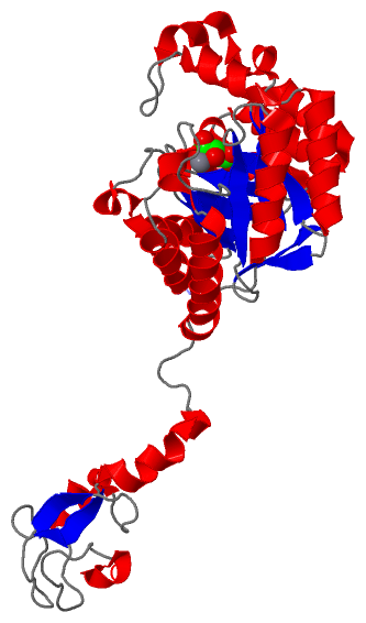
               SCOP domains d4ipja1 A:-1-281 automated matches                                                                                                                                                                                                                                                         d4ipja2 A:282-349 automated matches                                  SCOP domains
               CATH domains --------------------------------------------------------------------------------------------------------------------------------------------------------------------------------------------------------------------------------------------------------------------------------------------------------------------------------------------------------------- CATH domains
               Pfam domains --------------------------------------------------------------------------------------------------------------------------------------------------------------------------------------------------------------------------------------------------------------------------------------------------------------------------------------------------------------- Pfam domains
         Sec.struct. author .......eeee..eeee.....eeeeeee.....hhhhhhhhhhhhhhh...eeeeee.hhhhhhhhhhhhh.......hhhhhhhhhh.hhhhhhhhhhhhhhh..eeeeee.hhhhhhhhhhhh...eeehhhhh.hhhhhhhhhh....eeee....hhhhhhhhhhhhhhhh..eeeee.......hhhhhhhhhhhhhhhhh...eeeee.....hhhhhhhhhhh..eeeee..........hhhhh.hhhhhhhhhhhhhhhhhhh.......hhhhhhhhhhhh.eeee..............eeee.......hhhhhhhhh..............hhhhh. Sec.struct. author
                 SAPs(SNPs) --------------------------------------------------------------------------------------------------------------------------------------------------------------------------------------------------------------------------------------------------------------------------------------------------------------------------------------------------------------- SAPs(SNPs)
                    PROSITE --------------------------------------------------------------------------------------------------------------------------------------------------------------------------------------------------------------------------------------------------------------------------------------------------------------------------------------------------------------- PROSITE
                 Transcript --------------------------------------------------------------------------------------------------------------------------------------------------------------------------------------------------------------------------------------------------------------------------------------------------------------------------------------------------------------- Transcript
                 4ipj A  -1 GAMQNNNEFKIGNRSVGYNHEPLIICEIGINHEGSLKTAFEMVDAAYNAGAEVVKHQTHIVEDEMSDEAKQVIPGNADVSIYEIMERCALNEEDEIKLKEYVESKGMIFISTPFSRAAALRLQRMDIPAYKIGSGECNNYPLIKLVASFGKPIILSTGMNSIESIKKSVEIIREAGVPYALLHCTNIYPTPYEDVRLGGMNDLSEAFPDAIIGLSDHTLDNYACLGAVALGGSILERHFTDRMDRPGPDIVCSMNPDTFKELKQGAHALKLARGGKKDTIIAGEKPTKDFAFASVVADKDIKKGELLSGDNLWVKKPGNGDFSVNEYETLFGKVAACNIRKGAQIKKTDIE 349
                                     8        18        28        38        48        58        68        78        88        98       108       118       128       138       148       158       168       178       188       198       208       218       228       238       248       258       268       278       288       298       308       318       328       338       348 

   Legend:   → Mismatch (orange background)
  - → Gap (green background, '-', border residues have a numbering label)
    → Modified Residue (blue background, lower-case, 'x' indicates undefined single-letter code, labelled with number + name)
  x → Chemical Group (purple background, 'x', labelled with number + name, e.g. ACE or NH2)
  extra numbering lines below/above indicate numbering irregularities and modified residue names etc., number ends below/above '|'

 Classification and Annotation

(-) SCOP Domains  (2, 2)

Asymmetric Unit

(-) CATH Domains  (0, 0)

(no "CATH Domain" information available for 4IPJ)

(-) Pfam Domains  (0, 0)

(no "Pfam Domain" information available for 4IPJ)

(-) Gene Ontology  (3, 5)

Asymmetric Unit(hide GO term definitions)

 Visualization

(-) Interactive Views

Asymmetric Unit
  Complete Structure
    Jena3D(integrated viewing of ligand, site, SAP, PROSITE, SCOP information)
    WebMol | AstexViewer[tm]@PDBe
(Java Applets, require no local installation except for Java; loading may be slow)
    STRAP
(Java WebStart application, automatic local installation, requires Java; full application with system access!)
    RasMol
(require local installation)
    Molscript (VRML)
(requires installation of a VRML viewer; select preferred view via VRML and generate a mono or stereo PDF format file)
 
  Ligands, Modified Residues, Ions
    LMR  [ RasMol | Jena3D ]  +environment [ RasMol | Jena3D ]
    MN  [ RasMol | Jena3D ]  +environment [ RasMol | Jena3D ]
 
  Sites
    AC1  [ RasMol ]  +environment [ RasMol ]
    AC2  [ RasMol ]  +environment [ RasMol ]
 
  Cis Peptide Bonds
    Lys A:314 - Pro A:315   [ RasMol ]  
    Tyr A:186 - Pro A:187   [ RasMol ]  
 
Biological Unit
  Complete Structure
    Biological Unit 1  [ Jena3D ]

(-) Still Images

Jmol
  protein: cartoon or spacefill or dots and stick; nucleic acid: cartoon and stick; ligands: spacefill; active site: stick
Molscript
  protein, nucleic acid: cartoon; ligands: spacefill; active site: ball and stick

 Databases and Analysis Tools

(-) Databases

Access by PDB/NDB ID
  4ipj
    Family and Domain InformationProDom | SYSTERS
    General Structural InformationGlycoscienceDB | MMDB | NDB | OCA | PDB | PDBe | PDBj | PDBsum | PDBWiki | PQS | PROTEOPEDIA
    Orientation in MembranesOPM
    Protein SurfaceSURFACE
    Secondary StructureDSSP (structure derived) | HSSP (homology derived)
    Structural GenomicsGeneCensus
    Structural NeighboursCE | VAST
    Structure ClassificationCATH | Dali | SCOP
    Validation and Original DataBMRB Data View | BMRB Restraints Grid | EDS | PROCHECK | RECOORD | WHAT_CHECK
 
Access by UniProt ID/Accession number
  Q57265_NEIME | Q57265
    Comparative Protein Structure ModelsModBase
    Genomic InformationEnsembl
    Protein-protein InteractionDIP
    Sequence, Family and Domain InformationInterPro | Pfam | SMART | UniProtKB/TrEMBL
  Q7DDU0_NEIMB | Q7DDU0
    Comparative Protein Structure ModelsModBase
    Genomic InformationEnsembl
    Protein-protein InteractionDIP
    Sequence, Family and Domain InformationInterPro | Pfam | SMART | UniProtKB/SwissProt
 
Access by Enzyme Classificator   (EC Number)
  2.5.1.56
    General Enzyme InformationBRENDA | EC-PDB | Enzyme | IntEnz
    PathwayKEGG | MetaCyc
 
Access by Disease Identifier   (MIM ID)
  (no 'MIM ID' available)
    Disease InformationOMIM
 
Access by GenAge ID
  (no 'GenAge ID' available)
    Age Related InformationGenAge

(-) Analysis Tools

Access by PDB/NDB ID
    Domain InformationXDom
    Interatomic Contacts of Structural UnitsCSU
    Ligand-protein ContactsLPC
    Protein CavitiescastP
    Sequence and Secondary StructurePDBCartoon
    Structure AlignmentSTRAP(Java WebStart application, automatic local installation, requires Java; full application with system access!)
    Structure and Sequence BrowserSTING
 
Access by UniProt ID/Accession number
  Q57265_NEIME | Q57265
    Protein Disorder PredictionDisEMBL | FoldIndex | GLOBPLOT (for more information see DisProt)
  Q7DDU0_NEIMB | Q7DDU0
    Protein Disorder PredictionDisEMBL | FoldIndex | GLOBPLOT (for more information see DisProt)

 Related Entries

(-) Entries Sharing at Least One Protein Chain (UniProt ID)

UniProtKB/Swiss-Prot
        Q7DDU0_NEIMB | Q7DDU04ipi
UniProtKB/TrEMBL
        Q57265_NEIME | Q572651xuu 1xuz 2zdr 3cm4 4ipi
        Q7DDU0_NEIMB | Q7DDU02wqp

(-) Related Entries Specified in the PDB File

1xuu MALATE-BOUND WILD-TYPE ENZYME
1xuz SUBSTRATE-BOUND WILD-TYPE ENZYME
4ipi