Show PDB file:   
         Plain Text   HTML   (compressed file size)
QuickSearch:   
by PDB,NDB,UniProt,PROSITE Code or Search Term(s)  
(-)Asymmetric Unit
(-)Asym. Unit - sites
(-)Biological Unit 1
(-)Biol. Unit 1 - sites
(-)Biological Unit 2
(-)Biological Unit 3
collapse expand < >
Image Asymmetric Unit
Asymmetric Unit  (Jmol Viewer)
Image Asym. Unit - sites
Asym. Unit - sites  (Jmol Viewer)
Image Biological Unit 1
Biological Unit 1  (Jmol Viewer)
Image Biol. Unit 1 - sites
Biol. Unit 1 - sites  (Jmol Viewer)
Image Biological Unit 2
Biological Unit 2  (Jmol Viewer)
Image Biological Unit 3
Biological Unit 3  (Jmol Viewer)

(-) Description

Title :  CRYSTAL STRUCTURE OF BENM_DBD/CATB SITE 1 DNA COMPLEX
 
Authors :  A. Alanazi, C. Momany, E. L. Neidle
Date :  19 Dec 12  (Deposition) - 10 Jul 13  (Release) - 15 Jan 14  (Revision)
Method :  X-RAY DIFFRACTION
Resolution :  3.10
Chains :  Asym. Unit :  A,B,C,D,E,F,G,H
Biol. Unit 1:  A,B,E,F  (1x)
Biol. Unit 2:  C,D,G,H  (1x)
Biol. Unit 3:  A,B,C,D,E,F,G,H  (1x)
Keywords :  Whth, Hth, Transcriptional Regulator, Catb Promoter, Transcription- Dna Complex (Keyword Search: [Gene Ontology, PubMed, Web (Google))
 
Reference :  A. M. Alanazi, E. L. Neidle, C. Momany
The Dna-Binding Domain Of Benm Reveals The Structural Basis For The Recognition Of A T-N11-A Sequence Motif By Lysr-Typ Transcriptional Regulators.
Acta Crystallogr. , Sect. D V. 69 1995 2013
PubMed-ID: 24100318  |  Reference-DOI: 10.1107/S0907444913017320
(for further references see the PDB file header)

(-) Compounds

Molecule 1 - HTH-TYPE TRANSCRIPTIONAL REGULATOR BENM
    ChainsA, B, C, D
    EngineeredYES
    Expression SystemESCHERICHIA COLI
    Expression System PlasmidPET21B
    Expression System StrainBL21(DE3)RIL
    Expression System Taxid469008
    Expression System Vector TypePLASMID
    FragmentUNP RESIDUES 1-87
    GeneACIAD1435, BENM, BENR
    Organism ScientificACINETOBACTER SP.
    Organism Taxid62977
    StrainADP1
    SynonymBEN AND CAT OPERON TRANSCRIPTIONAL REGULATOR, TRANSCRIPTIONAL REGULATOR BENM DNA BINDING DOMAIN
 
Molecule 2 - CATB SITE 1 DNA
    ChainsE, G
    EngineeredYES
    Other DetailsSYNTHETIC DNA PURCHASED FROM IDT
    SyntheticYES
 
Molecule 3 - CATB SITE 1 DNA - COMPLEMENT
    ChainsF, H
    EngineeredYES
    Other DetailsSYNTHETIC DNA PURCHASED FROM IDT
    SyntheticYES

 Structural Features

(-) Chains, Units

  12345678
Asymmetric Unit ABCDEFGH
Biological Unit 1 (1x)AB  EF  
Biological Unit 2 (1x)  CD  GH
Biological Unit 3 (1x)ABCDEFGH

Summary Information (see also Sequences/Alignments below)

(-) Ligands, Modified Residues, Ions  (2, 3)

Asymmetric Unit (2, 3)
No.NameCountTypeFull Name
1MLI2Ligand/IonMALONATE ION
2NA1Ligand/IonSODIUM ION
Biological Unit 1 (1, 1)
No.NameCountTypeFull Name
1MLI1Ligand/IonMALONATE ION
2NA-1Ligand/IonSODIUM ION
Biological Unit 2 (1, 1)
No.NameCountTypeFull Name
1MLI1Ligand/IonMALONATE ION
2NA-1Ligand/IonSODIUM ION
Biological Unit 3 (1, 2)
No.NameCountTypeFull Name
1MLI2Ligand/IonMALONATE ION
2NA-1Ligand/IonSODIUM ION

(-) Sites  (3, 3)

Asymmetric Unit (3, 3)
No.NameEvidenceResiduesDescription
1AC1SOFTWAREASP A:76 , VAL A:79 , SER A:80 , HOH A:205 , ARG C:7 , MLI C:101 , NA C:102 , MET D:1 , GLU D:42BINDING SITE FOR RESIDUE MLI A 101
2AC2SOFTWAREARG A:7 , MLI A:101 , HOH A:205 , MET B:1 , ASP C:76 , SER C:80 , LYS C:83 , NA C:102 , HOH C:203BINDING SITE FOR RESIDUE MLI C 101
3AC3SOFTWAREMLI A:101 , ASP C:76 , MLI C:101BINDING SITE FOR RESIDUE NA C 102

(-) SS Bonds  (0, 0)

(no "SS Bond" information available for 4IHS)

(-) Cis Peptide Bonds  (3, 3)

Asymmetric Unit
No.Residues
1Gly A:51 -Ser A:52
2Arg A:53 -Pro A:54
3Arg C:53 -Pro C:54

 Sequence-Structure Mapping

(-) SAPs(SNPs)/Variants  (0, 0)

(no "SAP(SNP)/Variant" information available for 4IHS)

(-) PROSITE Motifs  (0, 0)

(no "PROSITE Motif" information available for 4IHS)

(-) Exons   (0, 0)

(no "Exon" information available for 4IHS)

(-) Sequences/Alignments

Asymmetric Unit
   Reformat: Number of residues per line =  ('0' or empty: single-line sequence representation)
  Number of residues per labelling interval =   
  UniProt sequence: complete  aligned part    
   Show mapping: SCOP domains CATH domains Pfam domains Secondary structure (by author)
SAPs(SNPs) PROSITE motifs Exons
(details for a mapped element are shown in a popup box when the mouse pointer rests over it)
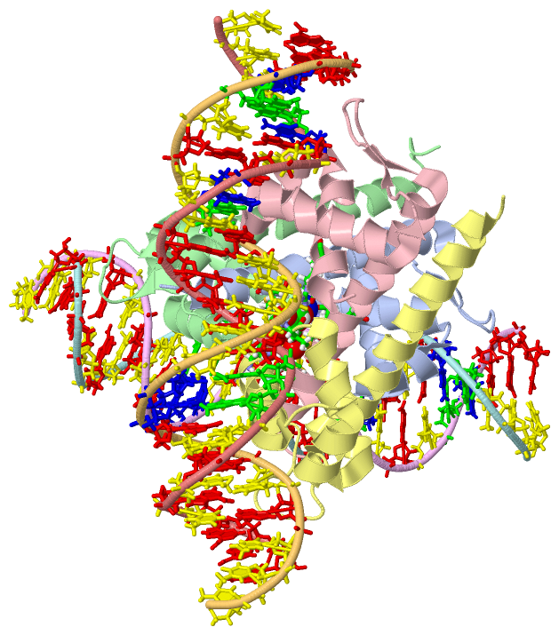
Chain A from PDB  Type:PROTEIN  Length:89
                                                                                                                        
               SCOP domains ----------------------------------------------------------------------------------------- SCOP domains
               CATH domains ----------------------------------------------------------------------------------------- CATH domains
               Pfam domains ----------------------------------------------------------------------------------------- Pfam domains
         Sec.struct. author .hhhhhhhhhhhhhh.hhhhhhhhhh.hhhhhhhhhhhhhhhhh...ee......eehhhhhhhhhhhhhhhhhhhhhhhhhhhhhh.. Sec.struct. author
                 SAPs(SNPs) ----------------------------------------------------------------------------------------- SAPs(SNPs)
                    PROSITE ----------------------------------------------------------------------------------------- PROSITE
                 Transcript ----------------------------------------------------------------------------------------- Transcript
                  4ihs A  1 MELRHLRYFVAVVEEQSFTKAADKLCIAQPPLSRQIQNLEEELGIQLLERGSRPVKTTPEGHFFYQYAIKLLSNVDQMVSMTKRIASGH 89
                                    10        20        30        40        50        60        70        80         

Chain B from PDB  Type:PROTEIN  Length:93
                                                                                                                            
               SCOP domains --------------------------------------------------------------------------------------------- SCOP domains
               CATH domains --------------------------------------------------------------------------------------------- CATH domains
               Pfam domains --------------------------------------------------------------------------------------------- Pfam domains
         Sec.struct. author .hhhhhhhhhhhhhhhhhhhhhhhh..hhhhhhhhhhhhhhhhh...ee......eehhhhhhhhhhhhhhhhhhhhhhhhhhhhhh...... Sec.struct. author
                 SAPs(SNPs) --------------------------------------------------------------------------------------------- SAPs(SNPs)
                    PROSITE --------------------------------------------------------------------------------------------- PROSITE
                 Transcript --------------------------------------------------------------------------------------------- Transcript
                  4ihs B  1 MELRHLRYFVAVVEEQSFTKAADKLCIAQPPLSRQIQNLEEELGIQLLERGSRPVKTTPEGHFFYQYAIKLLSNVDQMVSMTKRIASGHHHHH 93
                                    10        20        30        40        50        60        70        80        90   

Chain C from PDB  Type:PROTEIN  Length:90
                                                                                                                         
               SCOP domains ------------------------------------------------------------------------------------------ SCOP domains
               CATH domains ------------------------------------------------------------------------------------------ CATH domains
               Pfam domains ------------------------------------------------------------------------------------------ Pfam domains
         Sec.struct. author .hhhhhhhhhhhhhhhhhhhhhhhhh.hhhhhhhhhhhhhhhhh...ee......eehhhhhhhhhhhhhhhhhhhhhhhhhhhhhh... Sec.struct. author
                 SAPs(SNPs) ------------------------------------------------------------------------------------------ SAPs(SNPs)
                    PROSITE ------------------------------------------------------------------------------------------ PROSITE
                 Transcript ------------------------------------------------------------------------------------------ Transcript
                  4ihs C  1 MELRHLRYFVAVVEEQSFTKAADKLCIAQPPLSRQIQNLEEELGIQLLERGSRPVKTTPEGHFFYQYAIKLLSNVDQMVSMTKRIASGHH 90
                                    10        20        30        40        50        60        70        80        90

Chain D from PDB  Type:PROTEIN  Length:89
                                                                                                                        
               SCOP domains ----------------------------------------------------------------------------------------- SCOP domains
               CATH domains ----------------------------------------------------------------------------------------- CATH domains
               Pfam domains ----------------------------------------------------------------------------------------- Pfam domains
         Sec.struct. author .hhhhhhhhhhhhhhhhhhhhhhhh..hhhhhhhhhhhhhhhhh.............hhhhhhhhhhhhhhhhhhhhhhhhhhhhhh.. Sec.struct. author
                 SAPs(SNPs) ----------------------------------------------------------------------------------------- SAPs(SNPs)
                    PROSITE ----------------------------------------------------------------------------------------- PROSITE
                 Transcript ----------------------------------------------------------------------------------------- Transcript
                  4ihs D  1 MELRHLRYFVAVVEEQSFTKAADKLCIAQPPLSRQIQNLEEELGIQLLERGSRPVKTTPEGHFFYQYAIKLLSNVDQMVSMTKRIASGH 89
                                    10        20        30        40        50        60        70        80         

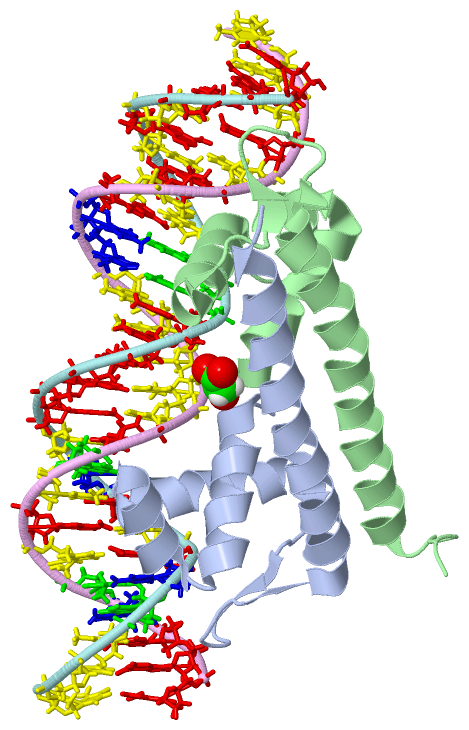
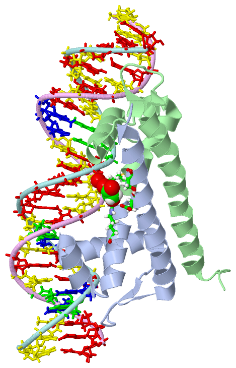
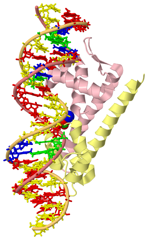
Chain E from PDB  Type:DNA  Length:25
                                                        
                  4ihs E  1 TTTATATACCTTTTTAGTATGCAAA 25
                                    10        20     

Chain F from PDB  Type:DNA  Length:25
                                                        
                  4ihs F  1 TTTGCATACTAAAAAGGTATATAAA 25
                                    10        20     

Chain G from PDB  Type:DNA  Length:25
                                                        
                  4ihs G  1 TTTATATACCTTTTTAGTATGCAAA 25
                                    10        20     

Chain H from PDB  Type:DNA  Length:25
                                                        
                  4ihs H  1 TTTGCATACTAAAAAGGTATATAAA 25
                                    10        20     

   Legend:   → Mismatch (orange background)
  - → Gap (green background, '-', border residues have a numbering label)
    → Modified Residue (blue background, lower-case, 'x' indicates undefined single-letter code, labelled with number + name)
  x → Chemical Group (purple background, 'x', labelled with number + name, e.g. ACE or NH2)
  extra numbering lines below/above indicate numbering irregularities and modified residue names etc., number ends below/above '|'

 Classification and Annotation

(-) SCOP Domains  (0, 0)

(no "SCOP Domain" information available for 4IHS)

(-) CATH Domains  (0, 0)

(no "CATH Domain" information available for 4IHS)

(-) Pfam Domains  (0, 0)

(no "Pfam Domain" information available for 4IHS)

(-) Gene Ontology  (5, 5)

Asymmetric Unit(hide GO term definitions)

 Visualization

(-) Interactive Views

Asymmetric Unit
  Complete Structure
    Jena3D(integrated viewing of ligand, site, SAP, PROSITE, SCOP information)
    WebMol | AstexViewer[tm]@PDBe
(Java Applets, require no local installation except for Java; loading may be slow)
    STRAP
(Java WebStart application, automatic local installation, requires Java; full application with system access!)
    RasMol
(require local installation)
    Molscript (VRML)
(requires installation of a VRML viewer; select preferred view via VRML and generate a mono or stereo PDF format file)
 
  Ligands, Modified Residues, Ions
    MLI  [ RasMol | Jena3D ]  +environment [ RasMol | Jena3D ]
    NA  [ RasMol | Jena3D ]  +environment [ RasMol | Jena3D ]
 
  Sites
    AC1  [ RasMol ]  +environment [ RasMol ]
    AC2  [ RasMol ]  +environment [ RasMol ]
    AC3  [ RasMol ]  +environment [ RasMol ]
 
  Cis Peptide Bonds
    Arg A:53 - Pro A:54   [ RasMol ]  
    Arg C:53 - Pro C:54   [ RasMol ]  
    Gly A:51 - Ser A:52   [ RasMol ]  
 
Biological Units
  Complete Structure
    Biological Unit 1  [ Jena3D ]
    Biological Unit 2  [ Jena3D ]
    Biological Unit 3  [ Jena3D ]

(-) Still Images

Jmol
  protein: cartoon or spacefill or dots and stick; nucleic acid: cartoon and stick; ligands: spacefill; active site: stick
Molscript
  protein, nucleic acid: cartoon; ligands: spacefill; active site: ball and stick

 Databases and Analysis Tools

(-) Databases

Access by PDB/NDB ID
  4ihs
    Family and Domain InformationProDom | SYSTERS
    General Structural InformationGlycoscienceDB | MMDB | NDB | OCA | PDB | PDBe | PDBj | PDBsum | PDBWiki | PQS | PROTEOPEDIA
    Orientation in MembranesOPM
    Protein SurfaceSURFACE
    Secondary StructureDSSP (structure derived) | HSSP (homology derived)
    Structural GenomicsGeneCensus
    Structural NeighboursCE | VAST
    Structure ClassificationCATH | Dali | SCOP
    Validation and Original DataBMRB Data View | BMRB Restraints Grid | EDS | PROCHECK | RECOORD | WHAT_CHECK
 
Access by UniProt ID/Accession number
  BENM_ACIAD | O68014
    Comparative Protein Structure ModelsModBase
    Genomic InformationEnsembl
    Protein-protein InteractionDIP
    Sequence, Family and Domain InformationInterPro | Pfam | SMART | UniProtKB/SwissProt
 
Access by Enzyme Classificator   (EC Number)
  (no 'Enzyme Classificator' available)
    General Enzyme InformationBRENDA | EC-PDB | Enzyme | IntEnz
    PathwayKEGG | MetaCyc
 
Access by Disease Identifier   (MIM ID)
  (no 'MIM ID' available)
    Disease InformationOMIM
 
Access by GenAge ID
  (no 'GenAge ID' available)
    Age Related InformationGenAge

(-) Analysis Tools

Access by PDB/NDB ID
    Domain InformationXDom
    Interatomic Contacts of Structural UnitsCSU
    Ligand-protein ContactsLPC
    Protein CavitiescastP
    Sequence and Secondary StructurePDBCartoon
    Structure AlignmentSTRAP(Java WebStart application, automatic local installation, requires Java; full application with system access!)
    Structure and Sequence BrowserSTING
 
Access by UniProt ID/Accession number
  BENM_ACIAD | O68014
    Protein Disorder PredictionDisEMBL | FoldIndex | GLOBPLOT (for more information see DisProt)

 Related Entries

(-) Entries Sharing at Least One Protein Chain (UniProt ID)

UniProtKB/Swiss-Prot
        BENM_ACIAD | O680142f6g 2f6p 2f78 2f7a 2f8d 2f97 2h99 2h9b 3k1m 3k1n 3k1p 3m1e 4iht

(-) Related Entries Specified in the PDB File

3m1e CRYSTAL STRUCTURE OF BENM DNA BINDING DOMAIN
4iht