Show PDB file:   
         Plain Text   HTML   (compressed file size)
QuickSearch:   
by PDB,NDB,UniProt,PROSITE Code or Search Term(s)  
(-)Asymmetric Unit
(-)Asym. Unit - sites
(-)Biological Unit 1
(-)Biol. Unit 1 - sites
collapse expand < >
Image Asymmetric Unit
Asymmetric Unit  (Jmol Viewer)
Image Asym. Unit - sites
Asym. Unit - sites  (Jmol Viewer)
Image Biological Unit 1
Biological Unit 1  (Jmol Viewer)
Image Biol. Unit 1 - sites
Biol. Unit 1 - sites  (Jmol Viewer)

(-) Description

Title :  CRYSTAL STRUCTURE OF THE KAINATE RECEPTOR GLUK3 LIGAND-BINDING DOMAIN IN COMPLEX WITH THE AGONIST ZA302
 
Authors :  A. P. Larsen, R. Venskutonyte, M. Gajhede, J. S. Kastrup, K. Frydenvang
Date :  18 Dec 12  (Deposition) - 06 Mar 13  (Release) - 03 Sep 14  (Revision)
Method :  X-RAY DIFFRACTION
Resolution :  2.65
Chains :  Asym. Unit :  A
Biol. Unit 1:  A  (2x)
Keywords :  Membrane Protein, Ionotropic Glutamate Receptor, Kainate Receptor, Ligand-Binding Domain, Agonist (Keyword Search: [Gene Ontology, PubMed, Web (Google))
 
Reference :  Z. Assaf, A. P. Larsen, R. Venskutonyte, L. Han, B. Abrahamsen, B. Nielsen, M. Gajhede, J. S. Kastrup, A. A. Jensen, D. S. Pickering, K. Frydenvang, T. Gefflaut, L. Bunch
Chemoenzymatic Synthesis Of New 2, 4-Syn-Functionalized (S)-Glutamate Analogues And Structure-Activity Relationship Studies At Ionotropic Glutamate Receptors And Excitatory Amino Acid Transporters.
J. Med. Chem. V. 56 1614 2013
PubMed-ID: 23414088  |  Reference-DOI: 10.1021/JM301433M
(for further references see the PDB file header)

(-) Compounds

Molecule 1 - GLUTAMATE RECEPTOR, IONOTROPIC KAINATE 3
    ChainsA
    EngineeredYES
    Expression SystemESCHERICHIA COLI
    Expression System PlasmidPOPINJ
    Expression System StrainORIGAMI 2
    Expression System Taxid562
    Expression System Vector TypePLASMID
    FragmentLIGAND-BINDING DOMAIN, UNP RESIDUES 432-546, 669-806
    GeneGLUR7, GRIK3
    Organism CommonRAT
    Organism ScientificRATTUS NORVEGICUS
    Organism Taxid10116
    SynonymGLUTAMATE RECEPTOR 7, GLUR-7, GLUR7

 Structural Features

(-) Chains, Units

  1
Asymmetric Unit A
Biological Unit 1 (2x)A

Summary Information (see also Sequences/Alignments below)
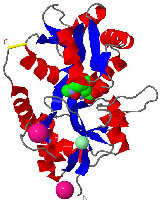
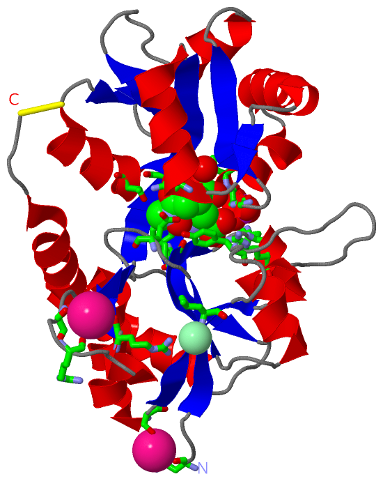
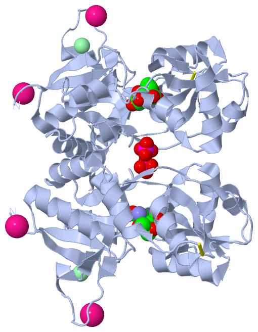
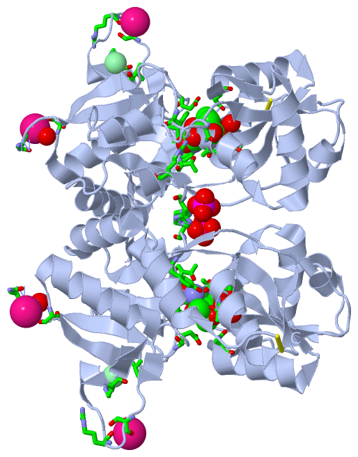
(-) Ligands, Modified Residues, Ions  (4, 5)

Asymmetric Unit (4, 5)
No.NameCountTypeFull Name
13ZA1Ligand/Ion(4R)-4-{3-[HYDROXY(METHYL)AMINO]-3-OXOPROPYL}-L-GLUTAMIC ACID
2CL1Ligand/IonCHLORIDE ION
3K2Ligand/IonPOTASSIUM ION
4PO41Ligand/IonPHOSPHATE ION
Biological Unit 1 (2, 4)
No.NameCountTypeFull Name
13ZA2Ligand/Ion(4R)-4-{3-[HYDROXY(METHYL)AMINO]-3-OXOPROPYL}-L-GLUTAMIC ACID
2CL-1Ligand/IonCHLORIDE ION
3K-1Ligand/IonPOTASSIUM ION
4PO42Ligand/IonPHOSPHATE ION

(-) Sites  (5, 5)

Asymmetric Unit (5, 5)
No.NameEvidenceResiduesDescription
1AC1SOFTWAREGLU A:443 , TYR A:491 , PRO A:518 , THR A:520 , ARG A:525 , GLY A:690 , ALA A:691 , THR A:692 , ASN A:722 , GLU A:739 , THR A:742 , TYR A:765 , HOH A:1047 , HOH A:1048BINDING SITE FOR RESIDUE 3ZA A 901
2AC2SOFTWAREASN A:433 , SER A:480 , HOH A:1005BINDING SITE FOR RESIDUE K A 902
3AC3SOFTWARESER A:452 , ARG A:454BINDING SITE FOR RESIDUE K A 903
4AC4SOFTWAREARG A:461 , LEU A:485BINDING SITE FOR RESIDUE CL A 904
5AC5SOFTWAREHIS A:523BINDING SITE FOR RESIDUE PO4 A 905

(-) SS Bonds  (1, 1)

Asymmetric Unit
No.Residues
1A:751 -A:805

(-) Cis Peptide Bonds  (1, 1)

Asymmetric Unit
No.Residues
1Glu A:444 -Pro A:445

 Sequence-Structure Mapping

(-) SAPs(SNPs)/Variants  (0, 0)

(no "SAP(SNP)/Variant" information available for 4IGR)

(-) PROSITE Motifs  (0, 0)

(no "PROSITE Motif" information available for 4IGR)

(-) Exons   (0, 0)

(no "Exon" information available for 4IGR)

(-) Sequences/Alignments

Asymmetric Unit
   Reformat: Number of residues per line =  ('0' or empty: single-line sequence representation)
  Number of residues per labelling interval =   
  UniProt sequence: complete  aligned part    
   Show mapping: SCOP domains CATH domains Pfam domains Secondary structure (by author)
SAPs(SNPs) PROSITE motifs Exons
(details for a mapped element are shown in a popup box when the mouse pointer rests over it)
Chain A from PDB  Type:PROTEIN  Length:253
                                                                                                                                                                                                                                                                                             
               SCOP domains d4igra_ A: automated matches                                                                                                                                                                                                                                  SCOP domains
               CATH domains ------------------------------------------------------------------------------------------------------------------------------------------------------------------------------------------------------------------------------------------------------------- CATH domains
               Pfam domains ------------------------------------------------------------------------------------------------------------------------------------------------------------------------------------------------------------------------------------------------------------- Pfam domains
         Sec.struct. author ...eeeee.......ee.......hhhh.eehhhhhhhhhhhhhhh..eeeee...............hhhhhhhhh....eeeeeee...hhhhh.ee....eee.eeeeee......hhhhhhh....eeeee..hhhhhhhhhh.hhhhhhhhhhhhhh...ee.hhhhhhhhhhhh.eeeeeehhhhhhhhhhh..eeee......eeeeeee.....hhhhhhhhhhhhhhhhhhhhhhhhhhh.... Sec.struct. author
                 SAPs(SNPs) ------------------------------------------------------------------------------------------------------------------------------------------------------------------------------------------------------------------------------------------------------------- SAPs(SNPs)
                    PROSITE ------------------------------------------------------------------------------------------------------------------------------------------------------------------------------------------------------------------------------------------------------------- PROSITE
                 Transcript ------------------------------------------------------------------------------------------------------------------------------------------------------------------------------------------------------------------------------------------------------------- Transcript
                 4igr A 433 NRSLIVTTLLEEPFVMFRKSDRTLYGNDRFEGYCIDLLKELAHILGFSYEIRLVEDGKYGAQDDKGQWNGMVKELIDHKADLAVAPLTITHVREKAIDFSKPFMTLGVSILYRKGTPIDSADDLAKQTKIEYGAVKDGATMTFFKKSKISTFEKMWAFMSSKPSALVKNNEEGIQRTLTADYALLMESTTIEYITQRNCNLTQIGGLIDSKGYGIGTPMGSPYRDKITIAILQLQEEDKLHIMKEKWWRGSGC 805
                                   442       452       462       472       482       492       502       512       522       532       542     ||672       682       692       702       712       722       732       742       752       762       772       782       792       802   
                                                                                                                                             548|                                                                                                                                        
                                                                                                                                              669                                                                                                                                        

   Legend:   → Mismatch (orange background)
  - → Gap (green background, '-', border residues have a numbering label)
    → Modified Residue (blue background, lower-case, 'x' indicates undefined single-letter code, labelled with number + name)
  x → Chemical Group (purple background, 'x', labelled with number + name, e.g. ACE or NH2)
  extra numbering lines below/above indicate numbering irregularities and modified residue names etc., number ends below/above '|'

 Classification and Annotation

(-) SCOP Domains  (1, 1)

Asymmetric Unit

(-) CATH Domains  (0, 0)

(no "CATH Domain" information available for 4IGR)

(-) Pfam Domains  (0, 0)

(no "Pfam Domain" information available for 4IGR)

(-) Gene Ontology  (24, 24)

Asymmetric Unit(hide GO term definitions)

 Visualization

(-) Interactive Views

Asymmetric Unit
  Complete Structure
    Jena3D(integrated viewing of ligand, site, SAP, PROSITE, SCOP information)
    WebMol | AstexViewer[tm]@PDBe
(Java Applets, require no local installation except for Java; loading may be slow)
    STRAP
(Java WebStart application, automatic local installation, requires Java; full application with system access!)
    RasMol
(require local installation)
    Molscript (VRML)
(requires installation of a VRML viewer; select preferred view via VRML and generate a mono or stereo PDF format file)
 
  Ligands, Modified Residues, Ions
    3ZA  [ RasMol | Jena3D ]  +environment [ RasMol | Jena3D ]
    CL  [ RasMol | Jena3D ]  +environment [ RasMol | Jena3D ]
    K  [ RasMol | Jena3D ]  +environment [ RasMol | Jena3D ]
    PO4  [ RasMol | Jena3D ]  +environment [ RasMol | Jena3D ]
 
  Sites
    AC1  [ RasMol ]  +environment [ RasMol ]
    AC2  [ RasMol ]  +environment [ RasMol ]
    AC3  [ RasMol ]  +environment [ RasMol ]
    AC4  [ RasMol ]  +environment [ RasMol ]
    AC5  [ RasMol ]  +environment [ RasMol ]
 
  Cis Peptide Bonds
    Glu A:444 - Pro A:445   [ RasMol ]  
 
Biological Unit
  Complete Structure
    Biological Unit 1  [ Jena3D ]

(-) Still Images

Jmol
  protein: cartoon or spacefill or dots and stick; nucleic acid: cartoon and stick; ligands: spacefill; active site: stick
Molscript
  protein, nucleic acid: cartoon; ligands: spacefill; active site: ball and stick

 Databases and Analysis Tools

(-) Databases

Access by PDB/NDB ID
  4igr
    Family and Domain InformationProDom | SYSTERS
    General Structural InformationGlycoscienceDB | MMDB | NDB | OCA | PDB | PDBe | PDBj | PDBsum | PDBWiki | PQS | PROTEOPEDIA
    Orientation in MembranesOPM
    Protein SurfaceSURFACE
    Secondary StructureDSSP (structure derived) | HSSP (homology derived)
    Structural GenomicsGeneCensus
    Structural NeighboursCE | VAST
    Structure ClassificationCATH | Dali | SCOP
    Validation and Original DataBMRB Data View | BMRB Restraints Grid | EDS | PROCHECK | RECOORD | WHAT_CHECK
 
Access by UniProt ID/Accession number
  GRIK3_RAT | P42264
    Comparative Protein Structure ModelsModBase
    Genomic InformationEnsembl
    Protein-protein InteractionDIP
    Sequence, Family and Domain InformationInterPro | Pfam | SMART | UniProtKB/SwissProt
 
Access by Enzyme Classificator   (EC Number)
  (no 'Enzyme Classificator' available)
    General Enzyme InformationBRENDA | EC-PDB | Enzyme | IntEnz
    PathwayKEGG | MetaCyc
 
Access by Disease Identifier   (MIM ID)
  (no 'MIM ID' available)
    Disease InformationOMIM
 
Access by GenAge ID
  (no 'GenAge ID' available)
    Age Related InformationGenAge

(-) Analysis Tools

Access by PDB/NDB ID
    Domain InformationXDom
    Interatomic Contacts of Structural UnitsCSU
    Ligand-protein ContactsLPC
    Protein CavitiescastP
    Sequence and Secondary StructurePDBCartoon
    Structure AlignmentSTRAP(Java WebStart application, automatic local installation, requires Java; full application with system access!)
    Structure and Sequence BrowserSTING
 
Access by UniProt ID/Accession number
  GRIK3_RAT | P42264
    Protein Disorder PredictionDisEMBL | FoldIndex | GLOBPLOT (for more information see DisProt)

 Related Entries

(-) Entries Sharing at Least One Protein Chain (UniProt ID)

UniProtKB/Swiss-Prot
        GRIK3_RAT | P422643olz 3s9e 3u92 3u93 3u94 4e0w 4g8n 4mh5 4nwc 4nwd

(-) Related Entries Specified in the PDB File

3s9e 4igt