|
|
|
|
 Description
|
|
Compounds
|
||||||||||||||||||||||||||||||||||||||||||||||||||||||||
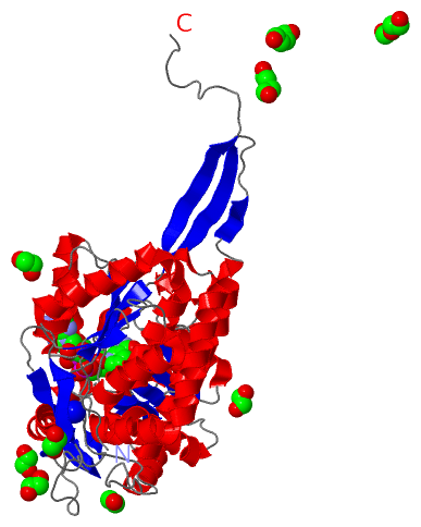
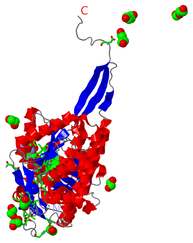
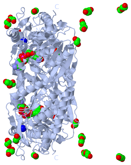
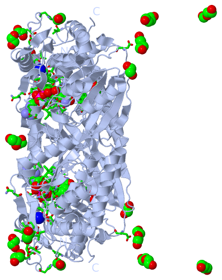
Chains, Units
Summary Information (see also Sequences/Alignments below) |
Ligands, Modified Residues, Ions (4, 12)| Asymmetric Unit (4, 12) Biological Unit 1 (3, 22) |
Sites (8, 8)
Asymmetric Unit (8, 8)
|
SS Bonds (0, 0)| (no "SS Bond" information available for 4I8Q) |
Cis Peptide Bonds (0, 0)| (no "Cis Peptide Bond" information available for 4I8Q) |
SAPs(SNPs)/Variants (0, 0)| (no "SAP(SNP)/Variant" information available for 4I8Q) |
PROSITE Motifs (0, 0)| (no "PROSITE Motif" information available for 4I8Q) |
Exons (0, 0)| (no "Exon" information available for 4I8Q) |
Sequences/Alignments
Asymmetric Unit
Chain A from PDB Type:PROTEIN Length:495
SCOP domains d4i8qa_ A: automated matches SCOP domains
CATH domains --------------------------------------------------------------------------------------------------------------------------------------------------------------------------------------------------------------------------------------------------------------------------------------------------------------------------------------------------------------------------------------------------------------------------------------------------------------------------------------------------------------- CATH domains
Pfam domains --------------------------------------------------------------------------------------------------------------------------------------------------------------------------------------------------------------------------------------------------------------------------------------------------------------------------------------------------------------------------------------------------------------------------------------------------------------------------------------------------------------- Pfam domains
SAPs(SNPs) --------------------------------------------------------------------------------------------------------------------------------------------------------------------------------------------------------------------------------------------------------------------------------------------------------------------------------------------------------------------------------------------------------------------------------------------------------------------------------------------------------------- SAPs(SNPs)
PROSITE --------------------------------------------------------------------------------------------------------------------------------------------------------------------------------------------------------------------------------------------------------------------------------------------------------------------------------------------------------------------------------------------------------------------------------------------------------------------------------------------------------------- PROSITE
Transcript --------------------------------------------------------------------------------------------------------------------------------------------------------------------------------------------------------------------------------------------------------------------------------------------------------------------------------------------------------------------------------------------------------------------------------------------------------------------------------------------------------------- Transcript
4i8q A 7 PIPRRQLYIGGEWREPVKKNRIPIINPATEEIIGDIPAATAEDVDIAVEAARKAIARDDWGSTTGAQRAKYLRAIAAKVLEKKSVLATLESLDSGKTLYESAADMDDVAGCFEYYAGLAEALDSRRMTPVNLNSDSYKSYVLREPLGVVGLITPWNYPLLMAIWKVAPALAAGCAAILKPSELASITCLELGEICREIGLPSGALNILTGLGPEAGGPLASHPHVDKISFTGSGPTGSKIMTAAAQLVKPVSLALGGKSPIVVFDDIDNLDIAAEWTLFGIFANTGQVCSATSRLIVQENIASAFMDRLLKWTKNIKISDPLEEDCKLGPVVSAGQYEKVLKFISNAKSEGATILCGGERPQHLKKGYYVQPTIITDVNTSMEIWKEEVFGPVLCVKTFKTEEQAIELANDTKYGLGAAVMSKDVKRCERFTKAFQTGIIWINCSQPTFNELPWGGKKRSGFGRDLGKWGLENFLNIKQVTEYTSAEPLAFYKSP 501
16 26 36 46 56 66 76 86 96 106 116 126 136 146 156 166 176 186 196 206 216 226 236 246 256 266 276 286 296 306 316 326 336 346 356 366 376 386 396 406 416 426 436 446 456 466 476 486 496
|
||||||||||||||||||||
SCOP Domains (1, 1)
Asymmetric Unit
 |
CATH Domains (0, 0)| (no "CATH Domain" information available for 4I8Q) |
Pfam Domains (0, 0)| (no "Pfam Domain" information available for 4I8Q) |
Gene Ontology (6, 6)|
Asymmetric Unit(hide GO term definitions) |
Interactive Views
|
||||||||||||||||||||||||||||||||||||||||||||||||||||||||||||||||||||||||||||||||||||||||||||||||||||||||||||||||||||||||||||||||||||||||||||||||||||||||||||||||||||||||||||||||||||||||||||||||||||||||||||||
Still Images
|
||||||||||||||||
Databases
|
||||||||||||||||||||||||||||||||||||||||||||||||||||||||||||||||||||||||||||||||||||||||||||||||||||||||||||||||||||||||||||||||||||||||||||||||||||||||||||||||
Analysis Tools
|
|||||||||||||||||||||||||||||||||||||||||||||||||||||||||||||
Entries Sharing at Least One Protein Chain (UniProt ID)
Related Entries Specified in the PDB File
|
|