Show PDB file:   
         Plain Text   HTML   (compressed file size)
QuickSearch:   
by PDB,NDB,UniProt,PROSITE Code or Search Term(s)  
(-)Asym./Biol. Unit
(-)Asym./Biol. Unit - sites
collapse expand < >
Image Asym./Biol. Unit
Asym./Biol. Unit  (Jmol Viewer)
Image Asym./Biol. Unit - sites
Asym./Biol. Unit - sites  (Jmol Viewer)

(-) Description

Title :  STRUCTURE OF PARKIN E3 LIGASE
 
Authors :  J. C. Lougheed, E. Brecht, N. H. Yao
Date :  20 Nov 12  (Deposition) - 19 Jun 13  (Release) - 03 Jul 13  (Revision)
Method :  X-RAY DIFFRACTION
Resolution :  2.00
Chains :  Asym./Biol. Unit :  A
Keywords :  Rbr E3 Ubiquitin Ligase, Ligase (Keyword Search: [Gene Ontology, PubMed, Web (Google))
 
Reference :  B. E. Riley, J. C. Lougheed, K. Callaway, M. Velasquez, E. Brecht, L. Nguyen, T. Shaler, D. Walker, Y. Yang, K. Regnstrom, L. Diep, Z. Zhang, S. Chiou, M. Bova, D. R. Artis, N. Yao, J. Baker, T. Yednock, J. A. Johnston
Structure And Function Of Parkin E3 Ubiquitin Ligase Reveal Aspects Of Ring And Hect Ligases.
Nat Commun V. 4 1982 2013
PubMed-ID: 23770887  |  Reference-DOI: 10.1038/NCOMMS2982

(-) Compounds

Molecule 1 - E3 UBIQUITIN-PROTEIN LIGASE PARKIN
    ChainsA
    EC Number6.3.2.-
    EngineeredYES
    Expression SystemESCHERICHIA COLI
    Expression System Taxid562
    FragmentR0RBR (UNP RESIDUES 141-465)
    GenePARK2, PRKN
    Organism CommonHUMAN
    Organism ScientificHOMO SAPIENS
    Organism Taxid9606
    SynonymPARKINSON JUVENILE DISEASE PROTEIN 2, PARKINSON DISEASE PROTEIN 2

 Structural Features

(-) Chains, Units

  1
Asymmetric/Biological Unit A

Summary Information (see also Sequences/Alignments below)

(-) Ligands, Modified Residues, Ions  (1, 8)

Asymmetric/Biological Unit (1, 8)
No.NameCountTypeFull Name
1ZN8Ligand/IonZINC ION

(-) Sites  (8, 8)

Asymmetric Unit (8, 8)
No.NameEvidenceResiduesDescription
1AC1SOFTWARECYS A:150 , CYS A:154 , CYS A:212 , HIS A:215BINDING SITE FOR RESIDUE ZN A 501
2AC2SOFTWARECYS A:166 , CYS A:169 , CYS A:196 , CYS A:201BINDING SITE FOR RESIDUE ZN A 502
3AC3SOFTWARECYS A:238 , CYS A:241 , CYS A:260 , CYS A:263BINDING SITE FOR RESIDUE ZN A 503
4AC4SOFTWARECYS A:253 , HIS A:257 , CYS A:289 , CYS A:293BINDING SITE FOR RESIDUE ZN A 504
5AC5SOFTWARECYS A:332 , CYS A:337 , CYS A:352 , CYS A:360BINDING SITE FOR RESIDUE ZN A 505
6AC6SOFTWARECYS A:365 , CYS A:368 , HIS A:373 , CYS A:377BINDING SITE FOR RESIDUE ZN A 506
7AC7SOFTWARECYS A:418 , CYS A:421 , CYS A:436 , CYS A:441BINDING SITE FOR RESIDUE ZN A 507
8AC8SOFTWARECYS A:446 , CYS A:449 , CYS A:457 , HIS A:461BINDING SITE FOR RESIDUE ZN A 508

(-) SS Bonds  (0, 0)

(no "SS Bond" information available for 4I1H)

(-) Cis Peptide Bonds  (2, 2)

Asymmetric/Biological Unit
No.Residues
1Gly A:152 -Pro A:153
2Ser A:246 -Pro A:247

 Sequence-Structure Mapping

(-) SAPs(SNPs)/Variants  (0, 0)

(no "SAP(SNP)/Variant" information available for 4I1H)

(-) PROSITE Motifs  (0, 0)

(no "PROSITE Motif" information available for 4I1H)

(-) Exons   (0, 0)

(no "Exon" information available for 4I1H)

(-) Sequences/Alignments

Asymmetric/Biological Unit
   Reformat: Number of residues per line =  ('0' or empty: single-line sequence representation)
  Number of residues per labelling interval =   
  UniProt sequence: complete  aligned part    
   Show mapping: SCOP domains CATH domains Pfam domains Secondary structure (by author)
SAPs(SNPs) PROSITE motifs Exons
(details for a mapped element are shown in a popup box when the mouse pointer rests over it)
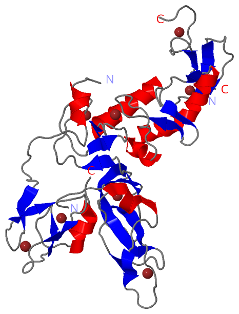
Chain A from PDB  Type:PROTEIN  Length:306
                                                                                                                                                                                                                                                                                                                                                  
               SCOP domains ------------------------------------------------------------------------------------------------------------------------------------------------------------------------------------------------------------------------------------------------------------------------------------------------------------------ SCOP domains
               CATH domains ------------------------------------------------------------------------------------------------------------------------------------------------------------------------------------------------------------------------------------------------------------------------------------------------------------------ CATH domains
               Pfam domains ------------------------------------------------------------------------------------------------------------------------------------------------------------------------------------------------------------------------------------------------------------------------------------------------------------------ Pfam domains
         Sec.struct. author ......eeee.....eeeeeeeeeee.......eee.....hhhhhhh.....eee.........eeeeeee...........ee...ee.................eee.......eeehhhhhhhhhhhhhh...eee...eee.............hhhhhhhhhhhhhhhhhhhhhhhhhhhh..ee........ee.......eee......eee................hhhhhh.............eee......eee......eee........eee.......hhhhhhhhh... Sec.struct. author
                 SAPs(SNPs) ------------------------------------------------------------------------------------------------------------------------------------------------------------------------------------------------------------------------------------------------------------------------------------------------------------------ SAPs(SNPs)
                    PROSITE ------------------------------------------------------------------------------------------------------------------------------------------------------------------------------------------------------------------------------------------------------------------------------------------------------------------ PROSITE
                 Transcript ------------------------------------------------------------------------------------------------------------------------------------------------------------------------------------------------------------------------------------------------------------------------------------------------------------------ Transcript
                 4i1h A 141 SIYNSFYVYCKGPCQRVQPGKLRVQCSTCRQATLTLTQGPSCWDDVLIPNRMSGECQSPHCPGTSAEFFFKCGAHPTSDKETSVALHLIATNSRNITCITCTDVRSPVLVFQCNSRHVICLDCFHLYCVTRLNDRQFVHDPQLGYSLPCVAGCPNSLIKELHHFRILGEEQYNRYQQYGAEECVLQMGGVLCPRPGCGAGLLPEPDQRKVTCEGCGFAFCRECKEAYHEGECSVDERAAEQARWEAASKETIKKTTKPCPRCHVPVEKNGGCMHMKCPQPQCRLEWCWNCGCEWNRVCMGDHWFDV 465
                                   150       160       170       180       190       200       210       220       230       240       250       260       270       280       290       300       310       320       330       340       350  ||   365       375  ||   399       409       419       429       439       449       459      
                                                                                                                                                                                                                                              353|                378|                                                                        
                                                                                                                                                                                                                                               359                 393                                                                        

   Legend:   → Mismatch (orange background)
  - → Gap (green background, '-', border residues have a numbering label)
    → Modified Residue (blue background, lower-case, 'x' indicates undefined single-letter code, labelled with number + name)
  x → Chemical Group (purple background, 'x', labelled with number + name, e.g. ACE or NH2)
  extra numbering lines below/above indicate numbering irregularities and modified residue names etc., number ends below/above '|'

 Classification and Annotation

(-) SCOP Domains  (0, 0)

(no "SCOP Domain" information available for 4I1H)

(-) CATH Domains  (0, 0)

(no "CATH Domain" information available for 4I1H)

(-) Pfam Domains  (0, 0)

(no "Pfam Domain" information available for 4I1H)

(-) Gene Ontology  (146, 146)

Asymmetric/Biological Unit(hide GO term definitions)

 Visualization

(-) Interactive Views

Asymmetric/Biological Unit
  Complete Structure
    Jena3D(integrated viewing of ligand, site, SAP, PROSITE, SCOP information)
    WebMol | AstexViewer[tm]@PDBe
(Java Applets, require no local installation except for Java; loading may be slow)
    STRAP
(Java WebStart application, automatic local installation, requires Java; full application with system access!)
    RasMol
(require local installation)
    Molscript (VRML)
(requires installation of a VRML viewer; select preferred view via VRML and generate a mono or stereo PDF format file)
 
  Ligands, Modified Residues, Ions
    ZN  [ RasMol | Jena3D ]  +environment [ RasMol | Jena3D ]
 
  Sites
    AC1  [ RasMol ]  +environment [ RasMol ]
    AC2  [ RasMol ]  +environment [ RasMol ]
    AC3  [ RasMol ]  +environment [ RasMol ]
    AC4  [ RasMol ]  +environment [ RasMol ]
    AC5  [ RasMol ]  +environment [ RasMol ]
    AC6  [ RasMol ]  +environment [ RasMol ]
    AC7  [ RasMol ]  +environment [ RasMol ]
    AC8  [ RasMol ]  +environment [ RasMol ]
 
  Cis Peptide Bonds
    Gly A:152 - Pro A:153   [ RasMol ]  
    Ser A:246 - Pro A:247   [ RasMol ]  
 

(-) Still Images

Jmol
  protein: cartoon or spacefill or dots and stick; nucleic acid: cartoon and stick; ligands: spacefill; active site: stick
Molscript
  protein, nucleic acid: cartoon; ligands: spacefill; active site: ball and stick

 Databases and Analysis Tools

(-) Databases

Access by PDB/NDB ID
  4i1h
    Family and Domain InformationProDom | SYSTERS
    General Structural InformationGlycoscienceDB | MMDB | NDB | OCA | PDB | PDBe | PDBj | PDBsum | PDBWiki | PQS | PROTEOPEDIA
    Orientation in MembranesOPM
    Protein SurfaceSURFACE
    Secondary StructureDSSP (structure derived) | HSSP (homology derived)
    Structural GenomicsGeneCensus
    Structural NeighboursCE | VAST
    Structure ClassificationCATH | Dali | SCOP
    Validation and Original DataBMRB Data View | BMRB Restraints Grid | EDS | PROCHECK | RECOORD | WHAT_CHECK
 
Access by UniProt ID/Accession number
  PRKN_HUMAN | O60260
    Comparative Protein Structure ModelsModBase
    Genomic InformationEnsembl
    Protein-protein InteractionDIP
    Sequence, Family and Domain InformationInterPro | Pfam | SMART | UniProtKB/SwissProt
 
Access by Enzyme Classificator   (EC Number)
  6.3.2.-
    General Enzyme InformationBRENDA | EC-PDB | Enzyme | IntEnz
    PathwayKEGG | MetaCyc
 
Access by Disease Identifier   (MIM ID)
  (no 'MIM ID' available)
    Disease InformationOMIM
 
Access by GenAge ID
  (no 'GenAge ID' available)
    Age Related InformationGenAge

(-) Analysis Tools

Access by PDB/NDB ID
    Domain InformationXDom
    Interatomic Contacts of Structural UnitsCSU
    Ligand-protein ContactsLPC
    Protein CavitiescastP
    Sequence and Secondary StructurePDBCartoon
    Structure AlignmentSTRAP(Java WebStart application, automatic local installation, requires Java; full application with system access!)
    Structure and Sequence BrowserSTING
 
Access by UniProt ID/Accession number
  PRKN_HUMAN | O60260
    Protein Disorder PredictionDisEMBL | FoldIndex | GLOBPLOT (for more information see DisProt)

 Related Entries

(-) Entries Sharing at Least One Protein Chain (UniProt ID)

UniProtKB/Swiss-Prot
        PRKN_HUMAN | O602601iyf 2jmo 4bm9 4i1f 5c1z 5c23 5c9v 5n2w 5n38 5tr5

(-) Related Entries Specified in the PDB File

4i1f STRUCTURE OF PARKIN R0RBR-P223S