|
|
|
|
 Description
|
|
Compounds
|
||||||||||||||||||||||||||||||||||||
Chains, Units
Summary Information (see also Sequences/Alignments below) |
Ligands, Modified Residues, Ions (4, 20)
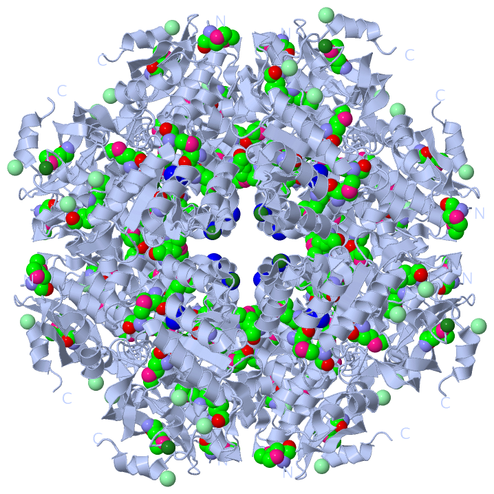
Asymmetric Unit (4, 20)
|
Sites (9, 9)
Asymmetric Unit (9, 9)
|
SS Bonds (0, 0)| (no "SS Bond" information available for 4HCD) |
Cis Peptide Bonds (4, 4)
Asymmetric Unit
|
||||||||||||||||||||
SAPs(SNPs)/Variants (0, 0)| (no "SAP(SNP)/Variant" information available for 4HCD) |
PROSITE Motifs (0, 0)| (no "PROSITE Motif" information available for 4HCD) |
Exons (0, 0)| (no "Exon" information available for 4HCD) |
Sequences/Alignments
Asymmetric Unit
Chain A from PDB Type:PROTEIN Length:384
SCOP domains d4hcda1 A:-1-126 automated matches d4hcda2 A:127-382 automated matches SCOP domains
CATH domains ------------------------------------------------------------------------------------------------------------------------------------------------------------------------------------------------------------------------------------------------------------------------------------------------------------------------------------------------------------------------------------------------ CATH domains
Pfam domains ------------------------------------------------------------------------------------------------------------------------------------------------------------------------------------------------------------------------------------------------------------------------------------------------------------------------------------------------------------------------------------------------ Pfam domains
SAPs(SNPs) ------------------------------------------------------------------------------------------------------------------------------------------------------------------------------------------------------------------------------------------------------------------------------------------------------------------------------------------------------------------------------------------------ SAPs(SNPs)
PROSITE ------------------------------------------------------------------------------------------------------------------------------------------------------------------------------------------------------------------------------------------------------------------------------------------------------------------------------------------------------------------------------------------------ PROSITE
Transcript ------------------------------------------------------------------------------------------------------------------------------------------------------------------------------------------------------------------------------------------------------------------------------------------------------------------------------------------------------------------------------------------------ Transcript
4hcd A -1 SHmIITDVEVRVFRTTTRRHSDSAGHAHPGPAHQVEQAmLTVRTEDGQEGHSFTAPEIVRPHVIEKFVKKVLIGEDHRDRERLWQDLAHWQRGSAAQLTDRTLAVVDCALWDLAGRSLGQPVYKLIGGYRDKVLAYGSImCGDELEGGLATPEDYGRFAETLVKRGYKGIKLHTWmPPVSWAPDVKmDLKACAAVREAVGPDIRLmIDAFHWYSRTDALALGRGLEKLGFDWIEEPmDEQSLSSYKWLSDNLDIPVVGPESAAGKHWHRAEWIKAGACDILRTGVNDVGGITPALKTmHLAEAFGmECEVHGNTAmNLHVVAATKNCRWYERGLLHPFLEYDDGHDYLKSLSDPmDRDGFVHVPDRPGLGEDIDFTFIDNNRVR 382
| 8 18 28 38 48 58 68 78 88 98 108 118 128 138 148 158 168 | 178 |188 198 | 208 218 228 |238 248 258 268 278 288 298 | 308 | 318 328 338 348 | 358 368 378
| 37-MSE 138-MSE 174-MSE 185-MSE 204-MSE 235-MSE 296-MSE 304-MSE 314-MSE 353-MSE
1-MSE
|
||||||||||||||||||||
SCOP Domains (2, 2)
Asymmetric Unit

|
CATH Domains (0, 0)| (no "CATH Domain" information available for 4HCD) |
Pfam Domains (0, 0)| (no "Pfam Domain" information available for 4HCD) |
Gene Ontology (4, 4)|
Asymmetric Unit(hide GO term definitions) |
Interactive Views
|
||||||||||||||||||||||||||||||||||||||||||||||||||||||||||||||||||||||||||||||||||||||||||||||||||||||||||||||||||||||||||||||||||||||||||||||||||||||||||||||||||||||||||||||||||||||||||||||||||||||||||||||||||||||||||||||||||||||||||||||||
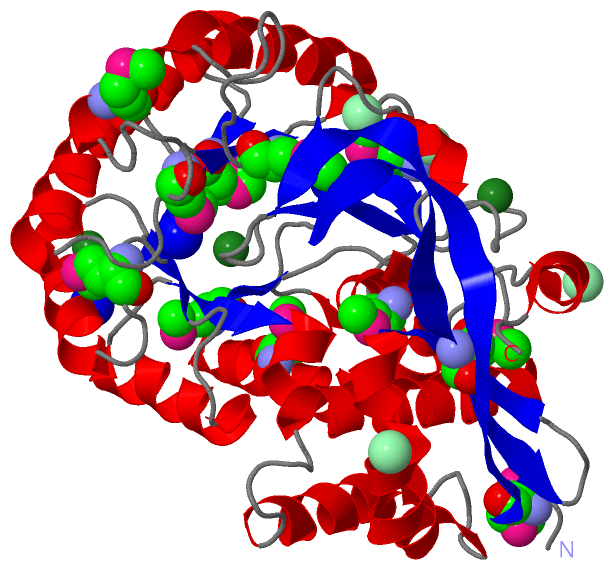
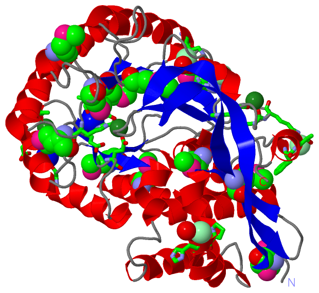
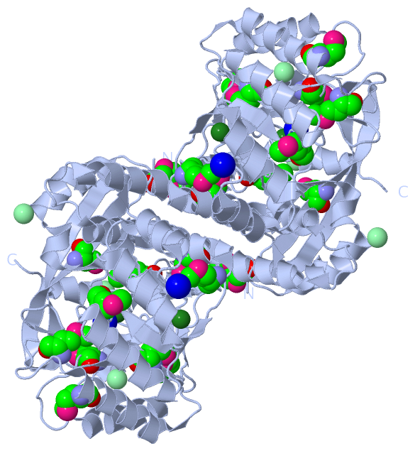
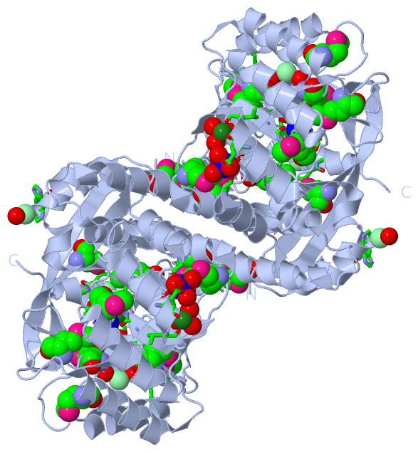
Still Images
|
||||||||||||||||
Databases
|
||||||||||||||||||||||||||||||||||||||||||||||||||||||||||||||||||||||||||||||||||||||||||||||||||||||||||||||||||||||||||||||||||||||||||||||||||||||||||||||||
Analysis Tools
|
|||||||||||||||||||||||||||||||||||||||||||||||||||||||||||||
Entries Sharing at Least One Protein Chain (UniProt ID)
Related Entries Specified in the PDB File
|
|