Show PDB file:   
         Plain Text   HTML   (compressed file size)
QuickSearch:   
by PDB,NDB,UniProt,PROSITE Code or Search Term(s)  
(-)Asym./Biol. Unit
collapse expand < >
Image Asym./Biol. Unit
Asym./Biol. Unit  (Jmol Viewer)

(-) Description

Title :  THE VON WILLEBRAND FACTOR A3 DOMAIN BINDING REGION OF TYPE III COLLAGEN STABILIZED BY THE CYSTEINE KNOT
 
Authors :  S. P. Boudko, H. P. Bachinger
Date :  05 Sep 12  (Deposition) - 17 Oct 12  (Release) - 16 Jan 13  (Revision)
Method :  X-RAY DIFFRACTION
Resolution :  1.49
Chains :  Asym./Biol. Unit :  A,B,C
Keywords :  Collagen Triple Helix, Type Iii Collagen Cysteine Knot, Blood Clotting, Von Willebrand Factor A3 Domain, 4-Hydroxylation, Extracellular Matrix, Structural Protein (Keyword Search: [Gene Ontology, PubMed, Web (Google))
 
Reference :  S. P. Boudko, H. P. Bachinger
The Nc2 Domain Of Type Ix Collagen Determines The Chain Register Of The Triple Helix.
J. Biol. Chem. V. 287 44536 2012
PubMed-ID: 23132862  |  Reference-DOI: 10.1074/JBC.M112.417543

(-) Compounds

Molecule 1 - TYPE III COLLAGEN FRAGMENT IN A HOST PEPTIDE STABILIZED BY THE CYSTEINE KNOT
    ChainsA, B, C
    EngineeredYES
    FragmentVON WILLEBRAND FACTOR BINDING FRAGMENT
    Organism CommonHUMAN
    Organism ScientificHOMO SAPIENS
    Organism Taxid9606
    Other DetailsTHIS SEQUENCE CONTAINS TWO FRAGMENTS OF HUMAN TYPE III COLLAGEN: THE VON WILLEBRAND FACTOR A3 DOMAIN BINDING REGION AND THE CYSTEINE KNOT
    SyntheticYES

 Structural Features

(-) Chains, Units

  123
Asymmetric/Biological Unit ABC

Summary Information (see also Sequences/Alignments below)

(-) Ligands, Modified Residues, Ions  (1, 18)

Asymmetric/Biological Unit (1, 18)
No.NameCountTypeFull Name
1HYP18Mod. Amino Acid4-HYDROXYPROLINE

(-) Sites  (0, 0)

(no "Site" information available for 4GYX)

(-) SS Bonds  (1, 1)

Asymmetric/Biological Unit
No.Residues
1A:27 -B:28

(-) Cis Peptide Bonds  (0, 0)

(no "Cis Peptide Bond" information available for 4GYX)

 Sequence-Structure Mapping

(-) SAPs(SNPs)/Variants  (0, 0)

(no "SAP(SNP)/Variant" information available for 4GYX)

(-) PROSITE Motifs  (0, 0)

(no "PROSITE Motif" information available for 4GYX)

(-) Exons   (0, 0)

(no "Exon" information available for 4GYX)

(-) Sequences/Alignments

Asymmetric/Biological Unit
   Reformat: Number of residues per line =  ('0' or empty: single-line sequence representation)
  Number of residues per labelling interval =   
  UniProt sequence: complete  aligned part    
   Show mapping: SCOP domains CATH domains Pfam domains Secondary structure (by author)
SAPs(SNPs) PROSITE motifs Exons
(details for a mapped element are shown in a popup box when the mouse pointer rests over it)
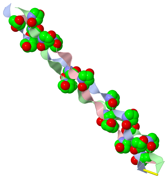
Chain A from PDB  Type:PROTEIN  Length:28
                                                           
               SCOP domains ---------------------------- SCOP domains
               CATH domains ---------------------------- CATH domains
               Pfam domains ---------------------------- Pfam domains
         Sec.struct. author ............................ Sec.struct. author
                 SAPs(SNPs) ---------------------------- SAPs(SNPs)
                    PROSITE ---------------------------- PROSITE
                 Transcript ---------------------------- Transcript
                  4gyx A  1 GPpGPpGPRGQpGVMGFpGPpGPpGPCC 28
                              |  |  10 |     |20|  |    
                              |  |     |     |  |  |    
                              3-HYP    |     |  |  |    
                                 6-HYP |     |  |  |    
                                      12-HYP |  |  |    
                                            18-HYP |    
                                               21-HYP   
                                                  24-HYP

Chain B from PDB  Type:PROTEIN  Length:28
                                                           
               SCOP domains ---------------------------- SCOP domains
               CATH domains ---------------------------- CATH domains
               Pfam domains ---------------------------- Pfam domains
         Sec.struct. author ............................ Sec.struct. author
                 SAPs(SNPs) ---------------------------- SAPs(SNPs)
                    PROSITE ---------------------------- PROSITE
                 Transcript ---------------------------- Transcript
                  4gyx B  1 GPpGPpGPRGQpGVMGFpGPpGPpGPCC 28
                              |  |  10 |     |20|  |    
                              3-HYP    |     |  |  |    
                                 6-HYP |     |  |  |    
                                      12-HYP |  |  |    
                                            18-HYP |    
                                               21-HYP   
                                                  24-HYP

Chain C from PDB  Type:PROTEIN  Length:26
                                                         
               SCOP domains -------------------------- SCOP domains
               CATH domains -------------------------- CATH domains
               Pfam domains -------------------------- Pfam domains
         Sec.struct. author .......................... Sec.struct. author
                 SAPs(SNPs) -------------------------- SAPs(SNPs)
                    PROSITE -------------------------- PROSITE
                 Transcript -------------------------- Transcript
                  4gyx C  1 GPpGPpGPRGQpGVMGFpGPpGPpGP 26
                              |  |  10 |     |20|  |  
                              3-HYP    |     |  |  |  
                                 6-HYP |     |  |  |  
                                      12-HYP |  |  |  
                                            18-HYP |  
                                               21-HYP 
                                                  24-HYP

   Legend:   → Mismatch (orange background)
  - → Gap (green background, '-', border residues have a numbering label)
    → Modified Residue (blue background, lower-case, 'x' indicates undefined single-letter code, labelled with number + name)
  x → Chemical Group (purple background, 'x', labelled with number + name, e.g. ACE or NH2)
  extra numbering lines below/above indicate numbering irregularities and modified residue names etc., number ends below/above '|'

 Classification and Annotation

(-) SCOP Domains  (0, 0)

(no "SCOP Domain" information available for 4GYX)

(-) CATH Domains  (0, 0)

(no "CATH Domain" information available for 4GYX)

(-) Pfam Domains  (0, 0)

(no "Pfam Domain" information available for 4GYX)

(-) Gene Ontology  (38, 38)

Asymmetric/Biological Unit(hide GO term definitions)

 Visualization

(-) Interactive Views

Asymmetric/Biological Unit
  Complete Structure
    Jena3D(integrated viewing of ligand, site, SAP, PROSITE, SCOP information)
    WebMol | AstexViewer[tm]@PDBe
(Java Applets, require no local installation except for Java; loading may be slow)
    STRAP
(Java WebStart application, automatic local installation, requires Java; full application with system access!)
    RasMol
(require local installation)
    Molscript (VRML)
(requires installation of a VRML viewer; select preferred view via VRML and generate a mono or stereo PDF format file)
 
  Ligands, Modified Residues, Ions
    HYP  [ RasMol | Jena3D ]  +environment [ RasMol | Jena3D ]
 
  Sites
(no "Sites" information available for 4gyx)
 
  Cis Peptide Bonds
(no "Cis Peptide Bonds" information available for 4gyx)
 

(-) Still Images

Jmol
  protein: cartoon or spacefill or dots and stick; nucleic acid: cartoon and stick; ligands: spacefill; active site: stick
Molscript
  protein, nucleic acid: cartoon; ligands: spacefill; active site: ball and stick

 Databases and Analysis Tools

(-) Databases

Access by PDB/NDB ID
  4gyx
    Family and Domain InformationProDom | SYSTERS
    General Structural InformationGlycoscienceDB | MMDB | NDB | OCA | PDB | PDBe | PDBj | PDBsum | PDBWiki | PQS | PROTEOPEDIA
    Orientation in MembranesOPM
    Protein SurfaceSURFACE
    Secondary StructureDSSP (structure derived) | HSSP (homology derived)
    Structural GenomicsGeneCensus
    Structural NeighboursCE | VAST
    Structure ClassificationCATH | Dali | SCOP
    Validation and Original DataBMRB Data View | BMRB Restraints Grid | EDS | PROCHECK | RECOORD | WHAT_CHECK
 
Access by UniProt ID/Accession number
  CO3A1_HUMAN | P02461
    Comparative Protein Structure ModelsModBase
    Genomic InformationEnsembl
    Protein-protein InteractionDIP
    Sequence, Family and Domain InformationInterPro | Pfam | SMART | UniProtKB/SwissProt
 
Access by Enzyme Classificator   (EC Number)
  (no 'Enzyme Classificator' available)
    General Enzyme InformationBRENDA | EC-PDB | Enzyme | IntEnz
    PathwayKEGG | MetaCyc
 
Access by Disease Identifier   (MIM ID)
  (no 'MIM ID' available)
    Disease InformationOMIM
 
Access by GenAge ID
  (no 'GenAge ID' available)
    Age Related InformationGenAge

(-) Analysis Tools

Access by PDB/NDB ID
    Domain InformationXDom
    Interatomic Contacts of Structural UnitsCSU
    Ligand-protein ContactsLPC
    Protein CavitiescastP
    Sequence and Secondary StructurePDBCartoon
    Structure AlignmentSTRAP(Java WebStart application, automatic local installation, requires Java; full application with system access!)
    Structure and Sequence BrowserSTING
 
Access by UniProt ID/Accession number
  CO3A1_HUMAN | P02461
    Protein Disorder PredictionDisEMBL | FoldIndex | GLOBPLOT (for more information see DisProt)

 Related Entries

(-) Entries Sharing at Least One Protein Chain (UniProt ID)

UniProtKB/Swiss-Prot
        CO3A1_HUMAN | P024612v53 3dmw 4ae2 4aej 4ak3

(-) Related Entries Specified in the PDB File

(no "Related Entries Specified in the PDB File" available for 4GYX)