Show PDB file:   
         Plain Text   HTML   (compressed file size)
QuickSearch:   
by PDB,NDB,UniProt,PROSITE Code or Search Term(s)  
(-)Asymmetric Unit
(-)Asym. Unit - sites
(-)Biological Unit 1
(-)Biol. Unit 1 - sites
(-)Biological Unit 2
collapse expand < >
Image Asymmetric Unit
Asymmetric Unit  (Jmol Viewer)
Image Asym. Unit - sites
Asym. Unit - sites  (Jmol Viewer)
Image Biological Unit 1
Biological Unit 1  (Jmol Viewer)
Image Biol. Unit 1 - sites
Biol. Unit 1 - sites  (Jmol Viewer)
Image Biological Unit 2
Biological Unit 2  (Jmol Viewer)

(-) Description

Title :  CRYSTAL STRUCTURE OF AMIDOTRANSFERASE HISH FROM VIBRIO CHOLERAE
 
Authors :  N. Maltseva, Y. Kim, S. Shatsman, W. F. Anderson, A. Joachimiak, Center Structural Genomics Of Infectious Diseases (Csgid)
Date :  29 Aug 12  (Deposition) - 12 Sep 12  (Release) - 12 Sep 12  (Revision)
Method :  X-RAY DIFFRACTION
Resolution :  1.91
Chains :  Asym. Unit :  A,B
Biol. Unit 1:  A  (1x)
Biol. Unit 2:  B  (1x)
Keywords :  Structural Genomics, Niaid, National Institute Of Allergy And Infectious Diseases, Center For Structural Genomics Of Infectious Diseases, Csgid, Alpha-Beta Sandwich, Cytosol, Transferase (Keyword Search: [Gene Ontology, PubMed, Web (Google))
 
Reference :  N. Maltseva, Y. Kim, S. Shatsman, W. F. Anderson, A. Joachimiak, Center For Structural Genomics Of Infectious Diseases (Csgid)
Crystal Structure Of Amidotransferase Hish From Vibrio Cholerae.
To Be Published 2013
PubMed: search

(-) Compounds

Molecule 1 - IMIDAZOLE GLYCEROL PHOSPHATE SYNTHASE SUBUNIT HISH
    ChainsA, B
    EC Number2.4.2.-
    EngineeredYES
    Expression SystemESCHERICHIA COLI
    Expression System PlasmidPMCSG28
    Expression System StrainBL21 MAGIC
    Expression System Taxid511693
    Expression System Vector TypePLASMID
    GeneHISH, VC_1136
    Organism ScientificVIBRIO CHOLERAE
    Organism Taxid243277
    StrainO1 BIOVAR EL TOR STR. N16961
    SynonymIGP SYNTHASE GLUTAMINE AMIDOTRANSFERASE SUBUNIT, IGP SYNTHASE SUBUNIT HISH, IMGP SYNTHASE SUBUNIT HISH, IGPS SUBUNIT HISH

 Structural Features

(-) Chains, Units

  12
Asymmetric Unit AB
Biological Unit 1 (1x)A 
Biological Unit 2 (1x) B

Summary Information (see also Sequences/Alignments below)

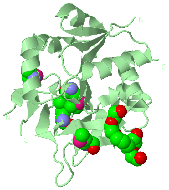
(-) Ligands, Modified Residues, Ions  (5, 12)

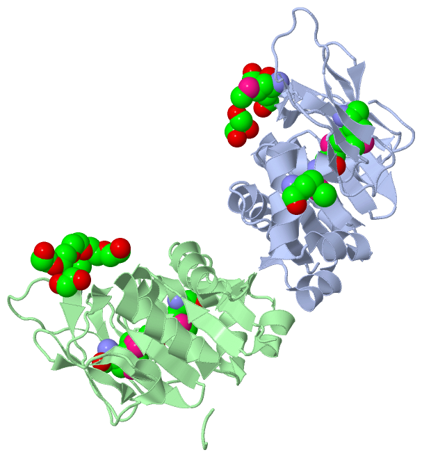
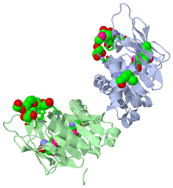
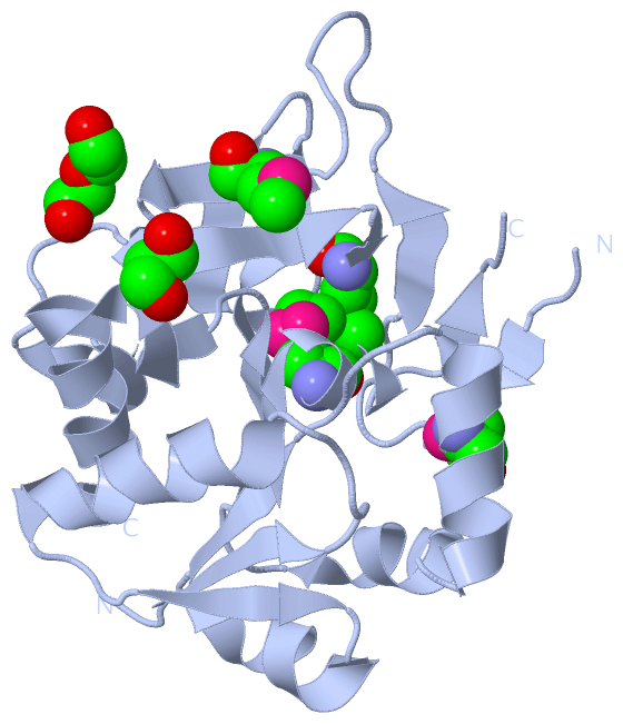
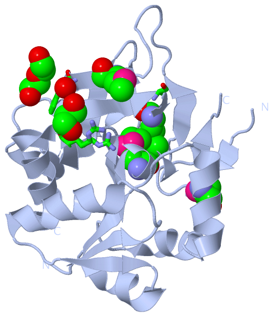
Asymmetric Unit (5, 12)
No.NameCountTypeFull Name
11PE1Ligand/IonPENTAETHYLENE GLYCOL
2EDO1Ligand/Ion1,2-ETHANEDIOL
3GOL1Ligand/IonGLYCEROL
4MSE8Mod. Amino AcidSELENOMETHIONINE
5PEG1Ligand/IonDI(HYDROXYETHYL)ETHER
Biological Unit 1 (3, 6)
No.NameCountTypeFull Name
11PE-1Ligand/IonPENTAETHYLENE GLYCOL
2EDO-1Ligand/Ion1,2-ETHANEDIOL
3GOL1Ligand/IonGLYCEROL
4MSE4Mod. Amino AcidSELENOMETHIONINE
5PEG1Ligand/IonDI(HYDROXYETHYL)ETHER
Biological Unit 2 (3, 6)
No.NameCountTypeFull Name
11PE1Ligand/IonPENTAETHYLENE GLYCOL
2EDO1Ligand/Ion1,2-ETHANEDIOL
3GOL-1Ligand/IonGLYCEROL
4MSE4Mod. Amino AcidSELENOMETHIONINE
5PEG-1Ligand/IonDI(HYDROXYETHYL)ETHER

(-) Sites  (4, 4)

Asymmetric Unit (4, 4)
No.NameEvidenceResiduesDescription
1AC1SOFTWARETRP A:125 , ARG A:188 , HOH A:426BINDING SITE FOR RESIDUE GOL A 301
2AC2SOFTWARETRP A:125 , SER B:151BINDING SITE FOR RESIDUE PEG A 302
3AC3SOFTWARESER A:151 , TRP B:125 , TYR B:147 , ARG B:188 , EDO B:302BINDING SITE FOR RESIDUE 1PE B 301
4AC4SOFTWARE1PE B:301BINDING SITE FOR RESIDUE EDO B 302

(-) SS Bonds  (0, 0)

(no "SS Bond" information available for 4GUD)

(-) Cis Peptide Bonds  (2, 2)

Asymmetric Unit
No.Residues
1Leu A:120 -Pro A:121
2Leu B:120 -Pro B:121

 Sequence-Structure Mapping

(-) SAPs(SNPs)/Variants  (0, 0)

(no "SAP(SNP)/Variant" information available for 4GUD)

(-) PROSITE Motifs  (0, 0)

(no "PROSITE Motif" information available for 4GUD)

(-) Exons   (0, 0)

(no "Exon" information available for 4GUD)

(-) Sequences/Alignments

Asymmetric Unit
   Reformat: Number of residues per line =  ('0' or empty: single-line sequence representation)
  Number of residues per labelling interval =   
  UniProt sequence: complete  aligned part    
   Show mapping: SCOP domains CATH domains Pfam domains Secondary structure (by author)
SAPs(SNPs) PROSITE motifs Exons
(details for a mapped element are shown in a popup box when the mouse pointer rests over it)
Chain A from PDB  Type:PROTEIN  Length:197
                                                                                                                                                                                                                                     
               SCOP domains d4guda_ A: automated matches                                                                                                                                                                          SCOP domains
               CATH domains ----------------------------------------------------------------------------------------------------------------------------------------------------------------------------------------------------- CATH domains
               Pfam domains ----------------------------------------------------------------------------------------------------------------------------------------------------------------------------------------------------- Pfam domains
         Sec.struct. author ..eeee.....hhhhhhhhhhhh...eeee.hhhhhhhh.eeee....hhhhhhhhhhhh.hhhhhhhh...eeeehhhhhh...ee......ee.......eeee.........eeeee..ee...hhhhh.......eeeee.ee......eeeeee...eeeeeeee..eeee..hhhhhhhhhhhhhhhhhhh Sec.struct. author
                 SAPs(SNPs) ----------------------------------------------------------------------------------------------------------------------------------------------------------------------------------------------------- SAPs(SNPs)
                    PROSITE ----------------------------------------------------------------------------------------------------------------------------------------------------------------------------------------------------- PROSITE
                 Transcript ----------------------------------------------------------------------------------------------------------------------------------------------------------------------------------------------------- Transcript
                 4gud A   3 QNVVIIDTGCANISSVKFAIERLGYAVTISRDPQVVLAADKLFLPGVGTASEAmKNLTERDLIELVKRVEKPLLGICLGmQLLGKLSEEKDEIVQCLGLVDGEVRLLQTGDLPLPHmGWNTVQVKEGHPLFNGIEPDAYFYFVHSFAmPVGDYTIAQCEYGQPFSAAIQAGNYYGVQFHPERSSKAGARLIQNFLEL 203
                                    12        22        32        42        52   |    62        72        82        92|      106       116      |126       136       146       156       166       176       186       196       
                                                                                56-MSE                    82-MSE    92|                       123-MSE                        154-MSE                                             
                                                                                                                     97                                                                                                          

Chain B from PDB  Type:PROTEIN  Length:201
                                                                                                                                                                                                                                         
               SCOP domains d4gudb_ B: automated matches                                                                                                                                                                              SCOP domains
               CATH domains --------------------------------------------------------------------------------------------------------------------------------------------------------------------------------------------------------- CATH domains
               Pfam domains --------------------------------------------------------------------------------------------------------------------------------------------------------------------------------------------------------- Pfam domains
         Sec.struct. author ...eeee.....hhhhhhhhhhhh....eee.hhhhhhhh.eeee....hhhhhhhhhhhh.hhhhhhhh...eeeehhhhhh...ee.....ee.......eeee.........eeeee..ee...hhhhh.......eeeee.ee......eeeeee...eeeeeeee..eeee..hhhhhhhhhhhhhhhhhh..... Sec.struct. author
                 SAPs(SNPs) --------------------------------------------------------------------------------------------------------------------------------------------------------------------------------------------------------- SAPs(SNPs)
                    PROSITE --------------------------------------------------------------------------------------------------------------------------------------------------------------------------------------------------------- PROSITE
                 Transcript --------------------------------------------------------------------------------------------------------------------------------------------------------------------------------------------------------- Transcript
                 4gud B   2 TQNVVIIDTGCANISSVKFAIERLGYAVTISRDPQVVLAADKLFLPGVGTASEAmKNLTERDLIELVKRVEKPLLGICLGmQLLGKLSEEKEIVQCLGLVDGEVRLLQTGDLPLPHmGWNTVQVKEGHPLFNGIEPDAYFYFVHSFAmPVGDYTIAQCEYGQPFSAAIQAGNYYGVQFHPERSSKAGARLIQNFLELNLYF 209
                                    11        21        31        41        51    |   61        71        81|       91||     106       116      |126       136       146       156       166       176       186       196      |208 
                                                                                 56-MSE                    82-MSE    92|                      123-MSE                        154-MSE                                          203|   
                                                                                                                      98                                                                                                       206   

   Legend:   → Mismatch (orange background)
  - → Gap (green background, '-', border residues have a numbering label)
    → Modified Residue (blue background, lower-case, 'x' indicates undefined single-letter code, labelled with number + name)
  x → Chemical Group (purple background, 'x', labelled with number + name, e.g. ACE or NH2)
  extra numbering lines below/above indicate numbering irregularities and modified residue names etc., number ends below/above '|'

 Classification and Annotation

(-) SCOP Domains  (1, 2)

Asymmetric Unit

(-) CATH Domains  (0, 0)

(no "CATH Domain" information available for 4GUD)

(-) Pfam Domains  (0, 0)

(no "Pfam Domain" information available for 4GUD)

(-) Gene Ontology  (8, 8)

Asymmetric Unit(hide GO term definitions)

 Visualization

(-) Interactive Views

Asymmetric Unit
  Complete Structure
    Jena3D(integrated viewing of ligand, site, SAP, PROSITE, SCOP information)
    WebMol | AstexViewer[tm]@PDBe
(Java Applets, require no local installation except for Java; loading may be slow)
    STRAP
(Java WebStart application, automatic local installation, requires Java; full application with system access!)
    RasMol
(require local installation)
    Molscript (VRML)
(requires installation of a VRML viewer; select preferred view via VRML and generate a mono or stereo PDF format file)
 
  Ligands, Modified Residues, Ions
    1PE  [ RasMol | Jena3D ]  +environment [ RasMol | Jena3D ]
    EDO  [ RasMol | Jena3D ]  +environment [ RasMol | Jena3D ]
    GOL  [ RasMol | Jena3D ]  +environment [ RasMol | Jena3D ]
    MSE  [ RasMol | Jena3D ]  +environment [ RasMol | Jena3D ]
    PEG  [ RasMol | Jena3D ]  +environment [ RasMol | Jena3D ]
 
  Sites
    AC1  [ RasMol ]  +environment [ RasMol ]
    AC2  [ RasMol ]  +environment [ RasMol ]
    AC3  [ RasMol ]  +environment [ RasMol ]
    AC4  [ RasMol ]  +environment [ RasMol ]
 
  Cis Peptide Bonds
    Leu A:120 - Pro A:121   [ RasMol ]  
    Leu B:120 - Pro B:121   [ RasMol ]  
 
Biological Units
  Complete Structure
    Biological Unit 1  [ Jena3D ]
    Biological Unit 2  [ Jena3D ]

(-) Still Images

Jmol
  protein: cartoon or spacefill or dots and stick; nucleic acid: cartoon and stick; ligands: spacefill; active site: stick
Molscript
  protein, nucleic acid: cartoon; ligands: spacefill; active site: ball and stick

 Databases and Analysis Tools

(-) Databases

Access by PDB/NDB ID
  4gud
    Family and Domain InformationProDom | SYSTERS
    General Structural InformationGlycoscienceDB | MMDB | NDB | OCA | PDB | PDBe | PDBj | PDBsum | PDBWiki | PQS | PROTEOPEDIA
    Orientation in MembranesOPM
    Protein SurfaceSURFACE
    Secondary StructureDSSP (structure derived) | HSSP (homology derived)
    Structural GenomicsGeneCensus
    Structural NeighboursCE | VAST
    Structure ClassificationCATH | Dali | SCOP
    Validation and Original DataBMRB Data View | BMRB Restraints Grid | EDS | PROCHECK | RECOORD | WHAT_CHECK
 
Access by UniProt ID/Accession number
  HIS5_VIBCH | Q9KSX0
    Comparative Protein Structure ModelsModBase
    Genomic InformationEnsembl
    Protein-protein InteractionDIP
    Sequence, Family and Domain InformationInterPro | Pfam | SMART | UniProtKB/SwissProt
 
Access by Enzyme Classificator   (EC Number)
  2.4.2.-
    General Enzyme InformationBRENDA | EC-PDB | Enzyme | IntEnz
    PathwayKEGG | MetaCyc
 
Access by Disease Identifier   (MIM ID)
  (no 'MIM ID' available)
    Disease InformationOMIM
 
Access by GenAge ID
  (no 'GenAge ID' available)
    Age Related InformationGenAge

(-) Analysis Tools

Access by PDB/NDB ID
    Domain InformationXDom
    Interatomic Contacts of Structural UnitsCSU
    Ligand-protein ContactsLPC
    Protein CavitiescastP
    Sequence and Secondary StructurePDBCartoon
    Structure AlignmentSTRAP(Java WebStart application, automatic local installation, requires Java; full application with system access!)
    Structure and Sequence BrowserSTING
 
Access by UniProt ID/Accession number
  HIS5_VIBCH | Q9KSX0
    Protein Disorder PredictionDisEMBL | FoldIndex | GLOBPLOT (for more information see DisProt)

 Related Entries

(-) Entries Sharing at Least One Protein Chain (UniProt ID)

(no "Entries Sharing at Least One Protein Chain" available for 4GUD)

(-) Related Entries Specified in the PDB File

(no "Related Entries Specified in the PDB File" available for 4GUD)