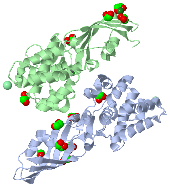
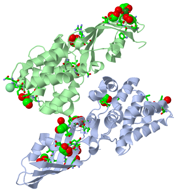
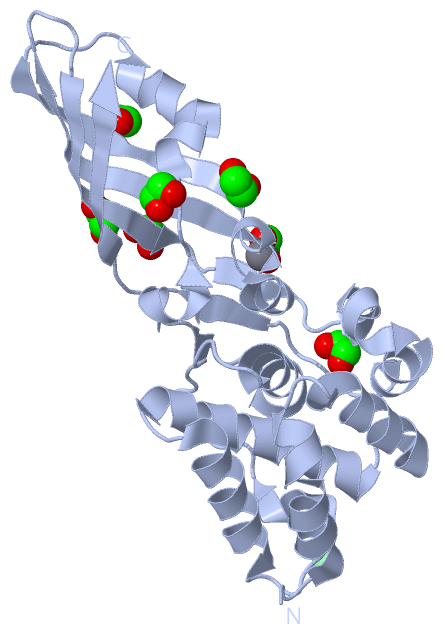
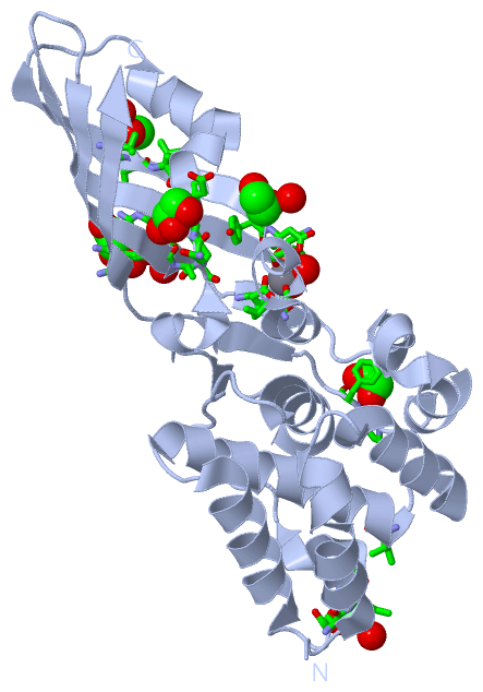
| No. | Name | Evidence | Residues | Description |
|---|
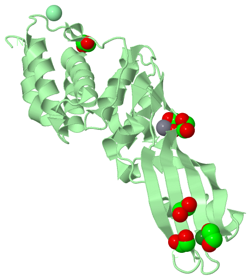
| 01 | AC1 | SOFTWARE | LEU A:202 , ASN A:204 , ASP A:232 , EDO A:302 , HOH A:427 , HOH A:436 | BINDING SITE FOR RESIDUE CA A 301 |
| 02 | AC2 | SOFTWARE | ASN A:204 , ASP A:232 , VAL A:233 , CA A:301 , HOH A:427 , HOH A:436 | BINDING SITE FOR RESIDUE EDO A 302 |
| 03 | AC3 | SOFTWARE | ARG A:208 , ARG A:228 , ALA A:229 , PHE A:230 , HOH A:581 | BINDING SITE FOR RESIDUE EDO A 303 |
| 04 | AC4 | SOFTWARE | ARG A:208 | BINDING SITE FOR RESIDUE EDO A 304 |
| 05 | AC5 | SOFTWARE | LEU A:183 , ASP A:184 , VAL A:185 , HOH A:419 | BINDING SITE FOR RESIDUE EDO A 305 |
| 06 | AC6 | SOFTWARE | ASN A:159 , VAL A:160 , ASP A:161 , HOH A:517 | BINDING SITE FOR RESIDUE EDO A 306 |
| 07 | AC7 | SOFTWARE | PRO A:257 , PHE A:258 | BINDING SITE FOR RESIDUE EDO A 307 |
| 08 | AC8 | SOFTWARE | GLN A:112 , ILE A:113 , ASN A:114 , HOH A:678 , ILE B:74 , EDO B:304 | BINDING SITE FOR RESIDUE CL A 308 |
| 09 | AC9 | SOFTWARE | TYR A:189 , ASP A:190 , HOH A:691 | BINDING SITE FOR RESIDUE EDO A 309 |
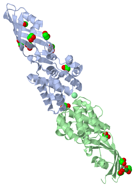
| 10 | BC1 | SOFTWARE | LEU B:202 , ASN B:204 , ASP B:232 , EDO B:303 , HOH B:439 , HOH B:516 | BINDING SITE FOR RESIDUE CA B 301 |
| 11 | BC2 | SOFTWARE | ASP B:214 , EDO B:308 , HOH B:522 , HOH B:528 , HOH B:529 | BINDING SITE FOR RESIDUE MG B 302 |
| 12 | BC3 | SOFTWARE | ASN B:204 , ASP B:232 , VAL B:233 , CA B:301 , HOH B:439 , HOH B:441 , HOH B:516 | BINDING SITE FOR RESIDUE EDO B 303 |
| 13 | BC4 | SOFTWARE | GLN A:112 , ASN A:114 , CL A:308 , HOH A:460 , GLY B:73 , LYS B:77 , GLU B:115 , HOH B:649 | BINDING SITE FOR RESIDUE EDO B 304 |
| 14 | BC5 | SOFTWARE | ARG B:208 , ARG B:228 , ALA B:229 , HOH B:527 | BINDING SITE FOR RESIDUE EDO B 305 |
| 15 | BC6 | SOFTWARE | ASP B:214 , ASN B:216 , ALA B:220 , TRP B:221 , THR B:222 | BINDING SITE FOR RESIDUE EDO B 306 |
| 16 | BC7 | SOFTWARE | THR B:212 , THR B:222 , SER B:224 , HOH B:599 | BINDING SITE FOR RESIDUE EDO B 307 |
| 17 | BC8 | SOFTWARE | THR B:180 , ASP B:214 , MG B:302 , HOH B:528 , HOH B:529 | BINDING SITE FOR RESIDUE EDO B 308 |
| 18 | BC9 | SOFTWARE | ILE A:74 , GLN B:112 , ILE B:113 , ASN B:114 | BINDING SITE FOR RESIDUE CL B 309 |
 Description
 Compounds