Show PDB file:   
         Plain Text   HTML   (compressed file size)
QuickSearch:   
by PDB,NDB,UniProt,PROSITE Code or Search Term(s)  
(-)Asymmetric Unit
(-)Asym. Unit - sites
(-)Biological Unit 1
(-)Biol. Unit 1 - sites
(-)Biological Unit 2
(-)Biological Unit 3
collapse expand < >
Image Asymmetric Unit
Asymmetric Unit  (Jmol Viewer)
Image Asym. Unit - sites
Asym. Unit - sites  (Jmol Viewer)
Image Biological Unit 1
Biological Unit 1  (Jmol Viewer)
Image Biol. Unit 1 - sites
Biol. Unit 1 - sites  (Jmol Viewer)
Image Biological Unit 2
Biological Unit 2  (Jmol Viewer)
Image Biological Unit 3
Biological Unit 3  (Jmol Viewer)

(-) Description

Title :  CRYSTAL STRUCTURE OF A RAS P21 PROTEIN ACTIVATOR (RASA1) FROM HOMO SAPIENS AT 2.25 A RESOLUTION
 
Authors :  Joint Center For Structural Genomics (Jcsg), Partnership For Biology (Tcell)
Date :  27 Jun 12  (Deposition) - 18 Jul 12  (Release) - 21 Oct 15  (Revision)
Method :  X-RAY DIFFRACTION
Resolution :  2.25
Chains :  Asym. Unit :  A,B,C
Biol. Unit 1:  A  (1x)
Biol. Unit 2:  B  (1x)
Biol. Unit 3:  C  (1x)
Keywords :  Gtpase Activating Protein, Sh3 Domain, Ras Signaling Pathway, Structural Genomics, Joint Center For Structural Genomics, Jcsg, Protein Structure Initiative, Psi-Biology, Signaling Protein (Keyword Search: [Gene Ontology, PubMed, Web (Google))
 
Reference :  Joint Center For Structural Genomics (Jcsg), Partnership For T-Cell Biology (Tcell)
Crystal Structure Of A Ras P21 Protein Activator (Rasa1) From Homo Sapiens At 2. 25 A Resolution
To Be Published
PubMed: search

(-) Compounds

Molecule 1 - RAS GTPASE-ACTIVATING PROTEIN 1
    ChainsA, B, C
    EngineeredYES
    Expression SystemESCHERICHIA COLI
    Expression System PlasmidSPEEDET
    Expression System StrainHK100
    Expression System Taxid562
    Expression System Vector TypePLASMID
    FragmentSH3 DOMAIN RESIDUES 281-341
    GeneBC033015, RASA, RASA1
    Organism CommonHUMAN
    Organism ScientificHOMO SAPIENS
    Organism Taxid9606
    SynonymGAP, GTPASE-ACTIVATING PROTEIN, RASGAP, RAS P21 PROTEIN ACTIVATOR, P120GAP

 Structural Features

(-) Chains, Units

  123
Asymmetric Unit ABC
Biological Unit 1 (1x)A  
Biological Unit 2 (1x) B 
Biological Unit 3 (1x)  C

Summary Information (see also Sequences/Alignments below)

(-) Ligands, Modified Residues, Ions  (4, 11)

Asymmetric Unit (4, 11)
No.NameCountTypeFull Name
1CL3Ligand/IonCHLORIDE ION
2GOL1Ligand/IonGLYCEROL
3MSE6Mod. Amino AcidSELENOMETHIONINE
4PO41Ligand/IonPHOSPHATE ION
Biological Unit 1 (2, 3)
No.NameCountTypeFull Name
1CL-1Ligand/IonCHLORIDE ION
2GOL-1Ligand/IonGLYCEROL
3MSE2Mod. Amino AcidSELENOMETHIONINE
4PO41Ligand/IonPHOSPHATE ION
Biological Unit 2 (1, 2)
No.NameCountTypeFull Name
1CL-1Ligand/IonCHLORIDE ION
2GOL-1Ligand/IonGLYCEROL
3MSE2Mod. Amino AcidSELENOMETHIONINE
4PO4-1Ligand/IonPHOSPHATE ION
Biological Unit 3 (2, 3)
No.NameCountTypeFull Name
1CL-1Ligand/IonCHLORIDE ION
2GOL1Ligand/IonGLYCEROL
3MSE2Mod. Amino AcidSELENOMETHIONINE
4PO4-1Ligand/IonPHOSPHATE ION

(-) Sites  (5, 5)

Asymmetric Unit (5, 5)
No.NameEvidenceResiduesDescription
1AC1SOFTWAREARG A:281 , HIS A:310 , ASP C:326BINDING SITE FOR RESIDUE PO4 A 1001
2AC2SOFTWARELYS A:292 , ASP A:295BINDING SITE FOR RESIDUE CL A 1002
3AC3SOFTWAREARG B:281BINDING SITE FOR RESIDUE CL B 401
4AC4SOFTWAREASP C:295BINDING SITE FOR RESIDUE CL C 401
5AC5SOFTWAREARG B:285 , VAL B:339 , GLY B:340 , ARG B:341 , LYS C:292 , GLU C:327BINDING SITE FOR RESIDUE GOL C 402

(-) SS Bonds  (0, 0)

(no "SS Bond" information available for 4FSS)

(-) Cis Peptide Bonds  (0, 0)

(no "Cis Peptide Bond" information available for 4FSS)

 Sequence-Structure Mapping

(-) SAPs(SNPs)/Variants  (0, 0)

(no "SAP(SNP)/Variant" information available for 4FSS)

(-) PROSITE Motifs  (0, 0)

(no "PROSITE Motif" information available for 4FSS)

(-) Exons   (0, 0)

(no "Exon" information available for 4FSS)

(-) Sequences/Alignments

Asymmetric Unit
   Reformat: Number of residues per line =  ('0' or empty: single-line sequence representation)
  Number of residues per labelling interval =   
  UniProt sequence: complete  aligned part    
   Show mapping: SCOP domains CATH domains Pfam domains Secondary structure (by author)
SAPs(SNPs) PROSITE motifs Exons
(details for a mapped element are shown in a popup box when the mouse pointer rests over it)
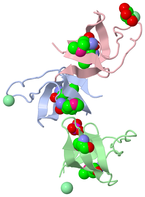
Chain A from PDB  Type:PROTEIN  Length:60
                                                                                            
               SCOP domains d4fssa_ A: automated matches                                 SCOP domains
               CATH domains ------------------------------------------------------------ CATH domains
               Pfam domains ------------------------------------------------------------ Pfam domains
         Sec.struct. author .eeeee...................eeeeeee....eeeeee.....eeeee...eee.. Sec.struct. author
                 SAPs(SNPs) ------------------------------------------------------------ SAPs(SNPs)
                    PROSITE ------------------------------------------------------------ PROSITE
                 Transcript ------------------------------------------------------------ Transcript
                 4fss A 281 RRRVRAILPYTKVPDTDEISFLKGDmFIVHNELEDGWmWVTNLRTDEQGLIVEDLVEEVG 340
                                   290       300     | 310       320       330       340
                                                   306-MSE     318-MSE                  

Chain B from PDB  Type:PROTEIN  Length:61
                                                                                             
               SCOP domains d4fssb_ B: automated matches                                  SCOP domains
               CATH domains ------------------------------------------------------------- CATH domains
               Pfam domains ------------------------------------------------------------- Pfam domains
         Sec.struct. author ..eeee...................eeeeeee....eeeeee.....eeeee...eee... Sec.struct. author
                 SAPs(SNPs) ------------------------------------------------------------- SAPs(SNPs)
                    PROSITE ------------------------------------------------------------- PROSITE
                 Transcript ------------------------------------------------------------- Transcript
                 4fss B 281 RRRVRAILPYTKVPDTDEISFLKGDmFIVHNELEDGWmWVTNLRTDEQGLIVEDLVEEVGR 341
                                   290       300     | 310       320       330       340 
                                                   306-MSE     318-MSE                   

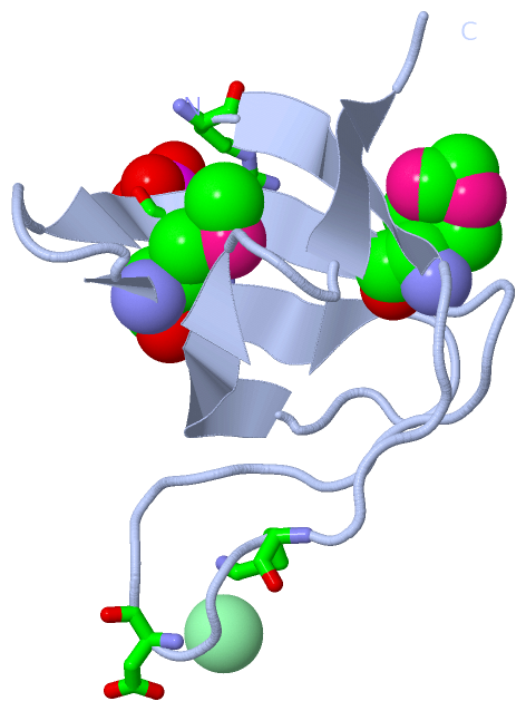
Chain C from PDB  Type:PROTEIN  Length:61
                                                                                             
               SCOP domains d4fssc_ C: automated matches                                  SCOP domains
               CATH domains ------------------------------------------------------------- CATH domains
               Pfam domains ------------------------------------------------------------- Pfam domains
         Sec.struct. author ..eeee...................eeeeeee....eeeeee....eeeeee...eeee.. Sec.struct. author
                 SAPs(SNPs) ------------------------------------------------------------- SAPs(SNPs)
                    PROSITE ------------------------------------------------------------- PROSITE
                 Transcript ------------------------------------------------------------- Transcript
                 4fss C 281 RRRVRAILPYTKVPDTDEISFLKGDmFIVHNELEDGWmWVTNLRTDEQGLIVEDLVEEVGR 341
                                   290       300     | 310       320       330       340 
                                                   306-MSE     318-MSE                   

   Legend:   → Mismatch (orange background)
  - → Gap (green background, '-', border residues have a numbering label)
    → Modified Residue (blue background, lower-case, 'x' indicates undefined single-letter code, labelled with number + name)
  x → Chemical Group (purple background, 'x', labelled with number + name, e.g. ACE or NH2)
  extra numbering lines below/above indicate numbering irregularities and modified residue names etc., number ends below/above '|'

 Classification and Annotation

(-) SCOP Domains  (1, 3)

Asymmetric Unit

(-) CATH Domains  (0, 0)

(no "CATH Domain" information available for 4FSS)

(-) Pfam Domains  (0, 0)

(no "Pfam Domain" information available for 4FSS)

(-) Gene Ontology  (27, 27)

Asymmetric Unit(hide GO term definitions)

 Visualization

(-) Interactive Views

Asymmetric Unit
  Complete Structure
    Jena3D(integrated viewing of ligand, site, SAP, PROSITE, SCOP information)
    WebMol | AstexViewer[tm]@PDBe
(Java Applets, require no local installation except for Java; loading may be slow)
    STRAP
(Java WebStart application, automatic local installation, requires Java; full application with system access!)
    RasMol
(require local installation)
    Molscript (VRML)
(requires installation of a VRML viewer; select preferred view via VRML and generate a mono or stereo PDF format file)
 
  Ligands, Modified Residues, Ions
    CL  [ RasMol | Jena3D ]  +environment [ RasMol | Jena3D ]
    GOL  [ RasMol | Jena3D ]  +environment [ RasMol | Jena3D ]
    MSE  [ RasMol | Jena3D ]  +environment [ RasMol | Jena3D ]
    PO4  [ RasMol | Jena3D ]  +environment [ RasMol | Jena3D ]
 
  Sites
    AC1  [ RasMol ]  +environment [ RasMol ]
    AC2  [ RasMol ]  +environment [ RasMol ]
    AC3  [ RasMol ]  +environment [ RasMol ]
    AC4  [ RasMol ]  +environment [ RasMol ]
    AC5  [ RasMol ]  +environment [ RasMol ]
 
  Cis Peptide Bonds
(no "Cis Peptide Bonds" information available for 4fss)
 
Biological Units
  Complete Structure
    Biological Unit 1  [ Jena3D ]
    Biological Unit 2  [ Jena3D ]
    Biological Unit 3  [ Jena3D ]

(-) Still Images

Jmol
  protein: cartoon or spacefill or dots and stick; nucleic acid: cartoon and stick; ligands: spacefill; active site: stick
Molscript
  protein, nucleic acid: cartoon; ligands: spacefill; active site: ball and stick

 Databases and Analysis Tools

(-) Databases

Access by PDB/NDB ID
  4fss
    Family and Domain InformationProDom | SYSTERS
    General Structural InformationGlycoscienceDB | MMDB | NDB | OCA | PDB | PDBe | PDBj | PDBsum | PDBWiki | PQS | PROTEOPEDIA
    Orientation in MembranesOPM
    Protein SurfaceSURFACE
    Secondary StructureDSSP (structure derived) | HSSP (homology derived)
    Structural GenomicsGeneCensus
    Structural NeighboursCE | VAST
    Structure ClassificationCATH | Dali | SCOP
    Validation and Original DataBMRB Data View | BMRB Restraints Grid | EDS | PROCHECK | RECOORD | WHAT_CHECK
 
Access by UniProt ID/Accession number
  RASA1_HUMAN | P20936
    Comparative Protein Structure ModelsModBase
    Genomic InformationEnsembl
    Protein-protein InteractionDIP
    Sequence, Family and Domain InformationInterPro | Pfam | SMART | UniProtKB/SwissProt
 
Access by Enzyme Classificator   (EC Number)
  (no 'Enzyme Classificator' available)
    General Enzyme InformationBRENDA | EC-PDB | Enzyme | IntEnz
    PathwayKEGG | MetaCyc
 
Access by Disease Identifier   (MIM ID)
  (no 'MIM ID' available)
    Disease InformationOMIM
 
Access by GenAge ID
  (no 'GenAge ID' available)
    Age Related InformationGenAge

(-) Analysis Tools

Access by PDB/NDB ID
    Domain InformationXDom
    Interatomic Contacts of Structural UnitsCSU
    Ligand-protein ContactsLPC
    Protein CavitiescastP
    Sequence and Secondary StructurePDBCartoon
    Structure AlignmentSTRAP(Java WebStart application, automatic local installation, requires Java; full application with system access!)
    Structure and Sequence BrowserSTING
 
Access by UniProt ID/Accession number
  RASA1_HUMAN | P20936
    Protein Disorder PredictionDisEMBL | FoldIndex | GLOBPLOT (for more information see DisProt)

 Related Entries

(-) Entries Sharing at Least One Protein Chain (UniProt ID)

UniProtKB/Swiss-Prot
        RASA1_HUMAN | P209361wer 1wq1 2gqi 2gsb 2j05 2j06 2m51

(-) Related Entries Specified in the PDB File

(no "Related Entries Specified in the PDB File" available for 4FSS)