|
|
|
|
 Description
|
|
Compounds
|
||||||||||||||||||||||||||||||||||||||||||||||||||||||||||||||||||||||||||||||||||||||||||||||||||||||||||||||||||||||||||||||||||||||||||||||||||||||||||||||||||||||||||||||||||||||||||||||||||||||||||||||||||||||||||||||||||||||||||||||||||
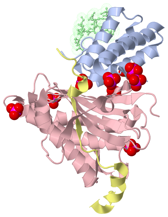
Chains, Units
Summary Information (see also Sequences/Alignments below) |
Ligands, Modified Residues, Ions (2, 9)| Asymmetric Unit (2, 9) Biological Unit 1 (1, 1) Biological Unit 2 (0, 0) Biological Unit 3 (1, 8) Biological Unit 4 (0, 0) |
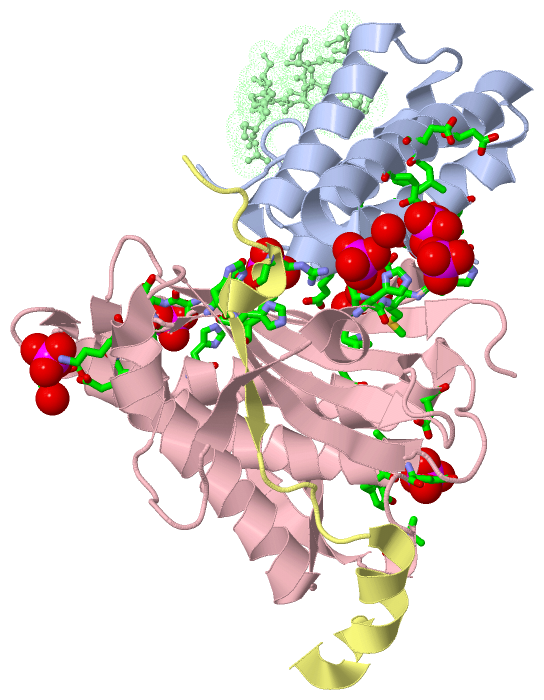
Sites (9, 9)
Asymmetric Unit (9, 9)
|
SS Bonds (0, 0)| (no "SS Bond" information available for 4FJO) |
Cis Peptide Bonds (0, 0)| (no "Cis Peptide Bond" information available for 4FJO) |
SAPs(SNPs)/Variants (0, 0)| (no "SAP(SNP)/Variant" information available for 4FJO) |
PROSITE Motifs (0, 0)| (no "PROSITE Motif" information available for 4FJO) |
Exons (0, 0)| (no "Exon" information available for 4FJO) |
Sequences/Alignments
Asymmetric Unit
Chain A from PDB Type:PROTEIN Length:97
SCOP domains ------------------------------------------------------------------------------------------------- SCOP domains
CATH domains ------------------------------------------------------------------------------------------------- CATH domains
Pfam domains ------------------------------------------------------------------------------------------------- Pfam domains
SAPs(SNPs) ------------------------------------------------------------------------------------------------- SAPs(SNPs)
PROSITE ------------------------------------------------------------------------------------------------- PROSITE
Transcript ------------------------------------------------------------------------------------------------- Transcript
4fjo A 1153 AAPNLAGAVEFSDVKTLLKEWITTISDPMEEDILQVVRYCTDLIEEKDLEKLDLVIKYMKRLMQQSVESVWNMAFDFILDNVQVVLQQTYGSTLKVT 1249
1162 1172 1182 1192 1202 1212 1222 1232 1242
Chain B from PDB Type:PROTEIN Length:10
SCOP domains ---------- SCOP domains
CATH domains ---------- CATH domains
Pfam domains ---------- Pfam domains
SAPs(SNPs) ---------- SAPs(SNPs)
PROSITE ---------- PROSITE
Transcript ---------- Transcript
4fjo B 565 SFFDKKRSER 574
574
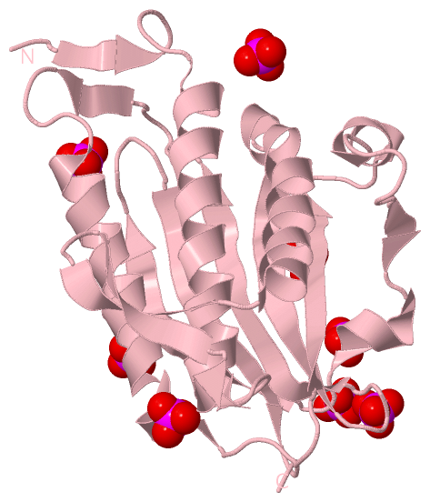
Chain C from PDB Type:PROTEIN Length:210
SCOP domains ------------------------------------------------------------------------------------------------------------------------------------------------------------------------------------------------------------------ SCOP domains
CATH domains ------------------------------------------------------------------------------------------------------------------------------------------------------------------------------------------------------------------ CATH domains
Pfam domains ------------------------------------------------------------------------------------------------------------------------------------------------------------------------------------------------------------------ Pfam domains
SAPs(SNPs) ------------------------------------------------------------------------------------------------------------------------------------------------------------------------------------------------------------------ SAPs(SNPs)
PROSITE ------------------------------------------------------------------------------------------------------------------------------------------------------------------------------------------------------------------ PROSITE
Transcript ------------------------------------------------------------------------------------------------------------------------------------------------------------------------------------------------------------------ Transcript
4fjo C 1 MTTLTRQDLNFGQVVADVLSEFLEVAVHLILYVREVYPVGIFQKRKKYNVPVQMSCHPELNQYIQDTLHCVKPLLEKNDVEKVVVVILDKEHRPVEKFVFEITQPPLLSINSDSLLSHVEQLLAAFILKISVCDAVLDHNPPGCTFTVLVHTREAATRNMEKIQVIKDFPWILADEQDVHMHDPRLIPLKTMTSDILKMQLYVEERAHKN 210
10 20 30 40 50 60 70 80 90 100 110 120 130 140 150 160 170 180 190 200 210
Chain D from PDB Type:PROTEIN Length:30
SCOP domains ------------------------------ SCOP domains
CATH domains ------------------------------ CATH domains
Pfam domains ------------------------------ Pfam domains
SAPs(SNPs) ------------------------------ SAPs(SNPs)
PROSITE ------------------------------ PROSITE
Transcript ------------------------------ Transcript
4fjo D 1865 GSFTPRTAHILKPLMSPPSREEIVATLLDH 1894
1874 1884 1894
|
||||||||||||||||||||
SCOP Domains (0, 0)| (no "SCOP Domain" information available for 4FJO) |
CATH Domains (0, 0)| (no "CATH Domain" information available for 4FJO) |
Pfam Domains (0, 0)| (no "Pfam Domain" information available for 4FJO) |
Gene Ontology (51, 73)|
Asymmetric Unit(hide GO term definitions) |
Interactive Views
|
||||||||||||||||||||||||||||||||||||||||||||||||||||||||||||||||||||||||||||||||||||||||||||||||||||||||||||||||||||||||||||||||||||||||||||||||||||||||||||||||||||||||||||||||||||||||||||||||||||||||||||||||||||||
Still Images
|
||||||||||||||||
Databases
|
||||||||||||||||||||||||||||||||||||||||||||||||||||||||||||||||||||||||||||||||||||||||||||||||||||||||||||||||||||||||||||||||||||||||||||||||||||||||||||||||||||||||||||||||||||||||||||||||||||||||||||||||||||||||||||||||||||||||||||||||||||||||||||||
Analysis Tools
|
||||||||||||||||||||||||||||||||||||||||||||||||||||||||||||||||||||||||||||||||||||||||||||||
Entries Sharing at Least One Protein Chain (UniProt ID)
Related Entries Specified in the PDB File
|
|