Show PDB file:   
         Plain Text   HTML   (compressed file size)
QuickSearch:   
by PDB,NDB,UniProt,PROSITE Code or Search Term(s)  
(-)Asym./Biol. Unit
(-)Asym./Biol. Unit - sites
collapse expand < >
Image Asym./Biol. Unit
Asym./Biol. Unit  (Jmol Viewer)
Image Asym./Biol. Unit - sites
Asym./Biol. Unit - sites  (Jmol Viewer)

(-) Description

Title :  X-RAY CRYSTAL STRUCTURE OF CORYNEBACTERIUM GLUTAMICUM NRDH-REDOXIN AT 1.5A
 
Authors :  J. Messens, V. T. Dufe, W. Khadija
Date :  11 Jun 12  (Deposition) - 06 Feb 13  (Release) - 03 Apr 13  (Revision)
Method :  X-RAY DIFFRACTION
Resolution :  1.50
Chains :  Asym./Biol. Unit :  A
Keywords :  Thioredoxin Fold, Oxidoreductase (Keyword Search: [Gene Ontology, PubMed, Web (Google))
 
Reference :  K. Van Laer, A. M. Dziewulska, M. Fislage, K. Wahni, A. Hbeddou, J. F. Collet, W. Versees, L. M. Mateos, V. Tamu Dufe, J. Messens
Nrdh-Redoxin Of Mycobacterium Tuberculosis And Corynebacterium Glutamicum Dimerizes At High Protein Concentration And Exclusively Receives Electrons From Thioredoxin Reductase.
J. Biol. Chem. V. 288 7942 2013
PubMed-ID: 23362277  |  Reference-DOI: 10.1074/JBC.M112.392688

(-) Compounds

Molecule 1 - PUTATIVE GLUTAREDOXIN NRDH
    ChainsA
    EC Number1.20.4.3
    EngineeredYES
    Expression SystemESCHERICHIA COLI
    Expression System Taxid562
    GeneNRDH
    Organism ScientificCORYNEBACTERIUM GLUTAMICUM
    Organism Taxid1718

 Structural Features

(-) Chains, Units

  1
Asymmetric/Biological Unit A

Summary Information (see also Sequences/Alignments below)
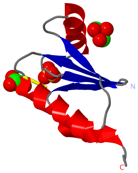
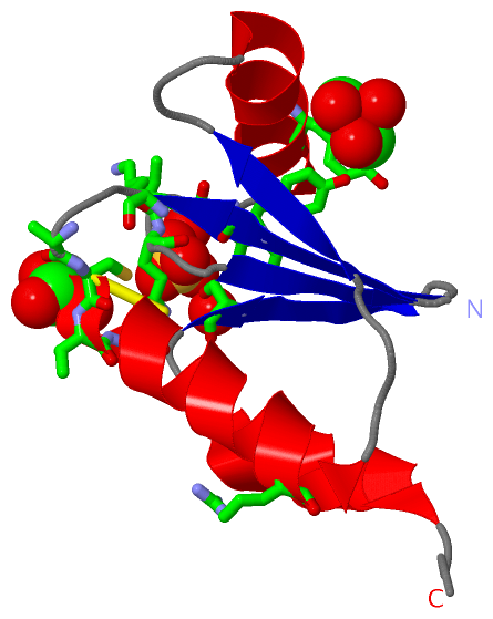
(-) Ligands, Modified Residues, Ions  (2, 3)

Asymmetric/Biological Unit (2, 3)
No.NameCountTypeFull Name
1GOL2Ligand/IonGLYCEROL
2SO41Ligand/IonSULFATE ION

(-) Sites  (3, 3)

Asymmetric Unit (3, 3)
No.NameEvidenceResiduesDescription
1AC1SOFTWARETYR A:48 , LEU A:49 , GLN A:50 , SER A:62 , HOH A:255 , HOH A:261BINDING SITE FOR RESIDUE SO4 A 101
2AC2SOFTWARELYS A:8 , ALA A:10 , CYS A:11 , VAL A:12 , GLN A:13 , LEU A:49BINDING SITE FOR RESIDUE GOL A 102
3AC3SOFTWARETYR A:6 , ARG A:23 , TYR A:42BINDING SITE FOR RESIDUE GOL A 103

(-) SS Bonds  (1, 1)

Asymmetric/Biological Unit
No.Residues
1A:11 -A:14

(-) Cis Peptide Bonds  (1, 1)

Asymmetric/Biological Unit
No.Residues
1Ala A:51 -Pro A:52

 Sequence-Structure Mapping

(-) SAPs(SNPs)/Variants  (0, 0)

(no "SAP(SNP)/Variant" information available for 4FIW)

(-) PROSITE Motifs  (0, 0)

(no "PROSITE Motif" information available for 4FIW)

(-) Exons   (0, 0)

(no "Exon" information available for 4FIW)

(-) Sequences/Alignments

Asymmetric/Biological Unit
   Reformat: Number of residues per line =  ('0' or empty: single-line sequence representation)
  Number of residues per labelling interval =   
  UniProt sequence: complete  aligned part    
   Show mapping: SCOP domains CATH domains Pfam domains Secondary structure (by author)
SAPs(SNPs) PROSITE motifs Exons
(details for a mapped element are shown in a popup box when the mouse pointer rests over it)
Chain A from PDB  Type:PROTEIN  Length:76
                                                                                                           
               SCOP domains d4fiwa_ A: Glutaredoxin-like NRDH-redoxin                                    SCOP domains
               CATH domains ---------------------------------------------------------------------------- CATH domains
               Pfam domains ---------------------------------------------------------------------------- Pfam domains
         Sec.struct. author .eeeee...hhhhhhhhhhhhhh....eeee...hhhhhhhhhhh......eeee..eeee..hhhhhhhhh.... Sec.struct. author
                 SAPs(SNPs) ---------------------------------------------------------------------------- SAPs(SNPs)
                    PROSITE ---------------------------------------------------------------------------- PROSITE
                 Transcript ---------------------------------------------------------------------------- Transcript
                  4fiw A  2 AITVYTKPACVQCNATKKALDRAGLEYDLVDISLDEEAREYVLALGYLQAPVVVADGSHWSGFRPERIREMATAAA 77
                                    11        21        31        41        51        61        71      

   Legend:   → Mismatch (orange background)
  - → Gap (green background, '-', border residues have a numbering label)
    → Modified Residue (blue background, lower-case, 'x' indicates undefined single-letter code, labelled with number + name)
  x → Chemical Group (purple background, 'x', labelled with number + name, e.g. ACE or NH2)
  extra numbering lines below/above indicate numbering irregularities and modified residue names etc., number ends below/above '|'

 Classification and Annotation

(-) SCOP Domains  (1, 1)

Asymmetric/Biological Unit

(-) CATH Domains  (0, 0)

(no "CATH Domain" information available for 4FIW)

(-) Pfam Domains  (0, 0)

(no "Pfam Domain" information available for 4FIW)

(-) Gene Ontology  (5, 5)

Asymmetric/Biological Unit(hide GO term definitions)

 Visualization

(-) Interactive Views

Asymmetric/Biological Unit
  Complete Structure
    Jena3D(integrated viewing of ligand, site, SAP, PROSITE, SCOP information)
    WebMol | AstexViewer[tm]@PDBe
(Java Applets, require no local installation except for Java; loading may be slow)
    STRAP
(Java WebStart application, automatic local installation, requires Java; full application with system access!)
    RasMol
(require local installation)
    Molscript (VRML)
(requires installation of a VRML viewer; select preferred view via VRML and generate a mono or stereo PDF format file)
 
  Ligands, Modified Residues, Ions
    GOL  [ RasMol | Jena3D ]  +environment [ RasMol | Jena3D ]
    SO4  [ RasMol | Jena3D ]  +environment [ RasMol | Jena3D ]
 
  Sites
    AC1  [ RasMol ]  +environment [ RasMol ]
    AC2  [ RasMol ]  +environment [ RasMol ]
    AC3  [ RasMol ]  +environment [ RasMol ]
 
  Cis Peptide Bonds
    Ala A:51 - Pro A:52   [ RasMol ]  
 

(-) Still Images

Jmol
  protein: cartoon or spacefill or dots and stick; nucleic acid: cartoon and stick; ligands: spacefill; active site: stick
Molscript
  protein, nucleic acid: cartoon; ligands: spacefill; active site: ball and stick

 Databases and Analysis Tools

(-) Databases

Access by PDB/NDB ID
  4fiw
    Family and Domain InformationProDom | SYSTERS
    General Structural InformationGlycoscienceDB | MMDB | NDB | OCA | PDB | PDBe | PDBj | PDBsum | PDBWiki | PQS | PROTEOPEDIA
    Orientation in MembranesOPM
    Protein SurfaceSURFACE
    Secondary StructureDSSP (structure derived) | HSSP (homology derived)
    Structural GenomicsGeneCensus
    Structural NeighboursCE | VAST
    Structure ClassificationCATH | Dali | SCOP
    Validation and Original DataBMRB Data View | BMRB Restraints Grid | EDS | PROCHECK | RECOORD | WHAT_CHECK
 
Access by UniProt ID/Accession number
  H7C6B5_CORGT | H7C6B5
    Comparative Protein Structure ModelsModBase
    Genomic InformationEnsembl
    Protein-protein InteractionDIP
    Sequence, Family and Domain InformationInterPro | Pfam | SMART | UniProtKB/TrEMBL
 
Access by Enzyme Classificator   (EC Number)
  1.20.4.3
    General Enzyme InformationBRENDA | EC-PDB | Enzyme | IntEnz
    PathwayKEGG | MetaCyc
 
Access by Disease Identifier   (MIM ID)
  (no 'MIM ID' available)
    Disease InformationOMIM
 
Access by GenAge ID
  (no 'GenAge ID' available)
    Age Related InformationGenAge

(-) Analysis Tools

Access by PDB/NDB ID
    Domain InformationXDom
    Interatomic Contacts of Structural UnitsCSU
    Ligand-protein ContactsLPC
    Protein CavitiescastP
    Sequence and Secondary StructurePDBCartoon
    Structure AlignmentSTRAP(Java WebStart application, automatic local installation, requires Java; full application with system access!)
    Structure and Sequence BrowserSTING
 
Access by UniProt ID/Accession number
  H7C6B5_CORGT | H7C6B5
    Protein Disorder PredictionDisEMBL | FoldIndex | GLOBPLOT (for more information see DisProt)

 Related Entries

(-) Entries Sharing at Least One Protein Chain (UniProt ID)

(no "Entries Sharing at Least One Protein Chain" available for 4FIW)

(-) Related Entries Specified in the PDB File

(no "Related Entries Specified in the PDB File" available for 4FIW)