|
|
|
|
 Description
|
|
Compounds
|
||||||||||||||||||||||||||||||||||||||||||||||||||||
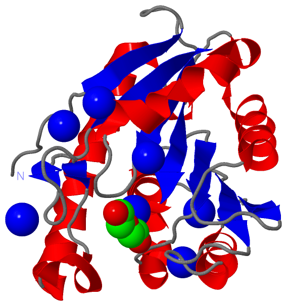
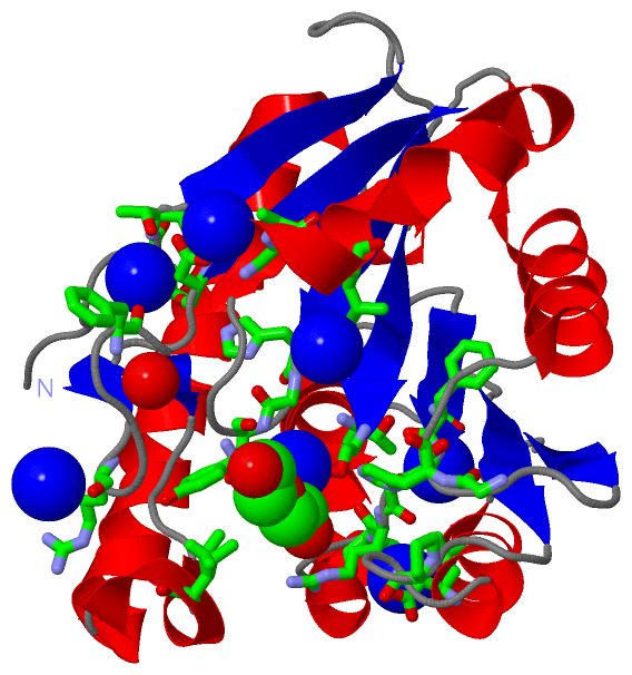
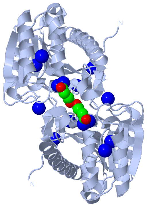
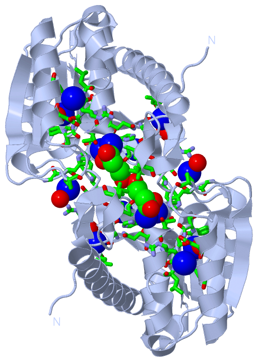
Chains, Units
Summary Information (see also Sequences/Alignments below) |
Ligands, Modified Residues, Ions (2, 8)| Asymmetric Unit (2, 8) Biological Unit 1 (1, 2) |
Sites (8, 8)
Asymmetric Unit (8, 8)
|
SS Bonds (0, 0)| (no "SS Bond" information available for 4FHZ) |
Cis Peptide Bonds (0, 0)| (no "Cis Peptide Bond" information available for 4FHZ) |
SAPs(SNPs)/Variants (0, 0)| (no "SAP(SNP)/Variant" information available for 4FHZ) |
PROSITE Motifs (0, 0)| (no "PROSITE Motif" information available for 4FHZ) |
Exons (0, 0)| (no "Exon" information available for 4FHZ) |
Sequences/Alignments
Asymmetric Unit
Chain A from PDB Type:PROTEIN Length:220
SCOP domains ---------------------------------------------------------------------------------------------------------------------------------------------------------------------------------------------------------------------------- SCOP domains
CATH domains ---------------------------------------------------------------------------------------------------------------------------------------------------------------------------------------------------------------------------- CATH domains
Pfam domains ---------------------------------------------------------------------------------------------------------------------------------------------------------------------------------------------------------------------------- Pfam domains
SAPs(SNPs) ---------------------------------------------------------------------------------------------------------------------------------------------------------------------------------------------------------------------------- SAPs(SNPs)
PROSITE ---------------------------------------------------------------------------------------------------------------------------------------------------------------------------------------------------------------------------- PROSITE
Transcript ---------------------------------------------------------------------------------------------------------------------------------------------------------------------------------------------------------------------------- Transcript
4fhz A 48 IMTRKLTFGRRGAAPGEATSLVVFLHGYGADGADLLGLAEPLAPHLPGTAFVAPDAPEPCRANGFGFQWFPIPWLDGSSETAAAEGMAAAARDLDAFLDERLAEEGLPPEALALVGFSQGTMMALHVAPRRAEEIAGIVGFSGRLLAPERLAEEARSKPPVLLVHGDADPVVPFADMSLAGEALAEAGFTTYGHVMKGTGHGIAPDGLSVALAFLKERLP 267
57 67 77 87 97 107 117 127 137 147 157 167 177 187 197 207 217 227 237 247 257 267
|
||||||||||||||||||||
SCOP Domains (0, 0)| (no "SCOP Domain" information available for 4FHZ) |
CATH Domains (0, 0)| (no "CATH Domain" information available for 4FHZ) |
Pfam Domains (0, 0)| (no "Pfam Domain" information available for 4FHZ) |
Gene Ontology (1, 1)|
Asymmetric Unit(hide GO term definitions) |
Interactive Views
|
||||||||||||||||||||||||||||||||||||||||||||||||||||||||||||||||||||||||||||||||||||||||||||||||||||||||||||||||||||||||||||||||||||||||||||||||||||||||||||||||||||||||||||||||||||||||||||||||
Still Images
|
||||||||||||||||
Databases
|
||||||||||||||||||||||||||||||||||||||||||||||||||||||||||||||||||||||||||||||||||||||||||||||||||||||||||||||||||||||||||||||||||||||||||||||||||||||||||||||||
Analysis Tools
|
|||||||||||||||||||||||||||||||||||||||||||||||||||||||||||||
Entries Sharing at Least One Protein Chain (UniProt ID)
Related Entries Specified in the PDB File
|
|