Show PDB file:   
         Plain Text   HTML   (compressed file size)
QuickSearch:   
by PDB,NDB,UniProt,PROSITE Code or Search Term(s)  
(-)Asymmetric Unit
(-)Asym. Unit - sites
(-)Biological Unit 1
(-)Biol. Unit 1 - sites
(-)Biological Unit 2
(-)Biological Unit 3
collapse expand < >
Image Asymmetric Unit
Asymmetric Unit  (Jmol Viewer)
Image Asym. Unit - sites
Asym. Unit - sites  (Jmol Viewer)
Image Biological Unit 1
Biological Unit 1  (Jmol Viewer)
Image Biol. Unit 1 - sites
Biol. Unit 1 - sites  (Jmol Viewer)
Image Biological Unit 2
Biological Unit 2  (Jmol Viewer)
Image Biological Unit 3
Biological Unit 3  (Jmol Viewer)

(-) Description

Title :  PLASMODIUM FALCIPARUM ATG8 IN COMPLEX WITH PLASMODIUM FALCIPARUM ATG3 PEPTIDE
 
Authors :  A. U. P. Hain, J. Bosch
Date :  16 Apr 12  (Deposition) - 20 Feb 13  (Release) - 20 Feb 13  (Revision)
Method :  X-RAY DIFFRACTION
Resolution :  2.22
Chains :  Asym. Unit :  A,B,C,D,E,F
Biol. Unit 1:  A,D  (1x)
Biol. Unit 2:  B,E  (1x)
Biol. Unit 3:  C,F  (1x)
Keywords :  Ubiquitin Fold, Transport Protein (Keyword Search: [Gene Ontology, PubMed, Web (Google))
 
Reference :  A. U. Hain, R. R. Weltzer, H. Hammond, B. Jayabalasingham, R. R. Dinglasan, D. R. Graham, D. R. Colquhoun, I. Coppens, J. Bosch
Structural Characterization And Inhibition Of The Plasmodiu Atg8-Atg3 Interaction.
J. Struct. Biol. V. 180 551 2012
PubMed-ID: 22982544  |  Reference-DOI: 10.1016/J.JSB.2012.09.001

(-) Compounds

Molecule 1 - MICROTUBULE-ASSOCIATED PROTEIN 1 LIGHT CHAIN 3
    ChainsA, B, C
    EngineeredYES
    Expression SystemESCHERICHIA COLI
    Expression System PlasmidMBP-PRSF-5
    Expression System StrainROSETTA 2
    Expression System Taxid562
    Expression System Vector TypePRSF-1B
    GenePF10_0193
    MutationYES
    Organism ScientificPLASMODIUM FALCIPARUM
    Organism Taxid36329
    Strain3D7
 
Molecule 2 - AUTOPHAGY-RELATED PROTEIN 3
    ChainsD, E, F
    EngineeredYES
    FragmentUNP RESIDUES 103-110
    Organism ScientificPLASMODIUM FALCIPARUM
    Organism Taxid36329
    Other DetailsPEPTIDE
    SyntheticYES

 Structural Features

(-) Chains, Units

  123456
Asymmetric Unit ABCDEF
Biological Unit 1 (1x)A  D  
Biological Unit 2 (1x) B  E 
Biological Unit 3 (1x)  C  F

Summary Information (see also Sequences/Alignments below)

(-) Ligands, Modified Residues, Ions  (4, 11)

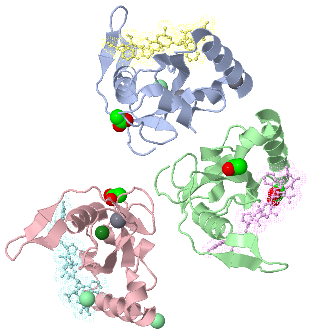
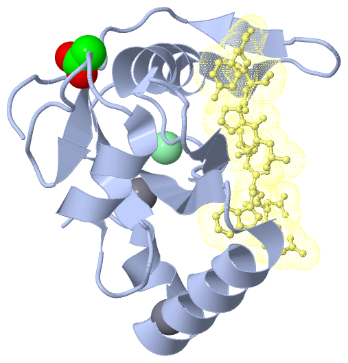
Asymmetric Unit (4, 11)
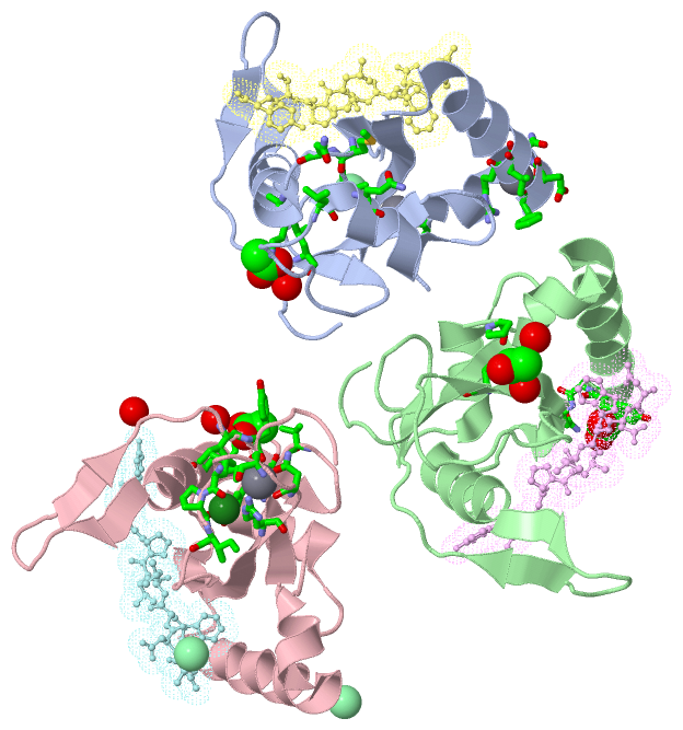
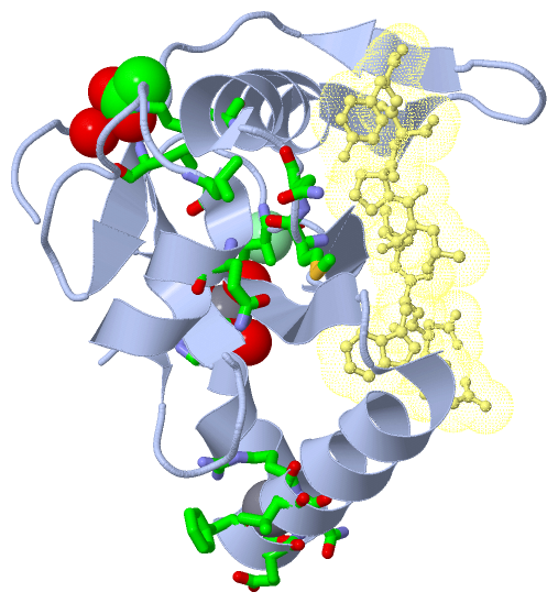
No.NameCountTypeFull Name
1ACT4Ligand/IonACETATE ION
2CA3Ligand/IonCALCIUM ION
3CL3Ligand/IonCHLORIDE ION
4MG1Ligand/IonMAGNESIUM ION
Biological Unit 1 (1, 1)
No.NameCountTypeFull Name
1ACT1Ligand/IonACETATE ION
2CA-1Ligand/IonCALCIUM ION
3CL-1Ligand/IonCHLORIDE ION
4MG-1Ligand/IonMAGNESIUM ION
Biological Unit 2 (1, 2)
No.NameCountTypeFull Name
1ACT2Ligand/IonACETATE ION
2CA-1Ligand/IonCALCIUM ION
3CL-1Ligand/IonCHLORIDE ION
4MG-1Ligand/IonMAGNESIUM ION
Biological Unit 3 (1, 1)
No.NameCountTypeFull Name
1ACT1Ligand/IonACETATE ION
2CA-1Ligand/IonCALCIUM ION
3CL-1Ligand/IonCHLORIDE ION
4MG-1Ligand/IonMAGNESIUM ION

(-) Sites  (9, 9)

Asymmetric Unit (9, 9)
No.NameEvidenceResiduesDescription
1AC1SOFTWAREPRO A:3 , GLU A:34 , HOH A:350 , HOH A:390 , HOH A:394BINDING SITE FOR RESIDUE CA A 201
2AC2SOFTWARELYS A:61 , LEU A:87 , HOH A:308BINDING SITE FOR RESIDUE ACT A 202
3AC3SOFTWARESER A:10 , PHE A:11 , GLU A:12 , ASN A:13 , ARG A:14 , VAL A:15BINDING SITE FOR RESIDUE CA A 203
4AC4SOFTWAREILE A:43 , MET A:53 , ASN A:54 , LEU A:99 , GLN A:101BINDING SITE FOR RESIDUE CL A 204
5AC5SOFTWARETYR B:25 , PRO B:26 , ASN B:27 , HOH F:407BINDING SITE FOR RESIDUE ACT B 201
6AC6SOFTWAREPRO B:3 , GLU B:34 , HOH B:363 , HOH B:367BINDING SITE FOR RESIDUE ACT B 202
7AC7SOFTWARELYS C:61 , ILE C:85 , TYR C:86 , LEU C:87 , VAL C:93 , HOH C:359BINDING SITE FOR RESIDUE ACT C 202
8AC8SOFTWAREARG C:35 , LEU C:41 , PRO C:42 , ILE C:43 , TYR C:118BINDING SITE FOR RESIDUE MG C 203
9AC9SOFTWAREARG C:35 , ASN C:37 , SER C:39BINDING SITE FOR RESIDUE CA C 204

(-) SS Bonds  (0, 0)

(no "SS Bond" information available for 4EOY)

(-) Cis Peptide Bonds  (0, 0)

(no "Cis Peptide Bond" information available for 4EOY)

 Sequence-Structure Mapping

(-) SAPs(SNPs)/Variants  (0, 0)

(no "SAP(SNP)/Variant" information available for 4EOY)

(-) PROSITE Motifs  (0, 0)

(no "PROSITE Motif" information available for 4EOY)

(-) Exons   (0, 0)

(no "Exon" information available for 4EOY)

(-) Sequences/Alignments

Asymmetric Unit
   Reformat: Number of residues per line =  ('0' or empty: single-line sequence representation)
  Number of residues per labelling interval =   
  UniProt sequence: complete  aligned part    
   Show mapping: SCOP domains CATH domains Pfam domains Secondary structure (by author)
SAPs(SNPs) PROSITE motifs Exons
(details for a mapped element are shown in a popup box when the mouse pointer rests over it)
Chain A from PDB  Type:PROTEIN  Length:124
                                                                                                                                                            
               SCOP domains ---------------------------------------------------------------------------------------------------------------------------- SCOP domains
               CATH domains ---------------------------------------------------------------------------------------------------------------------------- CATH domains
               Pfam domains ---------------------------------------------------------------------------------------------------------------------------- Pfam domains
         Sec.struct. author ...hhhhhhhhhhhhhhhhhhhhhh..eeeeeeee............eeeee...hhhhhhhhhhhhhheee.....eee.....eeee.........hhhhhhhhhh.....eeeeee..... Sec.struct. author
                 SAPs(SNPs) ---------------------------------------------------------------------------------------------------------------------------- SAPs(SNPs)
                    PROSITE ---------------------------------------------------------------------------------------------------------------------------- PROSITE
                 Transcript ---------------------------------------------------------------------------------------------------------------------------- Transcript
                 4eoy A   1 GMPSLKDEVSFENRVAETHKIRSKYPNRIPVVIERANRSNLPIIEKKKFLVPMNMLVGEFKFILHQHINQSAYGSNMKLFRERTIYLFVNNIVPKTGLLMQDLYEMYKDEDGYLYMEYSSESSL 124
                                    10        20        30        40        50        60        70        80        90       100       110       120    

Chain B from PDB  Type:PROTEIN  Length:123
                                                                                                                                                           
               SCOP domains --------------------------------------------------------------------------------------------------------------------------- SCOP domains
               CATH domains --------------------------------------------------------------------------------------------------------------------------- CATH domains
               Pfam domains --------------------------------------------------------------------------------------------------------------------------- Pfam domains
         Sec.struct. author ..hhhhhhhhhhhhhhhhhhhhhh..eeeeeeee............eeeee...hhhhhhhhhhhhhheee.....eee.....eee..........hhhhhhhhhh.....eeeeee..... Sec.struct. author
                 SAPs(SNPs) --------------------------------------------------------------------------------------------------------------------------- SAPs(SNPs)
                    PROSITE --------------------------------------------------------------------------------------------------------------------------- PROSITE
                 Transcript --------------------------------------------------------------------------------------------------------------------------- Transcript
                 4eoy B   2 MPSLKDEVSFENRVAETHKIRSKYPNRIPVVIERANRSNLPIIEKKKFLVPMNMLVGEFKFILHQHINQSAYGSNMKLFRERTIYLFVNNIVPKTGLLMQDLYEMYKDEDGYLYMEYSSESSL 124
                                    11        21        31        41        51        61        71        81        91       101       111       121   

Chain C from PDB  Type:PROTEIN  Length:123
                                                                                                                                                           
               SCOP domains --------------------------------------------------------------------------------------------------------------------------- SCOP domains
               CATH domains --------------------------------------------------------------------------------------------------------------------------- CATH domains
               Pfam domains --------------------------------------------------------------------------------------------------------------------------- Pfam domains
         Sec.struct. author ..hhhhhhhhhhhhhhhhhhhhhh..eeeeeeee............eeeee...hhhhhhhhhhhhhheee.....eee.....eeee.........hhhhhhhhhh.....eeeeee..... Sec.struct. author
                 SAPs(SNPs) --------------------------------------------------------------------------------------------------------------------------- SAPs(SNPs)
                    PROSITE --------------------------------------------------------------------------------------------------------------------------- PROSITE
                 Transcript --------------------------------------------------------------------------------------------------------------------------- Transcript
                 4eoy C   2 MPSLKDEVSFENRVAETHKIRSKYPNRIPVVIERANRSNLPIIEKKKFLVPMNMLVGEFKFILHQHINQSAYGSNMKLFRERTIYLFVNNIVPKTGLLMQDLYEMYKDEDGYLYMEYSSESSL 124
                                    11        21        31        41        51        61        71        81        91       101       111       121   

Chain D from PDB  Type:PROTEIN  Length:8
                                        
               SCOP domains -------- SCOP domains
               CATH domains -------- CATH domains
               Pfam domains -------- Pfam domains
         Sec.struct. author ........ Sec.struct. author
                 SAPs(SNPs) -------- SAPs(SNPs)
                    PROSITE -------- PROSITE
                 Transcript -------- Transcript
                 4eoy D 103 NDWLLPSY 110

Chain E from PDB  Type:PROTEIN  Length:8
                                        
               SCOP domains -------- SCOP domains
               CATH domains -------- CATH domains
               Pfam domains -------- Pfam domains
         Sec.struct. author ........ Sec.struct. author
                 SAPs(SNPs) -------- SAPs(SNPs)
                    PROSITE -------- PROSITE
                 Transcript -------- Transcript
                 4eoy E 103 NDWLLPSY 110

Chain F from PDB  Type:PROTEIN  Length:8
                                        
               SCOP domains -------- SCOP domains
               CATH domains -------- CATH domains
               Pfam domains -------- Pfam domains
         Sec.struct. author ........ Sec.struct. author
                 SAPs(SNPs) -------- SAPs(SNPs)
                    PROSITE -------- PROSITE
                 Transcript -------- Transcript
                 4eoy F 103 NDWLLPSY 110

   Legend:   → Mismatch (orange background)
  - → Gap (green background, '-', border residues have a numbering label)
    → Modified Residue (blue background, lower-case, 'x' indicates undefined single-letter code, labelled with number + name)
  x → Chemical Group (purple background, 'x', labelled with number + name, e.g. ACE or NH2)
  extra numbering lines below/above indicate numbering irregularities and modified residue names etc., number ends below/above '|'

 Classification and Annotation

(-) SCOP Domains  (0, 0)

(no "SCOP Domain" information available for 4EOY)

(-) CATH Domains  (0, 0)

(no "CATH Domain" information available for 4EOY)

(-) Pfam Domains  (0, 0)

(no "Pfam Domain" information available for 4EOY)

(-) Gene Ontology  (17, 20)

Asymmetric Unit(hide GO term definitions)

 Visualization

(-) Interactive Views

Asymmetric Unit
  Complete Structure
    Jena3D(integrated viewing of ligand, site, SAP, PROSITE, SCOP information)
    WebMol | AstexViewer[tm]@PDBe
(Java Applets, require no local installation except for Java; loading may be slow)
    STRAP
(Java WebStart application, automatic local installation, requires Java; full application with system access!)
    RasMol
(require local installation)
    Molscript (VRML)
(requires installation of a VRML viewer; select preferred view via VRML and generate a mono or stereo PDF format file)
 
  Ligands, Modified Residues, Ions
    ACT  [ RasMol | Jena3D ]  +environment [ RasMol | Jena3D ]
    CA  [ RasMol | Jena3D ]  +environment [ RasMol | Jena3D ]
    CL  [ RasMol | Jena3D ]  +environment [ RasMol | Jena3D ]
    MG  [ RasMol | Jena3D ]  +environment [ RasMol | Jena3D ]
 
  Sites
    AC1  [ RasMol ]  +environment [ RasMol ]
    AC2  [ RasMol ]  +environment [ RasMol ]
    AC3  [ RasMol ]  +environment [ RasMol ]
    AC4  [ RasMol ]  +environment [ RasMol ]
    AC5  [ RasMol ]  +environment [ RasMol ]
    AC6  [ RasMol ]  +environment [ RasMol ]
    AC7  [ RasMol ]  +environment [ RasMol ]
    AC8  [ RasMol ]  +environment [ RasMol ]
    AC9  [ RasMol ]  +environment [ RasMol ]
 
  Cis Peptide Bonds
(no "Cis Peptide Bonds" information available for 4eoy)
 
Biological Units
  Complete Structure
    Biological Unit 1  [ Jena3D ]
    Biological Unit 2  [ Jena3D ]
    Biological Unit 3  [ Jena3D ]

(-) Still Images

Jmol
  protein: cartoon or spacefill or dots and stick; nucleic acid: cartoon and stick; ligands: spacefill; active site: stick
Molscript
  protein, nucleic acid: cartoon; ligands: spacefill; active site: ball and stick

 Databases and Analysis Tools

(-) Databases

Access by PDB/NDB ID
  4eoy
    Family and Domain InformationProDom | SYSTERS
    General Structural InformationGlycoscienceDB | MMDB | NDB | OCA | PDB | PDBe | PDBj | PDBsum | PDBWiki | PQS | PROTEOPEDIA
    Orientation in MembranesOPM
    Protein SurfaceSURFACE
    Secondary StructureDSSP (structure derived) | HSSP (homology derived)
    Structural GenomicsGeneCensus
    Structural NeighboursCE | VAST
    Structure ClassificationCATH | Dali | SCOP
    Validation and Original DataBMRB Data View | BMRB Restraints Grid | EDS | PROCHECK | RECOORD | WHAT_CHECK
 
Access by UniProt ID/Accession number
  C0H519_PLAF7 | C0H519
    Comparative Protein Structure ModelsModBase
    Genomic InformationEnsembl
    Protein-protein InteractionDIP
    Sequence, Family and Domain InformationInterPro | Pfam | SMART | UniProtKB/TrEMBL
  Q8IJK2_PLAF7 | Q8IJK2
    Comparative Protein Structure ModelsModBase
    Genomic InformationEnsembl
    Protein-protein InteractionDIP
    Sequence, Family and Domain InformationInterPro | Pfam | SMART | UniProtKB/TrEMBL
 
Access by Enzyme Classificator   (EC Number)
  (no 'Enzyme Classificator' available)
    General Enzyme InformationBRENDA | EC-PDB | Enzyme | IntEnz
    PathwayKEGG | MetaCyc
 
Access by Disease Identifier   (MIM ID)
  (no 'MIM ID' available)
    Disease InformationOMIM
 
Access by GenAge ID
  (no 'GenAge ID' available)
    Age Related InformationGenAge

(-) Analysis Tools

Access by PDB/NDB ID
    Domain InformationXDom
    Interatomic Contacts of Structural UnitsCSU
    Ligand-protein ContactsLPC
    Protein CavitiescastP
    Sequence and Secondary StructurePDBCartoon
    Structure AlignmentSTRAP(Java WebStart application, automatic local installation, requires Java; full application with system access!)
    Structure and Sequence BrowserSTING
 
Access by UniProt ID/Accession number
  C0H519_PLAF7 | C0H519
    Protein Disorder PredictionDisEMBL | FoldIndex | GLOBPLOT (for more information see DisProt)
  Q8IJK2_PLAF7 | Q8IJK2
    Protein Disorder PredictionDisEMBL | FoldIndex | GLOBPLOT (for more information see DisProt)

 Related Entries

(-) Entries Sharing at Least One Protein Chain (UniProt ID)

(no "Entries Sharing at Least One Protein Chain" available for 4EOY)

(-) Related Entries Specified in the PDB File

(no "Related Entries Specified in the PDB File" available for 4EOY)