Show PDB file:   
         Plain Text   HTML   (compressed file size)
QuickSearch:   
by PDB,NDB,UniProt,PROSITE Code or Search Term(s)  
(-)Asymmetric Unit
(-)Asym. Unit - sites
(-)Biological Unit 1
(-)Biol. Unit 1 - sites
collapse expand < >
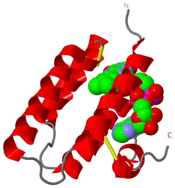
Image Asymmetric Unit
Asymmetric Unit  (Jmol Viewer)
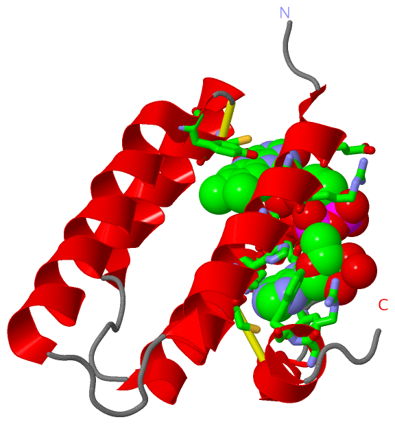
Image Asym. Unit - sites
Asym. Unit - sites  (Jmol Viewer)
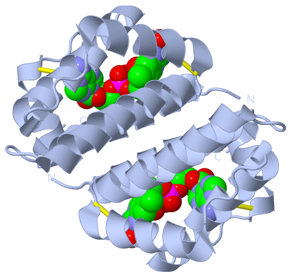
Image Biological Unit 1
Biological Unit 1  (Jmol Viewer)
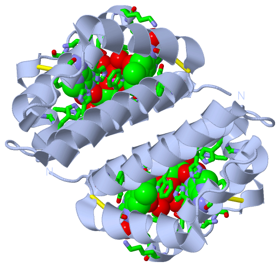
Image Biol. Unit 1 - sites
Biol. Unit 1 - sites  (Jmol Viewer)

(-) Description

Title :  CRYSTAL STRUCTURE OF FAD BINDING DOMAIN OF ERV1 FROM SACCHAROMYCES CEREVISIAE
 
Authors :  P. C. Guo, J. D. Ma, Y. L. Jiang, S. J. Wang, T. T. Hu, Y. X. Chen, C. Z. Zhou
Date :  04 Mar 12  (Deposition) - 29 Aug 12  (Release) - 30 Jan 13  (Revision)
Method :  X-RAY DIFFRACTION
Resolution :  2.00
Chains :  Asym. Unit :  A
Biol. Unit 1:  A  (2x)
Keywords :  Four-Helix Bundle, Flavin-Linked Sulfhydryl Oxidase, Fad Binding, Oxidation, Mitochondrial Intermembrane Space, Oxidoreductase (Keyword Search: [Gene Ontology, PubMed, Web (Google)] )
 
Reference :  P. C. Guo, J. D. Ma, Y. L. Jiang, S. J. Wang, Z. Z. Bao, X. J. Yu, Y. Chen, C. Z. Zhou
Structure Of Yeast Sulfhydryl Oxidase Erv1 Reveals Electron Transfer Of The Disulfide Relay System In The Mitochondrial Intermembrane Space
J. Biol. Chem. V. 287 34961 2012
PubMed-ID: 22910915  |  Reference-DOI: 10.1074/JBC.M112.394759

(-) Compounds

Molecule 1 - MITOCHONDRIAL FAD-LINKED SULFHYDRYL OXIDASE ERV1
    Chains: A
    EC Number: 1.8.3.2
    Engineered: YES
    Expression System: ESCHERICHIA COLI
    Expression System Plasmid: PET28
    Expression System Strain: BL21(DE3)
    Expression System Taxid: 562
    Expression System Vector Type: PLASMID
    Fragment: FAD BINDING DOMAIN
    Gene: ERV1
    Organism Common: YEAST
    Organism Scientific: SACCHAROMYCES CEREVISIAE
    Organism Taxid: 559292
    Strain: S288C
    Synonym: 14 KDA REGULATORY PROTEIN, ESSENTIAL FOR RESPIRATION AND VEGETATIVE GROWTH PROTEIN 1

 Structural Features

(-) Chains, Units

  1
Asymmetric Unit : A
Biological Unit 1 (2x): A

Summary Information (see also Sequences/Alignments below)

(-) Ligands, Modified Residues, Ions  (1, 1)

Asymmetric Unit (1, 1)
No.NameCountTypeFull Name
1FAD1Ligand/IonFLAVIN-ADENINE DINUCLEOTIDE
Biological Unit 1 (1, 2)
No.NameCountTypeFull Name
1FAD2Ligand/IonFLAVIN-ADENINE DINUCLEOTIDE

(-) Sites  (1, 1)

Asymmetric Unit (1, 1)
No.NameEvidenceResiduesDescription
1AC1SOFTWAREGLU A:88 , GLY A:91 , ARG A:92 , TRP A:95 , HIS A:99 , TYR A:128 , CYS A:133 , CYS A:159 , HIS A:162 , ASN A:163 , VAL A:165 , ASN A:166 , LYS A:168 , LEU A:169 , LYS A:171 , PHE A:174 , ARG A:182 , TRP A:183 , HOH A:304 , HOH A:305 , HOH A:320BINDING SITE FOR RESIDUE FAD A 201

(-) SS Bonds  (2, 2)

Asymmetric Unit
No.Residues
1A:130 -A:133
2A:159 -A:176

(-) Cis Peptide Bonds  (0, 0)

(no "Cis Peptide Bond" information available for 4E0H)

 Sequence-Structure Mapping

(-) SAPs(SNPs)/Variants  (0, 0)

(no "SAP(SNP)/Variant" information available for 4E0H)

(-) PROSITE Motifs  (0, 0)

(no "PROSITE Motif" information available for 4E0H)

(-) Exons   (0, 0)

(no "Exon" information available for 4E0H)

(-) Sequences/Alignments

Asymmetric Unit
   Reformat: Number of residues per line =  ('0' or empty: single-line sequence representation)
  Number of residues per labelling interval =   
  UniProt sequence: complete  aligned part    
   Show mapping: SCOP domains CATH domains Pfam domains Secondary structure (by author)
SAPs(SNPs) PROSITE motifs Exons
(details for a mapped element are shown in a popup box when the mouse pointer rests over it)
Chain A from PDB  Type:PROTEIN  Length:105
 aligned with ERV1_YEAST | P27882 from UniProtKB/Swiss-Prot  Length:189

    Alignment length:105
                                    93       103       113       123       133       143       153       163       173       183     
           ERV1_YEAST    84 PPDVEQLGRSSWTLLHSVAASYPAQPTDQQKGEMKQFLNIFSHIYPCNWCAKDFEKYIRENAPQVESREELGRWMCEAHNKVNKKLRKPKFDCNFWEKRWKDGWD 188
               SCOP domains d4e0ha_ A: automated matches                                                                              SCOP domains
               CATH domains --------------------------------------------------------------------------------------------------------- CATH domains
               Pfam domains --------------------------------------------------------------------------------------------------------- Pfam domains
         Sec.struct. author ..hhhhhhhhhhhhhhhhhhh.....hhhhhhhhhhhhhhhhhhh.hhhhhhhhhhhhhhh.....hhhhhhhhhhhhhhhhhhhh.....hhhhhhhhhh.... Sec.struct. author
                 SAPs(SNPs) --------------------------------------------------------------------------------------------------------- SAPs(SNPs)
                    PROSITE --------------------------------------------------------------------------------------------------------- PROSITE
                 Transcript --------------------------------------------------------------------------------------------------------- Transcript
                 4e0h A  84 HMDVEQLGRSSWTLLHSVAASYPAQPTDQQKGEMKQFLNIFSHIYPCNWCAKDFEKYIRENAPQVESREELGRWMCEAHNKVNKKLRKPKFDCNFWEKRWKDGWD 188
                                    93       103       113       123       133       143       153       163       173       183     

   Legend:   → Mismatch (orange background)
  - → Gap (green background, '-', border residues have a numbering label)
    → Modified Residue (blue background, lower-case, 'x' indicates undefined single-letter code, labelled with number + name)
  x → Chemical Group (purple background, 'x', labelled with number + name, e.g. ACE or NH2)
  extra numbering lines below/above indicate numbering irregularities and modified residue names etc., number ends below/above '|'

 Classification and Annotation

(-) SCOP Domains  (1, 1)

Asymmetric Unit

(-) CATH Domains  (0, 0)

(no "CATH Domain" information available for 4E0H)

(-) Pfam Domains  (0, 0)

(no "Pfam Domain" information available for 4E0H)

(-) Gene Ontology  (11, 11)

Asymmetric Unit(hide GO term definitions)
Chain A   (ERV1_YEAST | P27882)
molecular function
    GO:0016971    flavin-linked sulfhydryl oxidase activity    Catalysis of the formation of disulfide bridges.
    GO:0016491    oxidoreductase activity    Catalysis of an oxidation-reduction (redox) reaction, a reversible chemical reaction in which the oxidation state of an atom or atoms within a molecule is altered. One substrate acts as a hydrogen or electron donor and becomes oxidized, while the other acts as hydrogen or electron acceptor and becomes reduced.
    GO:0005515    protein binding    Interacting selectively and non-covalently with any protein or protein complex (a complex of two or more proteins that may include other nonprotein molecules).
    GO:0016972    thiol oxidase activity    Catalysis of the reaction: 4 R'C(R)SH + O2 = 2 R'C(R)S-S(R)CR' + 2 H2O.
biological process
    GO:0006879    cellular iron ion homeostasis    Any process involved in the maintenance of an internal steady state of iron ions at the level of a cell.
    GO:0034599    cellular response to oxidative stress    Any process that results in a change in state or activity of a cell (in terms of movement, secretion, enzyme production, gene expression, etc.) as a result of oxidative stress, a state often resulting from exposure to high levels of reactive oxygen species, e.g. superoxide anions, hydrogen peroxide (H2O2), and hydroxyl radicals.
    GO:0055114    oxidation-reduction process    A metabolic process that results in the removal or addition of one or more electrons to or from a substance, with or without the concomitant removal or addition of a proton or protons.
    GO:0045041    protein import into mitochondrial intermembrane space    The import of proteins into the space between the inner and outer mitochondrial membranes.
    GO:0006626    protein targeting to mitochondrion    The process of directing proteins towards and into the mitochondrion, usually mediated by mitochondrial proteins that recognize signals contained within the imported protein.
cellular component
    GO:0005758    mitochondrial intermembrane space    The region between the inner and outer lipid bilayers of the mitochondrial envelope.
    GO:0005739    mitochondrion    A semiautonomous, self replicating organelle that occurs in varying numbers, shapes, and sizes in the cytoplasm of virtually all eukaryotic cells. It is notably the site of tissue respiration.

 Visualization

(-) Interactive Views

Asymmetric Unit
  Complete Structure
    Jena3D(integrated viewing of ligand, site, SAP, PROSITE, SCOP information)
    WebMol | AstexViewer[tm]@PDBe
(Java Applets, require no local installation except for Java; loading may be slow)
    STRAP
(Java WebStart application, automatic local installation, requires Java; full application with system access!)
    RasMol
(require local installation)
    Molscript (VRML)
(requires installation of a VRML viewer; select preferred view via VRML and generate a mono or stereo PDF format file)
 
  Ligands, Modified Residues, Ions
    FAD  [ RasMol | Jena3D ]  +environment [ RasMol | Jena3D ]
 
  Sites
    AC1  [ RasMol ]  +environment [ RasMol ]
 
  Cis Peptide Bonds
(no "Cis Peptide Bonds" information available for 4e0h)
 
Biological Unit
  Complete Structure
    Biological Unit 1  [ Jena3D ]

(-) Still Images

Jmol
  protein: cartoon or spacefill or dots and stick; nucleic acid: cartoon and stick; ligands: spacefill; active site: stick
Molscript
  protein, nucleic acid: cartoon; ligands: spacefill; active site: ball and stick

 Databases and Analysis Tools

(-) Databases

Access by PDB/NDB ID
  4e0h
    Family and Domain Information: ProDom | SYSTERS
    General Structural Information: GlycoscienceDB | MMDB | NDB | OCA | PDB | PDBe | PDBj | PDBsum | PDBWiki | PQS | PROTEOPEDIA
    Orientation in Membranes: OPM
    Protein Surface: SURFACE
    Secondary Structure: DSSP (structure derived) | HSSP (homology derived)
    Structural Genomics: GeneCensus
    Structural Neighbours: CE | VAST
    Structure Classification: CATH | Dali | SCOP
    Validation and Original Data: BMRB Data View | BMRB Restraints Grid | EDS | PROCHECK | RECOORD | WHAT_CHECK
 
Access by UniProt ID/Accession number
  ERV1_YEAST | P27882
    Comparative Protein Structure Models: ModBase
    Genomic Information: Ensembl
    Protein-protein Interaction: DIP
    Sequence, Family and Domain Information: InterPro | Pfam | SMART | UniProtKB/SwissProt
 
Access by Enzyme Classificator   (EC Number)
  1.8.3.2
    General Enzyme Information: BRENDA | EC-PDB | Enzyme | IntEnz
    Pathway: KEGG | MetaCyc
 
Access by Disease Identifier   (MIM ID)
  (no 'MIM ID' available)
    Disease Information: OMIM
 
Access by GenAge ID
  (no 'GenAge ID' available)
    Age Related Information: GenAge

(-) Analysis Tools

Access by PDB/NDB ID
    Domain Information: XDom
    Interatomic Contacts of Structural Units: CSU
    Ligand-protein Contacts: LPC
    Protein Cavities: castP
    Sequence and Secondary Structure: PDBCartoon
    Structure Alignment: STRAP(Java WebStart application, automatic local installation, requires Java; full application with system access!)
    Structure and Sequence Browser: STING
 
Access by UniProt ID/Accession number
  ERV1_YEAST | P27882
    Protein Disorder Prediction: DisEMBL | FoldIndex | GLOBPLOT (for more information see DisProt)

 Related Entries

(-) Entries Sharing at Least One Protein Chain (UniProt ID)

UniProtKB/Swiss-Prot
        ERV1_YEAST | P27882: 3w4y 4e0i

(-) Related Entries Specified in the PDB File

1oqc 4e0i