Show PDB file:   
         Plain Text   HTML   (compressed file size)
QuickSearch:   
by PDB,NDB,UniProt,PROSITE Code or Search Term(s)  
(-)Asym./Biol. Unit
(-)Asym./Biol. Unit - sites
collapse expand < >
Image Asym./Biol. Unit
Asym./Biol. Unit  (Jmol Viewer)
Image Asym./Biol. Unit - sites
Asym./Biol. Unit - sites  (Jmol Viewer)

(-) Description

Title :  AQUALYSIN I: THE CRYSTAL STRUCTURE OF A SERINE PROTEASE FROM AN EXTREME THERMOPHILE, THERMUS AQUATICUS YT-1
 
Authors :  B. L. Barnett, P. R. Green, L. C. Strickland, J. D. Oliver, T. Rydel, J. F.
Date :  01 Mar 12  (Deposition) - 28 Mar 12  (Release) - 28 Mar 12  (Revision)
Method :  X-RAY DIFFRACTION
Resolution :  1.95
Chains :  Asym./Biol. Unit :  A
Keywords :  Serine Protease, Calcium Binding, Inhibitor, Hydrolase-Hydrolase Inhibitor Complex (Keyword Search: [Gene Ontology, PubMed, Web (Google))
 
Reference :  B. L. Barnett, P. R. Green, L. C. Strickland, J. D. Oliver, T. Rydel, J. F. Sullivan
Aqualysin I: The Crystal Structure Of A Serine Protease Fro An Extreme Thermophile, Thermus Aquaticus Yt-1
To Be Published
PubMed: search
(for further references see the PDB file header)

(-) Compounds

Molecule 1 - AQUALYSIN-1
    ChainsA
    EC Number3.4.21.111
    FragmentUNP RESIDUES 128-403
    Organism ScientificTHERMUS AQUATICUS
    Organism Taxid271
    StrainYT-1
    SynonymAQUALYSIN-I

 Structural Features

(-) Chains, Units

  1
Asymmetric/Biological Unit A

Summary Information (see also Sequences/Alignments below)
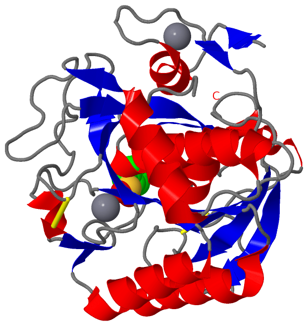
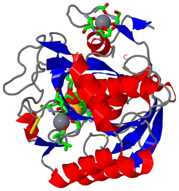
(-) Ligands, Modified Residues, Ions  (2, 3)

Asymmetric/Biological Unit (2, 3)
No.NameCountTypeFull Name
1CA2Ligand/IonCALCIUM ION
2PMS1Ligand/IonPHENYLMETHANESULFONIC ACID

(-) Sites  (3, 3)

Asymmetric Unit (3, 3)
No.NameEvidenceResiduesDescription
1AC1SOFTWAREHIS A:70 , SER A:128 , ALA A:154 , ASN A:157 , ASN A:219 , THR A:221 , SER A:222 , MET A:223BINDING SITE FOR RESIDUE PMS A 301
2AC2SOFTWAREASP A:11 , ASP A:14 , GLN A:15 , SER A:21 , SER A:23 , HOH A:1012BINDING SITE FOR RESIDUE CA A 302
3AC3SOFTWAREVAL A:170 , ALA A:171 , ALA A:173 , THR A:175 , ASP A:196 , HOH A:1074BINDING SITE FOR RESIDUE CA A 303

(-) SS Bonds  (2, 2)

Asymmetric/Biological Unit
No.Residues
1A:67 -A:99
2A:163 -A:194

(-) Cis Peptide Bonds  (4, 4)

Asymmetric/Biological Unit
No.Residues
1Ser A:4 -Pro A:5
2Leu A:18 -Pro A:19
3Arg A:121 -Pro A:122
4Ser A:166 -Pro A:167

 Sequence-Structure Mapping

(-) SAPs(SNPs)/Variants  (0, 0)

(no "SAP(SNP)/Variant" information available for 4DZT)

(-) PROSITE Motifs  (3, 3)

Asymmetric/Biological Unit (3, 3)
 PROSITEUniProtKBPDB
No.IDACDescriptionIDLocationCountLocation
1SUBTILASE_ASPPS00136 Serine proteases, subtilase family, aspartic acid active site.AQL1_THEAQ162-173  1A:35-46
2SUBTILASE_HISPS00137 Serine proteases, subtilase family, histidine active site.AQL1_THEAQ197-207  1A:70-80
3SUBTILASE_SERPS00138 Serine proteases, subtilase family, serine active site.AQL1_THEAQ347-357  1A:220-230

(-) Exons   (0, 0)

(no "Exon" information available for 4DZT)

(-) Sequences/Alignments

Asymmetric/Biological Unit
   Reformat: Number of residues per line =  ('0' or empty: single-line sequence representation)
  Number of residues per labelling interval =   
  UniProt sequence: complete  aligned part    
   Show mapping: SCOP domains CATH domains Pfam domains Secondary structure (by author)
SAPs(SNPs) PROSITE motifs Exons
(details for a mapped element are shown in a popup box when the mouse pointer rests over it)
Chain A from PDB  Type:PROTEIN  Length:276
 aligned with AQL1_THEAQ | P08594 from UniProtKB/Swiss-Prot  Length:513

    Alignment length:276
                                   137       147       157       167       177       187       197       207       217       227       237       247       257       267       277       287       297       307       317       327       337       347       357       367       377       387       397      
           AQL1_THEAQ   128 ATQSPAPWGLDRIDQRDLPLSNSYTYTATGRGVNVYVIDTGIRTTHREFGGRARVGYDALGGNGQDCNGHGTHVAGTIGGVTYGVAKAVNLYAVRVLDCNGSGSTSGVIAGVDWVTRNHRRPAVANMSLGGGVSTALDNAVKNSIAAGVVYAVAAGNDNANACNYSPARVAEALTVGATTSSDARASFSNYGSCVDLFAPGASIPSAWYTSDTATQTLNGTSMATPHVAGVAALYLEQNPSATPASVASAILNGATTGRLSGIGSGSPNRLLYSLL 403
               SCOP domains d4dzta_ A: automated matches                                                                                                                                                                                                                                                         SCOP domains
               CATH domains ------------------------------------------------------------------------------------------------------------------------------------------------------------------------------------------------------------------------------------------------------------------------------------ CATH domains
               Pfam domains ------------------------------------------------------------------------------------------------------------------------------------------------------------------------------------------------------------------------------------------------------------------------------------ Pfam domains
         Sec.struct. author .ee...hhhhhhhh.........ee........eeeeee......hhhhh..eeeeee..........hhhhhhhhhhhh.........eeeeee........hhhhhhhhhhhhhhhh...eeeee...ee.hhhhhhhhhhhhhh..eeeee......hhh.ee.......eeeeee.................eeee...eeee.......eeee.hhhhhhhhhhhhhhhhhhhh...hhhhhhhhhhhhhee............ee..... Sec.struct. author
                 SAPs(SNPs) ------------------------------------------------------------------------------------------------------------------------------------------------------------------------------------------------------------------------------------------------------------------------------------ SAPs(SNPs)
                    PROSITE ----------------------------------SUBTILASE_AS-----------------------SUBTILASE_H-------------------------------------------------------------------------------------------------------------------------------------------SUBTILASE_S---------------------------------------------- PROSITE
                 Transcript ------------------------------------------------------------------------------------------------------------------------------------------------------------------------------------------------------------------------------------------------------------------------------------ Transcript
                 4dzt A   1 ATQSPAPWGLDRIDQRDLPLSNSYTYTATGRGVNVYVIDTGIRTTHREFGGRARVGYDALGGNGQDCNGHGTHVAGTIGGVTYGVAKAVNLYAVRVLDCNGSGSTSGVIAGVDWVTRNHRRPAVANMSLGGGVSTALDNAVKNSIAAGVVYAVAAGNDNANACNYSPARVAEALTVGATTSSDARASFSNYGSCVDLFAPGASIPSAWYTSDTATQTLNGTSMATPHVAGVAALYLEQNPSATPASVASAILNGATTGRLSGIGSGSPNRLLYSLL 276
                                    10        20        30        40        50        60        70        80        90       100       110       120       130       140       150       160       170       180       190       200       210       220       230       240       250       260       270      

   Legend:   → Mismatch (orange background)
  - → Gap (green background, '-', border residues have a numbering label)
    → Modified Residue (blue background, lower-case, 'x' indicates undefined single-letter code, labelled with number + name)
  x → Chemical Group (purple background, 'x', labelled with number + name, e.g. ACE or NH2)
  extra numbering lines below/above indicate numbering irregularities and modified residue names etc., number ends below/above '|'

 Classification and Annotation

(-) SCOP Domains  (1, 1)

Asymmetric/Biological Unit

(-) CATH Domains  (0, 0)

(no "CATH Domain" information available for 4DZT)

(-) Pfam Domains  (0, 0)

(no "Pfam Domain" information available for 4DZT)

(-) Gene Ontology  (6, 6)

Asymmetric/Biological Unit(hide GO term definitions)
Chain A   (AQL1_THEAQ | P08594)
molecular function
    GO:0016787    hydrolase activity    Catalysis of the hydrolysis of various bonds, e.g. C-O, C-N, C-C, phosphoric anhydride bonds, etc. Hydrolase is the systematic name for any enzyme of EC class 3.
    GO:0008233    peptidase activity    Catalysis of the hydrolysis of a peptide bond. A peptide bond is a covalent bond formed when the carbon atom from the carboxyl group of one amino acid shares electrons with the nitrogen atom from the amino group of a second amino acid.
    GO:0004252    serine-type endopeptidase activity    Catalysis of the hydrolysis of internal, alpha-peptide bonds in a polypeptide chain by a catalytic mechanism that involves a catalytic triad consisting of a serine nucleophile that is activated by a proton relay involving an acidic residue (e.g. aspartate or glutamate) and a basic residue (usually histidine).
    GO:0008236    serine-type peptidase activity    Catalysis of the hydrolysis of peptide bonds in a polypeptide chain by a catalytic mechanism that involves a catalytic triad consisting of a serine nucleophile that is activated by a proton relay involving an acidic residue (e.g. aspartate or glutamate) and a basic residue (usually histidine).
biological process
    GO:0006508    proteolysis    The hydrolysis of proteins into smaller polypeptides and/or amino acids by cleavage of their peptide bonds.
cellular component
    GO:0005576    extracellular region    The space external to the outermost structure of a cell. For cells without external protective or external encapsulating structures this refers to space outside of the plasma membrane. This term covers the host cell environment outside an intracellular parasite.

 Visualization

(-) Interactive Views

Asymmetric/Biological Unit
  Complete Structure
    Jena3D(integrated viewing of ligand, site, SAP, PROSITE, SCOP information)
    WebMol | AstexViewer[tm]@PDBe
(Java Applets, require no local installation except for Java; loading may be slow)
    STRAP
(Java WebStart application, automatic local installation, requires Java; full application with system access!)
    RasMol
(require local installation)
    Molscript (VRML)
(requires installation of a VRML viewer; select preferred view via VRML and generate a mono or stereo PDF format file)
 
  Ligands, Modified Residues, Ions
    CA  [ RasMol | Jena3D ]  +environment [ RasMol | Jena3D ]
    PMS  [ RasMol | Jena3D ]  +environment [ RasMol | Jena3D ]
 
  Sites
    AC1  [ RasMol ]  +environment [ RasMol ]
    AC2  [ RasMol ]  +environment [ RasMol ]
    AC3  [ RasMol ]  +environment [ RasMol ]
 
  Cis Peptide Bonds
    Arg A:121 - Pro A:122   [ RasMol ]  
    Leu A:18 - Pro A:19   [ RasMol ]  
    Ser A:166 - Pro A:167   [ RasMol ]  
    Ser A:4 - Pro A:5   [ RasMol ]  
 

(-) Still Images

Jmol
  protein: cartoon or spacefill or dots and stick; nucleic acid: cartoon and stick; ligands: spacefill; active site: stick
Molscript
  protein, nucleic acid: cartoon; ligands: spacefill; active site: ball and stick

 Databases and Analysis Tools

(-) Databases

Access by PDB/NDB ID
  4dzt
    Family and Domain InformationProDom | SYSTERS
    General Structural InformationGlycoscienceDB | MMDB | NDB | OCA | PDB | PDBe | PDBj | PDBsum | PDBWiki | PQS | PROTEOPEDIA
    Orientation in MembranesOPM
    Protein SurfaceSURFACE
    Secondary StructureDSSP (structure derived) | HSSP (homology derived)
    Structural GenomicsGeneCensus
    Structural NeighboursCE | VAST
    Structure ClassificationCATH | Dali | SCOP
    Validation and Original DataBMRB Data View | BMRB Restraints Grid | EDS | PROCHECK | RECOORD | WHAT_CHECK
 
Access by UniProt ID/Accession number
  AQL1_THEAQ | P08594
    Comparative Protein Structure ModelsModBase
    Genomic InformationEnsembl
    Protein-protein InteractionDIP
    Sequence, Family and Domain InformationInterPro | Pfam | SMART | UniProtKB/SwissProt
 
Access by Enzyme Classificator   (EC Number)
  3.4.21.111
    General Enzyme InformationBRENDA | EC-PDB | Enzyme | IntEnz
    PathwayKEGG | MetaCyc
 
Access by Disease Identifier   (MIM ID)
  (no 'MIM ID' available)
    Disease InformationOMIM
 
Access by GenAge ID
  (no 'GenAge ID' available)
    Age Related InformationGenAge

(-) Analysis Tools

Access by PDB/NDB ID
    Domain InformationXDom
    Interatomic Contacts of Structural UnitsCSU
    Ligand-protein ContactsLPC
    Protein CavitiescastP
    Sequence and Secondary StructurePDBCartoon
    Structure AlignmentSTRAP(Java WebStart application, automatic local installation, requires Java; full application with system access!)
    Structure and Sequence BrowserSTING
 
Access by UniProt ID/Accession number
  AQL1_THEAQ | P08594
    Protein Disorder PredictionDisEMBL | FoldIndex | GLOBPLOT (for more information see DisProt)

 Related Entries

(-) Entries Sharing at Least One Protein Chain (UniProt ID)

(no "Entries Sharing at Least One Protein Chain" available for 4DZT)

(-) Related Entries Specified in the PDB File

1sup HOMOLOGOUS PROTEIN SUBTILISIN BPN'
1svn HOMOLOGOUS PROTEIN SAVINASE
2gko HOMOLOGOUS PROTEIN SUBTILISIN
2prk HOMOLOGOUS PROTEIN PROTEINASE K