|
|
|
|
 Description
|
|
Compounds
|
||||||||||||||||||||||||
Chains, Units
Summary Information (see also Sequences/Alignments below) |
Ligands, Modified Residues, Ions (2, 3)| Asymmetric Unit (2, 3) Biological Unit 1 (1, 2) |
Sites (4, 4)
Asymmetric Unit (4, 4)
|
SS Bonds (0, 0)| (no "SS Bond" information available for 4CPV) |
Cis Peptide Bonds (0, 0)| (no "Cis Peptide Bond" information available for 4CPV) |
SAPs(SNPs)/Variants (0, 0)| (no "SAP(SNP)/Variant" information available for 4CPV) |
PROSITE Motifs (2, 4)
Asymmetric Unit (2, 4)
|
||||||||||||||||||||||||||||||||||||||||||||||||||||||||||||||||
Exons (0, 0)| (no "Exon" information available for 4CPV) |
Sequences/Alignments
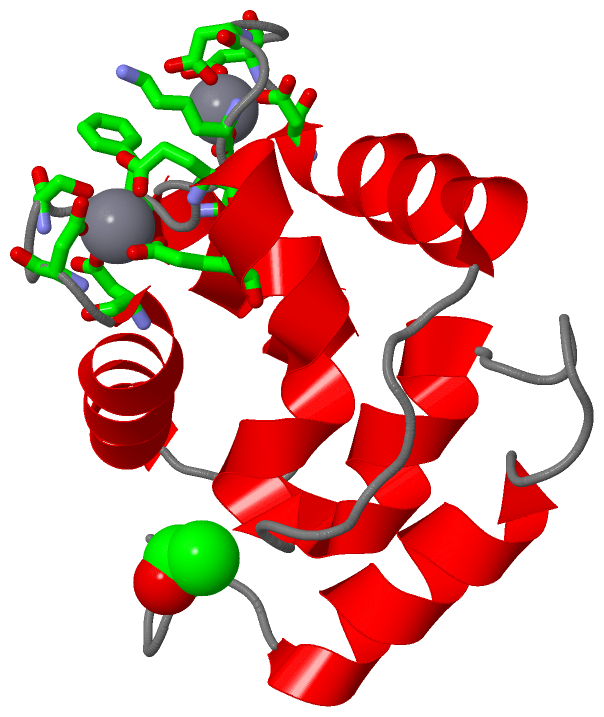
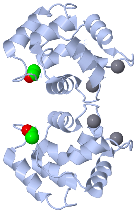
Asymmetric UnitChain A from PDB Type:PROTEIN Length:109 aligned with PRVB_CYPCA | P02618 from UniProtKB/Swiss-Prot Length:108 Alignment length:109 1 | 9 19 29 39 49 59 69 79 89 99 PRVB_CYPCA - -AFAGVLNDADIAAALEACKAADSFNHKAFFAKVGLTSKSADDVKKAFAIIDQDKSGFIEEDELKLFLQNFKADARALTDGETKTFLKAGDSDGDGKIGVDEFTALVKA 108 SCOP domains d4cpva_ A: Parvalbumin SCOP domains CATH domains -4cpvA00 A:1-108 EF-hand CATH domains Pfam domains ------------------------------------------------------------------------------------------------------------- Pfam domains SAPs(SNPs) ------------------------------------------------------------------------------------------------------------- SAPs(SNPs) PROSITE (1) --------------------------------------EF_HAND_2 PDB: A:38-73 ---EF_HAND_2 PDB: A:77-108 PROSITE (1) PROSITE (2) ---------------------------------------------------EF_HAND_1 --------------------------EF_HAND_1 ------ PROSITE (2) Transcript ------------------------------------------------------------------------------------------------------------- Transcript 4cpv A 0 xAFAGVLNDADIAAALEACKAADSFNHKAFFAKVGLTSKSADDVKKAFAIIDQDKSGFIEEDELKLFLQNFKADARALTDGETKTFLKAGDSDGDGKIGVDEFTALVKA 108 | 9 19 29 39 49 59 69 79 89 99 | 0-ACE
|
||||||||||||||||||||
SCOP Domains (1, 1)
Asymmetric Unit
 |
CATH Domains (1, 1)
Asymmetric Unit
|
Pfam Domains (0, 0)| (no "Pfam Domain" information available for 4CPV) |
Gene Ontology (2, 2)|
Asymmetric Unit(hide GO term definitions) Chain A (PRVB_CYPCA | P02618)
|
||||||||||||||||||
Interactive Views
|
||||||||||||||||||||||||||||||||||||||||||||||||||||||||||||||||||||||||||||||||||||||||||||||||||||||||||||||||||||||||||||||||||||||||||||||||||||||||||||||||||||
Still Images
|
||||||||||||||||
Databases
|
||||||||||||||||||||||||||||||||||||||||||||||||||||||||||||||||||||||||||||||||||||||||||||||||||||||||||||||||||||||||||||||||||||||||||||||||||||||||||||||||
Analysis Tools
|
|||||||||||||||||||||||||||||||||||||||||||||||||||||||||||||
Entries Sharing at Least One Protein Chain (UniProt ID)
Related Entries Specified in the PDB File
|
|