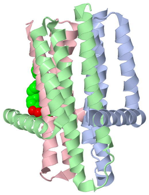
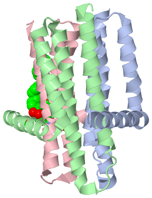
Show PDB file:   
         Plain Text   HTML   (compressed file size)
QuickSearch:   
by PDB,NDB,UniProt,PROSITE Code or Search Term(s)  
(-)Asym./Biol. Unit
(-)Asym./Biol. Unit - sites
collapse expand < >
Image Asym./Biol. Unit
Asym./Biol. Unit  (Jmol Viewer)
Image Asym./Biol. Unit - sites
Asym./Biol. Unit - sites  (Jmol Viewer)

(-) Description

Title :  CRYSTAL STRUCTURE OF THE INTEGRAL MEMBRANE DIACYLGLYCEROL KINASE DGKA-9.9, DELTA 4
 
Authors :  D. Li, D. Aragao, V. E. Pye, M. Caffrey
Date :  23 Dec 13  (Deposition) - 28 Jan 15  (Release) - 16 Nov 16  (Revision)
Method :  X-RAY DIFFRACTION
Resolution :  3.25
Chains :  Asym./Biol. Unit :  A,B,C
Keywords :  Transferase, In Meso Crystallisation, Lcp, Lipid Cubic Phase, Lipidic Mesophase, Lipid Metabolism, Membrane Protein, Microcrystal, Monoacylglycerol, Monoolein, 9. 9 Mag, Thermostable Mutant (Keyword Search: [Gene Ontology, PubMed, Web (Google))
 
Reference :  D. Li, M. Caffrey
Crystal Structure Of The Integral Membrane Diacylglycerol Kinase With Zn-Amppcp Bound And Its Catalytic Mechanism
To Be Published
PubMed: search

(-) Compounds

Molecule 1 - DIACYLGLYCEROL KINASE
    ChainsA, B, C
    EC Number2.7.1.107
    EngineeredYES
    Expression SystemESCHERICHIA COLI
    Expression System PlasmidPTRCHISB-DGKA-DELTA 4
    Expression System StrainWH1061
    Expression System Taxid562
    Expression System VectorPTRCHISB
    Expression System Vector TypePLASMID
    MutationYES
    Organism ScientificESCHERICHIA COLI
    Organism Taxid83333
    StrainK-12
    SynonymDAGK, DIGLYCERIDE KINASE, DGK, DIACYLGLYCEROL KINASE - DELTA 4

 Structural Features

(-) Chains, Units

  123
Asymmetric/Biological Unit ABC

Summary Information (see also Sequences/Alignments below)

(-) Ligands, Modified Residues, Ions  (1, 1)

Asymmetric/Biological Unit (1, 1)
No.NameCountTypeFull Name
1OLC1Ligand/Ion(2R)-2,3-DIHYDROXYPROPYL (9Z)-OCTADEC-9-ENOATE

(-) Sites  (1, 1)

Asymmetric Unit (1, 1)
No.NameEvidenceResiduesDescription
1AC1SOFTWAREALA B:13 , ALA C:30 , GLN C:33 , GLU C:69 , ASN C:72 , SER C:98 , VAL C:101 , ALA C:108 , TRP C:112BINDING SITE FOR RESIDUE OLC C1120

(-) SS Bonds  (0, 0)

(no "SS Bond" information available for 4CJZ)

(-) Cis Peptide Bonds  (0, 0)

(no "Cis Peptide Bond" information available for 4CJZ)

 Sequence-Structure Mapping

(-) SAPs(SNPs)/Variants  (0, 0)

(no "SAP(SNP)/Variant" information available for 4CJZ)

(-) PROSITE Motifs  (0, 0)

(no "PROSITE Motif" information available for 4CJZ)

(-) Exons   (0, 0)

(no "Exon" information available for 4CJZ)

(-) Sequences/Alignments

Asymmetric/Biological Unit
   Reformat: Number of residues per line =  ('0' or empty: single-line sequence representation)
  Number of residues per labelling interval =   
  UniProt sequence: complete  aligned part    
   Show mapping: SCOP domains CATH domains Pfam domains Secondary structure (by author)
SAPs(SNPs) PROSITE motifs Exons
(details for a mapped element are shown in a popup box when the mouse pointer rests over it)
Chain A from PDB  Type:PROTEIN  Length:114
                                                                                                                                                  
               SCOP domains ------------------------------------------------------------------------------------------------------------------ SCOP domains
               CATH domains ------------------------------------------------------------------------------------------------------------------ CATH domains
               Pfam domains ------------------------------------------------------------------------------------------------------------------ Pfam domains
         Sec.struct. author ..hhhhhhhhhhhhhhhhhhhhhhhhhhhhhhhhhhhhhhhh..hhhhhhhhhhhhhhhhhhhhhhhhhhhhhhhhhhhhh...hhhhhhhhhhhhhhhhhhhhhhhhhhhhhh Sec.struct. author
                 SAPs(SNPs) ------------------------------------------------------------------------------------------------------------------ SAPs(SNPs)
                    PROSITE ------------------------------------------------------------------------------------------------------------------ PROSITE
                 Transcript ------------------------------------------------------------------------------------------------------------------ Transcript
                 4cjz A   7 FTRIIKAAGYSWKGLRAAWINEAAFRQEGVAVLLAVVIACWLDVDACTRVLLISSVMLVMIVELLNSAIEAVVDRIGSEYHELSGRAKDLGSAAVLIAIIDAVITWCILLWSHF 120
                                    16        26        36        46        56        66        76        86        96       106       116    

Chain B from PDB  Type:PROTEIN  Length:117
                                                                                                                                                     
               SCOP domains --------------------------------------------------------------------------------------------------------------------- SCOP domains
               CATH domains --------------------------------------------------------------------------------------------------------------------- CATH domains
               Pfam domains --------------------------------------------------------------------------------------------------------------------- Pfam domains
         Sec.struct. author hhhhhhhhhhhhhhhhhhhhhhhhhhhhhhhhhhhhhhhhhhhh..hhhhhhhhhhhhhhhhhhhhhhhhhhhhhhhhhhhh....hhhhhhhhhhhhhhhhhhhhhhhhhhhhhh. Sec.struct. author
                 SAPs(SNPs) --------------------------------------------------------------------------------------------------------------------- SAPs(SNPs)
                    PROSITE --------------------------------------------------------------------------------------------------------------------- PROSITE
                 Transcript --------------------------------------------------------------------------------------------------------------------- Transcript
                 4cjz B   5 TGFTRIIKAAGYSWKGLRAAWINEAAFRQEGVAVLLAVVIACWLDVDACTRVLLISSVMLVMIVELLNSAIEAVVDRIGSEYHELSGRAKDLGSAAVLIAIIDAVITWCILLWSHFG 121
                                    14        24        34        44        54        64        74        84        94       104       114       

Chain C from PDB  Type:PROTEIN  Length:104
                                                                                                                                        
               SCOP domains -------------------------------------------------------------------------------------------------------- SCOP domains
               CATH domains -------------------------------------------------------------------------------------------------------- CATH domains
               Pfam domains -------------------------------------------------------------------------------------------------------- Pfam domains
         Sec.struct. author hhhhhhhhhhhhhhhhhhhhhhhhhhhhhhhhh..hhhhhhhhhhhhhhhhhhhhhhhhhhhhhhhhh...hhhhhhhhhhhhhhhhhhhhhhhhhhhhhhh.. Sec.struct. author
                 SAPs(SNPs) -------------------------------------------------------------------------------------------------------- SAPs(SNPs)
                    PROSITE -------------------------------------------------------------------------------------------------------- PROSITE
                 Transcript -------------------------------------------------------------------------------------------------------- Transcript
                 4cjz C  16 YSWKGLRAAWINEAAFRQEGVAVLLAVVIACWLDVDACTRVLLISSVMLVMIVELLNSAIEAVVDRIGSEYHELSGRAKDLGSAAVLIAIIDAVITWCILLWSH 119
                                    25        35        45        55        65        75        85        95       105       115    

   Legend:   → Mismatch (orange background)
  - → Gap (green background, '-', border residues have a numbering label)
    → Modified Residue (blue background, lower-case, 'x' indicates undefined single-letter code, labelled with number + name)
  x → Chemical Group (purple background, 'x', labelled with number + name, e.g. ACE or NH2)
  extra numbering lines below/above indicate numbering irregularities and modified residue names etc., number ends below/above '|'

 Classification and Annotation

(-) SCOP Domains  (0, 0)

(no "SCOP Domain" information available for 4CJZ)

(-) CATH Domains  (0, 0)

(no "CATH Domain" information available for 4CJZ)

(-) Pfam Domains  (0, 0)

(no "Pfam Domain" information available for 4CJZ)

(-) Gene Ontology  (12, 12)

Asymmetric/Biological Unit(hide GO term definitions)

 Visualization

(-) Interactive Views

Asymmetric/Biological Unit
  Complete Structure
    Jena3D(integrated viewing of ligand, site, SAP, PROSITE, SCOP information)
    WebMol | AstexViewer[tm]@PDBe
(Java Applets, require no local installation except for Java; loading may be slow)
    STRAP
(Java WebStart application, automatic local installation, requires Java; full application with system access!)
    RasMol
(require local installation)
    Molscript (VRML)
(requires installation of a VRML viewer; select preferred view via VRML and generate a mono or stereo PDF format file)
 
  Ligands, Modified Residues, Ions
    OLC  [ RasMol | Jena3D ]  +environment [ RasMol | Jena3D ]
 
  Sites
    AC1  [ RasMol ]  +environment [ RasMol ]
 
  Cis Peptide Bonds
(no "Cis Peptide Bonds" information available for 4cjz)
 

(-) Still Images

Jmol
  protein: cartoon or spacefill or dots and stick; nucleic acid: cartoon and stick; ligands: spacefill; active site: stick
Molscript
  protein, nucleic acid: cartoon; ligands: spacefill; active site: ball and stick

 Databases and Analysis Tools

(-) Databases

Access by PDB/NDB ID
  4cjz
    Family and Domain InformationProDom | SYSTERS
    General Structural InformationGlycoscienceDB | MMDB | NDB | OCA | PDB | PDBe | PDBj | PDBsum | PDBWiki | PQS | PROTEOPEDIA
    Orientation in MembranesOPM
    Protein SurfaceSURFACE
    Secondary StructureDSSP (structure derived) | HSSP (homology derived)
    Structural GenomicsGeneCensus
    Structural NeighboursCE | VAST
    Structure ClassificationCATH | Dali | SCOP
    Validation and Original DataBMRB Data View | BMRB Restraints Grid | EDS | PROCHECK | RECOORD | WHAT_CHECK
 
Access by UniProt ID/Accession number
  KDGL_ECOLI | P0ABN1
    Comparative Protein Structure ModelsModBase
    Genomic InformationEnsembl
    Protein-protein InteractionDIP
    Sequence, Family and Domain InformationInterPro | Pfam | SMART | UniProtKB/SwissProt
 
Access by Enzyme Classificator   (EC Number)
  2.7.1.107
    General Enzyme InformationBRENDA | EC-PDB | Enzyme | IntEnz
    PathwayKEGG | MetaCyc
 
Access by Disease Identifier   (MIM ID)
  (no 'MIM ID' available)
    Disease InformationOMIM
 
Access by GenAge ID
  (no 'GenAge ID' available)
    Age Related InformationGenAge

(-) Analysis Tools

Access by PDB/NDB ID
    Domain InformationXDom
    Interatomic Contacts of Structural UnitsCSU
    Ligand-protein ContactsLPC
    Protein CavitiescastP
    Sequence and Secondary StructurePDBCartoon
    Structure AlignmentSTRAP(Java WebStart application, automatic local installation, requires Java; full application with system access!)
    Structure and Sequence BrowserSTING
 
Access by UniProt ID/Accession number
  KDGL_ECOLI | P0ABN1
    Protein Disorder PredictionDisEMBL | FoldIndex | GLOBPLOT (for more information see DisProt)

 Related Entries

(-) Entries Sharing at Least One Protein Chain (UniProt ID)

UniProtKB/Swiss-Prot
        KDGL_ECOLI | P0ABN12kdc 3ze3 3ze4 3ze5 4bpd 4brb 4brr 4ck0 4d2e 4up6 4uxw 4uxx 4uxz 4uyo 5d56 5d57 5d6i 5dwk

(-) Related Entries Specified in the PDB File

4ck0 CRYSTAL STRUCTURE OF THE INTEGRAL MEMBRANE DIACYLGLYCEROL KINASE - FORM 2