|
|
|
|
 Description
|
|
Compounds
|
||||||||||||||||||||||||||||||||||||||||||||||||||||||||
Chains, Units
Summary Information (see also Sequences/Alignments below) |
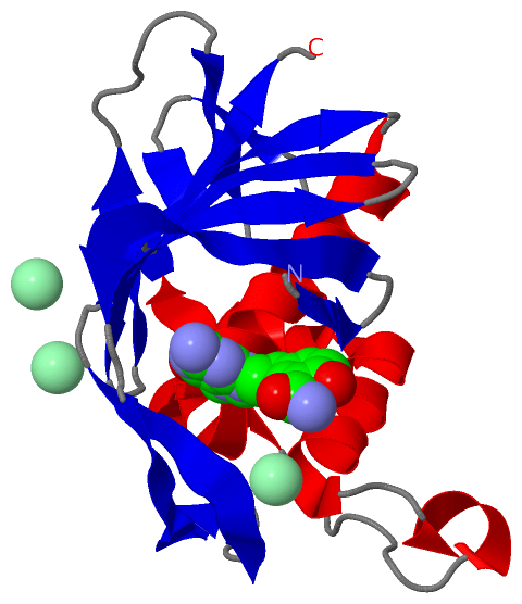
Ligands, Modified Residues, Ions (2, 4)| Asymmetric/Biological Unit (2, 4) |
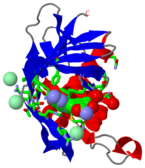
Sites (4, 4)
Asymmetric Unit (4, 4)
|
SS Bonds (0, 0)| (no "SS Bond" information available for 4CB5) |
Cis Peptide Bonds (0, 0)| (no "Cis Peptide Bond" information available for 4CB5) |
SAPs(SNPs)/Variants (0, 0)| (no "SAP(SNP)/Variant" information available for 4CB5) |
PROSITE Motifs (0, 0)| (no "PROSITE Motif" information available for 4CB5) |
Exons (0, 0)| (no "Exon" information available for 4CB5) |
Sequences/Alignments
Asymmetric/Biological UnitChain A from PDB Type:PROTEIN Length:162 aligned with Q2LG68_9INFA | Q2LG68 from UniProtKB/TrEMBL Length:759 Alignment length:162 331 341 351 361 371 381 391 401 411 421 431 441 451 461 471 481 Q2LG68_9INFA 322 SFSFGGFTFKRTSGSSVKKEEEVLTGNLQTLKIRVHEGYEEFTMVGRRATAILRKATRRLIQLIVSGRDEQSIAEAIIVAMVFSQEDCMIKAVRGDLNFVNRANQRLNPMHQLLRHFQKDAKVLFQNWGIEPIDNVMGMIGILPDMTPSTEMSLRGVRVSKM 483 SCOP domains ------------------------------------------------------------------------------------------------------------------------------------------------------------------ SCOP domains CATH domains ------------------------------------------------------------------------------------------------------------------------------------------------------------------ CATH domains Pfam domains ------------------------------------------------------------------------------------------------------------------------------------------------------------------ Pfam domains SAPs(SNPs) ------------------------------------------------------------------------------------------------------------------------------------------------------------------ SAPs(SNPs) PROSITE ------------------------------------------------------------------------------------------------------------------------------------------------------------------ PROSITE Transcript ------------------------------------------------------------------------------------------------------------------------------------------------------------------ Transcript 4cb5 A 322 SFSFGGFTFKRTSGSSVKKEEEVLTGNLQTLKIRVHEGYEEFTMVGRRATAILRKATRRLIQLIVSGRDEQSIAEAIIVAMVFSQEDCMIKAVRGDLNFVNRANQRLNPMHQLLRHFQKDAKVLFQNWGIEPIDNVMGMIGILPDMTPSTEMSLRGVRVSKM 483 331 341 351 361 371 381 391 401 411 421 431 441 451 461 471 481
|
||||||||||||||||||||
SCOP Domains (0, 0)| (no "SCOP Domain" information available for 4CB5) |
CATH Domains (0, 0)| (no "CATH Domain" information available for 4CB5) |
Pfam Domains (0, 0)| (no "Pfam Domain" information available for 4CB5) |
Gene Ontology (9, 9)|
Asymmetric/Biological Unit(hide GO term definitions) Chain A (Q2LG68_9INFA | Q2LG68)
|
||||||||||||||||||||||||||||||||||||||||||||||||||||||||||||||||||||||||
Interactive Views
|
||||||||||||||||||||||||||||||||||||||||||||||||||||||||||||||||||||||||||||||||||||||||||||||||||||||||||||||||||||||||||||||||||||||||||||||||||
Still Images
|
||||||||||||||||
Databases
|
||||||||||||||||||||||||||||||||||||||||||||||||||||||||||||||||||||||||||||||||||||||||||||||||||||||||||||||||||||||||||||||||||||||||||||||||||||||||||||||||
Analysis Tools
|
|||||||||||||||||||||||||||||||||||||||||||||||||||||||||||||
Entries Sharing at Least One Protein Chain (UniProt ID)
Related Entries Specified in the PDB File
|
|