Show PDB file:   
         Plain Text   HTML   (compressed file size)
QuickSearch:   
by PDB,NDB,UniProt,PROSITE Code or Search Term(s)  
(-)Asym./Biol. Unit
(-)Asym./Biol. Unit - sites
collapse expand < >
Image Asym./Biol. Unit
Asym./Biol. Unit  (Jmol Viewer)
Image Asym./Biol. Unit - sites
Asym./Biol. Unit - sites  (Jmol Viewer)

(-) Description

Title :  STRUCTURE OF THE EXTRACELLULAR PORTION OF MOUSE CD200R
 
Authors :  D. Hatherley, S. M. Lea, S. Johnson, A. N. Barclay
Date :  18 Mar 13  (Deposition) - 01 May 13  (Release) - 29 May 13  (Revision)
Method :  X-RAY DIFFRACTION
Resolution :  2.08
Chains :  Asym./Biol. Unit :  A
Keywords :  Immune System, Paired Receptor, Ig Domains, Viral Mimicry, Leukaemia (Keyword Search: [Gene Ontology, PubMed, Web (Google))
 
Reference :  D. Hatherley, S. M. Lea, S. Johnson, A. N. Barclay
Structures Of Cd200/Cd200 Receptor Family And Implications For Topology, Regulation, And Evolution
Structure V. 21 820 2013
PubMed-ID: 23602662  |  Reference-DOI: 10.1016/J.STR.2013.03.008

(-) Compounds

Molecule 1 - CELL SURFACE GLYCOPROTEIN CD200 RECEPTOR 1
    ChainsA
    EngineeredYES
    Expression SystemCRICETULUS GRISEUS
    Expression System Cell LineCHO
    Expression System CommonCHINESE HAMSTER
    Expression System PlasmidPEE14
    Expression System Taxid10029
    Expression System VariantLEC3.2.8.1
    Expression System Vector TypePLASMID
    FragmentEXTRACELLULAR DOMAIN, RESIDUES 26-228
    Organism CommonHOUSE MOUSE
    Organism ScientificMUS MUSCULUS
    Organism Taxid10090
    SynonymCD200 CELL SURFACE GLYCOPROTEIN RECEPTOR, CELL SURFACE GLYCOPROTEIN OX2 RECEPTOR 1

 Structural Features

(-) Chains, Units

  1
Asymmetric/Biological Unit A

Summary Information (see also Sequences/Alignments below)
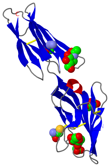
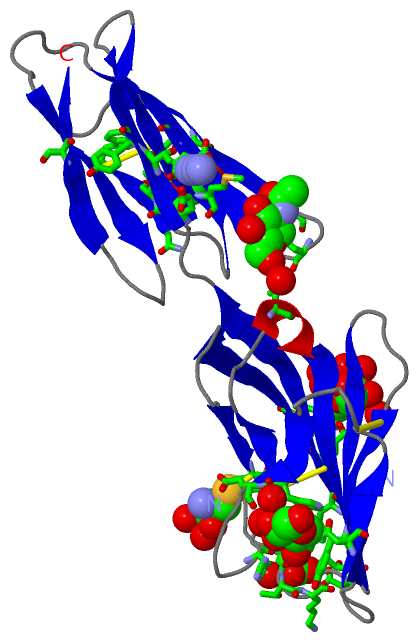
(-) Ligands, Modified Residues, Ions  (4, 7)

Asymmetric/Biological Unit (4, 7)
No.NameCountTypeFull Name
1ACT2Ligand/IonACETATE ION
2AZI1Ligand/IonAZIDE ION
3CYS1Mod. Amino AcidCYSTEINE
4NAG3Ligand/IonN-ACETYL-D-GLUCOSAMINE

(-) Sites  (7, 7)

Asymmetric Unit (7, 7)
No.NameEvidenceResiduesDescription
1AC1SOFTWARELEU A:40 , THR A:41 , LYS A:42 , THR A:109 , GLU A:111 , PHE A:114 , HOH A:2033BINDING SITE FOR RESIDUE ACT A1205
2AC2SOFTWAREGLN A:18 , GLN A:150 , ILE A:151 , HOH A:2121 , HOH A:2141BINDING SITE FOR RESIDUE ACT A1206
3AC3SOFTWAREGLU A:127 , THR A:129 , GLU A:141 , MET A:143BINDING SITE FOR RESIDUE AZI A1207
4AC4SOFTWARECYS A:34 , GLU A:89 , HOH A:2142 , HOH A:2143BINDING SITE FOR RESIDUE CYS A1208
5AC5SOFTWAREGLN A:18 , ASN A:20 , ASN A:117 , ILE A:151 , TRP A:153 , THR A:161 , SER A:175 , HOH A:2110 , HOH A:2144 , HOH A:2145 , HOH A:2146 , HOH A:2147 , HOH A:2148BINDING SITE FOR MONO-SACCHARIDE NAG A2000 BOUND TO ASN A 20
6AC6SOFTWARETYR A:62 , ASN A:69 , TRP A:80 , ALA A:81 , HIS A:86 , TYR A:130 , SER A:201 , HOH A:2079 , HOH A:2080BINDING SITE FOR MONO-SACCHARIDE NAG A 690 BOUND TO ASN A 69
7AC7SOFTWARELEU A:97 , HIS A:166 , ASN A:168 , HOH A:2085BINDING SITE FOR MONO-SACCHARIDE NAG A1680 BOUND TO ASN A 168

(-) SS Bonds  (5, 5)

Asymmetric/Biological Unit
No.Residues
1A:34 -A:1208
2A:35 -A:105
3A:58 -A:73
4A:140 -A:189
5A:159 -A:177

(-) Cis Peptide Bonds  (2, 2)

Asymmetric/Biological Unit
No.Residues
1Lys A:146 -Pro A:147
2Ser A:154 -Pro A:155

 Sequence-Structure Mapping

(-) SAPs(SNPs)/Variants  (0, 0)

(no "SAP(SNP)/Variant" information available for 4BFG)

(-) PROSITE Motifs  (0, 0)

(no "PROSITE Motif" information available for 4BFG)

(-) Exons   (0, 0)

(no "Exon" information available for 4BFG)

(-) Sequences/Alignments

Asymmetric/Biological Unit
   Reformat: Number of residues per line =  ('0' or empty: single-line sequence representation)
  Number of residues per labelling interval =   
  UniProt sequence: complete  aligned part    
   Show mapping: SCOP domains CATH domains Pfam domains Secondary structure (by author)
SAPs(SNPs) PROSITE motifs Exons
(details for a mapped element are shown in a popup box when the mouse pointer rests over it)
Chain A from PDB  Type:PROTEIN  Length:191
 aligned with MO2R1_MOUSE | Q9ES57 from UniProtKB/Swiss-Prot  Length:326

    Alignment length:217
                                    48        58        68        78        88        98       108       118       128       138       148       158       168       178       188       198       208       218       228       238       248       
         MO2R1_MOUSE     39 PLTQVNTTVSVQIGTKALLCCFSIPLTKAVLITWIIKLRGLPSCTIAYKVDTKTNETSCLGRNITWASTPDHSPELQISAVTLQHEGTYTCETVTPEGNFEKNYDLQVLVPPEVTYFPEKNRSAVCEAMAGKPAAQISWSPDGDCVTTSESHSNGTVTVRSTCHWEQNNVSDVSCIVSHLTGNQSLSIELSRGGNQSLRPYIPYIIPSIIILIIIGC  255
               SCOP domains ------------------------------------------------------------------------------------------------------------------------------------------------------------------------------------------------------------------------- SCOP domains
               CATH domains ------------------------------------------------------------------------------------------------------------------------------------------------------------------------------------------------------------------------- CATH domains
               Pfam domains ------------------------------------------------------------------------------------------------------------------------------------------------------------------------------------------------------------------------- Pfam domains
         Sec.struct. author ..eeeeeeeeee....eee...........eeeeeeee.....eeeeee....eeee......eee.........eee...hhhh.eeeeeeeee..eeeeeeeeeeee...eeeeeee...eeeeeeeee....eeeee...eeeeeeee.....eeeeeeee.......eeeeeee.....eeeeee.--------------------------. Sec.struct. author
                 SAPs(SNPs) ------------------------------------------------------------------------------------------------------------------------------------------------------------------------------------------------------------------------- SAPs(SNPs)
                    PROSITE ------------------------------------------------------------------------------------------------------------------------------------------------------------------------------------------------------------------------- PROSITE
                 Transcript ------------------------------------------------------------------------------------------------------------------------------------------------------------------------------------------------------------------------- Transcript
                4bfg A   15 PLTQVNTTVSVQIGTKALLCCFSIPLTKAVLITWIIKLRGLPSCTIAYKVDTKTNETSCLGRNITWASTPDHSPELQISAVTLQHEGTYTCETVTPEGNFEKNYDLQVLVPPEVTYFPEKNRSAVCEAMAGKPAAQISWSPDGDCVTTSESHSNGTVTVRSTCHWEQNNVSDVSCIVSHLTGNQSLSIEL--------------------------C 1208
                                    24        34        44        54        64        74        84        94       104       114       124       134       144       154       164       174       184       194       204         -         -      |
                                                                                                                                                                                                                       204                       1208

   Legend:   → Mismatch (orange background)
  - → Gap (green background, '-', border residues have a numbering label)
    → Modified Residue (blue background, lower-case, 'x' indicates undefined single-letter code, labelled with number + name)
  x → Chemical Group (purple background, 'x', labelled with number + name, e.g. ACE or NH2)
  extra numbering lines below/above indicate numbering irregularities and modified residue names etc., number ends below/above '|'

 Classification and Annotation

(-) SCOP Domains  (0, 0)

(no "SCOP Domain" information available for 4BFG)

(-) CATH Domains  (0, 0)

(no "CATH Domain" information available for 4BFG)

(-) Pfam Domains  (0, 0)

(no "Pfam Domain" information available for 4BFG)

(-) Gene Ontology  (7, 7)

Asymmetric/Biological Unit(hide GO term definitions)
Chain A   (MO2R1_MOUSE | Q9ES57)
molecular function
    GO:0005515    protein binding    Interacting selectively and non-covalently with any protein or protein complex (a complex of two or more proteins that may include other nonprotein molecules).
    GO:0004872    receptor activity    Combining with an extracellular or intracellular messenger to initiate a change in cell activity.
cellular component
    GO:0009897    external side of plasma membrane    The leaflet of the plasma membrane that faces away from the cytoplasm and any proteins embedded or anchored in it or attached to its surface.
    GO:0016021    integral component of membrane    The component of a membrane consisting of the gene products and protein complexes having at least some part of their peptide sequence embedded in the hydrophobic region of the membrane.
    GO:0016020    membrane    A lipid bilayer along with all the proteins and protein complexes embedded in it an attached to it.
    GO:0005886    plasma membrane    The membrane surrounding a cell that separates the cell from its external environment. It consists of a phospholipid bilayer and associated proteins.
    GO:0043235    receptor complex    Any protein complex that undergoes combination with a hormone, neurotransmitter, drug or intracellular messenger to initiate a change in cell function.

 Visualization

(-) Interactive Views

Asymmetric/Biological Unit
  Complete Structure
    Jena3D(integrated viewing of ligand, site, SAP, PROSITE, SCOP information)
    WebMol | AstexViewer[tm]@PDBe
(Java Applets, require no local installation except for Java; loading may be slow)
    STRAP
(Java WebStart application, automatic local installation, requires Java; full application with system access!)
    RasMol
(require local installation)
    Molscript (VRML)
(requires installation of a VRML viewer; select preferred view via VRML and generate a mono or stereo PDF format file)
 
  Ligands, Modified Residues, Ions
    ACT  [ RasMol | Jena3D ]  +environment [ RasMol | Jena3D ]
    AZI  [ RasMol | Jena3D ]  +environment [ RasMol | Jena3D ]
    CYS  [ RasMol | Jena3D ]  +environment [ RasMol | Jena3D ]
    NAG  [ RasMol | Jena3D ]  +environment [ RasMol | Jena3D ]
 
  Sites
    AC1  [ RasMol ]  +environment [ RasMol ]
    AC2  [ RasMol ]  +environment [ RasMol ]
    AC3  [ RasMol ]  +environment [ RasMol ]
    AC4  [ RasMol ]  +environment [ RasMol ]
    AC5  [ RasMol ]  +environment [ RasMol ]
    AC6  [ RasMol ]  +environment [ RasMol ]
    AC7  [ RasMol ]  +environment [ RasMol ]
 
  Cis Peptide Bonds
    Lys A:146 - Pro A:147   [ RasMol ]  
    Ser A:154 - Pro A:155   [ RasMol ]  
 

(-) Still Images

Jmol
  protein: cartoon or spacefill or dots and stick; nucleic acid: cartoon and stick; ligands: spacefill; active site: stick
Molscript
  protein, nucleic acid: cartoon; ligands: spacefill; active site: ball and stick

 Databases and Analysis Tools

(-) Databases

Access by PDB/NDB ID
  4bfg
    Family and Domain InformationProDom | SYSTERS
    General Structural InformationGlycoscienceDB | MMDB | NDB | OCA | PDB | PDBe | PDBj | PDBsum | PDBWiki | PQS | PROTEOPEDIA
    Orientation in MembranesOPM
    Protein SurfaceSURFACE
    Secondary StructureDSSP (structure derived) | HSSP (homology derived)
    Structural GenomicsGeneCensus
    Structural NeighboursCE | VAST
    Structure ClassificationCATH | Dali | SCOP
    Validation and Original DataBMRB Data View | BMRB Restraints Grid | EDS | PROCHECK | RECOORD | WHAT_CHECK
 
Access by UniProt ID/Accession number
  MO2R1_MOUSE | Q9ES57
    Comparative Protein Structure ModelsModBase
    Genomic InformationEnsembl
    Protein-protein InteractionDIP
    Sequence, Family and Domain InformationInterPro | Pfam | SMART | UniProtKB/SwissProt
 
Access by Enzyme Classificator   (EC Number)
  (no 'Enzyme Classificator' available)
    General Enzyme InformationBRENDA | EC-PDB | Enzyme | IntEnz
    PathwayKEGG | MetaCyc
 
Access by Disease Identifier   (MIM ID)
  (no 'MIM ID' available)
    Disease InformationOMIM
 
Access by GenAge ID
  (no 'GenAge ID' available)
    Age Related InformationGenAge

(-) Analysis Tools

Access by PDB/NDB ID
    Domain InformationXDom
    Interatomic Contacts of Structural UnitsCSU
    Ligand-protein ContactsLPC
    Protein CavitiescastP
    Sequence and Secondary StructurePDBCartoon
    Structure AlignmentSTRAP(Java WebStart application, automatic local installation, requires Java; full application with system access!)
    Structure and Sequence BrowserSTING
 
Access by UniProt ID/Accession number
  MO2R1_MOUSE | Q9ES57
    Protein Disorder PredictionDisEMBL | FoldIndex | GLOBPLOT (for more information see DisProt)

 Related Entries

(-) Entries Sharing at Least One Protein Chain (UniProt ID)

UniProtKB/Swiss-Prot
        MO2R1_MOUSE | Q9ES574bfi

(-) Related Entries Specified in the PDB File

4bfe STRUCTURE OF THE EXTRACELLULAR PORTION OF MOUSE CD200RLA
4bfi STRUCTURE OF THE COMPLEX OF THE EXTRACELLULAR PORTIONS OF MOUSE CD200R AND MOUSE CD200