Show PDB file:   
         Plain Text   HTML   (compressed file size)
QuickSearch:   
by PDB,NDB,UniProt,PROSITE Code or Search Term(s)  
(-)Asym./Biol. Unit
(-)Asym./Biol. Unit - sites
collapse expand < >
Image Asym./Biol. Unit
Asym./Biol. Unit  (Jmol Viewer)
Image Asym./Biol. Unit - sites
Asym./Biol. Unit - sites  (Jmol Viewer)

(-) Description

Title :  STRUCTURAL BASIS OF L-PHOSPHOSERINE BINDING TO BACILLUS ALCALOPHILUS PHOSPHOSERINE AMINOTRANSFERASE
 
Authors :  P. Battula, A. P. Dubnovitsky, A. C. Papageorgiou
Date :  26 Jun 12  (Deposition) - 01 May 13  (Release) - 15 May 13  (Revision)
Method :  X-RAY DIFFRACTION
Resolution :  1.50
Chains :  Asym./Biol. Unit :  A,B
Keywords :  Transferase (Keyword Search: [Gene Ontology, PubMed, Web (Google))
 
Reference :  P. Battula, A. P. Dubnovitsky, A. C. Papageorgiou
Structural Basis Of L-Phosphoserine Binding To Bacillus Alcalophilus Phosphoserine Aminotransferase
Acta Crystallogr. , Sect. D V. 69 804 2013
PubMed-ID: 23633589  |  Reference-DOI: 10.1107/S0907444913002096

(-) Compounds

Molecule 1 - PHOSPHOSERINE AMINOTRANSFERASE
    ChainsA, B
    EC Number2.6.1.52
    EngineeredYES
    Expression SystemESCHERICHIA COLI
    Expression System PlasmidPBALC-PSAT
    Expression System StrainBL21(DE3)
    Expression System Taxid469008
    Expression System Vector TypePLASMID
    Organism ScientificBACILLUS ALCALOPHILUS
    Organism Taxid1445
    Other DetailsCOVALENT BOND BETWEEN PLP AND L-PHOSPHOSERINE
    SynonymPHOSPHOHYDROXYTHREONINE AMINOTRANSFERASE, PSAT

 Structural Features

(-) Chains, Units

  12
Asymmetric/Biological Unit AB

Summary Information (see also Sequences/Alignments below)
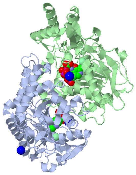
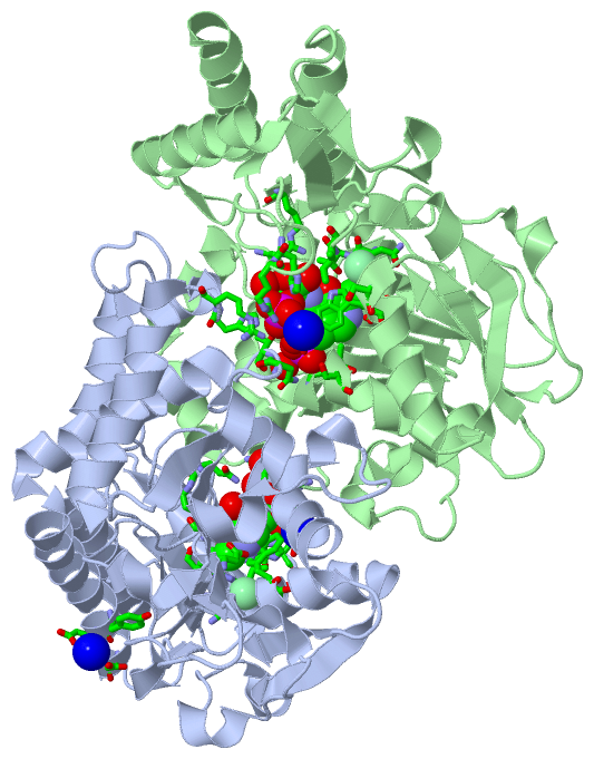
(-) Ligands, Modified Residues, Ions  (4, 9)

Asymmetric/Biological Unit (4, 9)
No.NameCountTypeFull Name
1CL2Ligand/IonCHLORIDE ION
2NA3Ligand/IonSODIUM ION
3PLP2Ligand/IonPYRIDOXAL-5'-PHOSPHATE
4SEP2Mod. ResiduePHOSPHOSERINE

(-) Sites  (9, 9)

Asymmetric Unit (9, 9)
No.NameEvidenceResiduesDescription
1AC1SOFTWAREALA A:9 , TRP A:102 , THR A:152 , ILE A:153 , LYS A:196 , HIS A:327 , ARG A:328 , ARG A:334 , PLP A:501 , NA A:1362 , HOH A:2564 , HOH A:2565 , HIS B:41 , ARG B:42BINDING SITE FOR RESIDUE SEP A 500
2AC2SOFTWAREGLY A:75 , ALA A:76 , SER A:77 , TRP A:102 , THR A:152 , ASP A:172 , SER A:174 , GLN A:195 , LYS A:196 , SEP A:500 , HOH A:2362 , HOH A:2565 , HOH A:2566 , ASN B:237 , THR B:238BINDING SITE FOR RESIDUE PLP A 501
3AC3SOFTWARESER A:101 , TRP A:102 , ASN A:151 , THR A:152 , ILE A:153BINDING SITE FOR RESIDUE CL A1361
4AC4SOFTWARETRP A:102 , ARG A:328 , SEP A:500 , HOH A:2260 , HOH A:2567BINDING SITE FOR RESIDUE NA A1362
5AC5SOFTWARETYR A:275 , ASP A:276 , ASP A:279 , HOH A:2569BINDING SITE FOR RESIDUE NA A1363
6AC6SOFTWAREHIS A:41 , ARG A:42 , ALA B:9 , TRP B:102 , THR B:152 , ILE B:153 , LYS B:196 , HIS B:327 , ARG B:328 , ARG B:334 , PLP B:501 , NA B:1362 , HOH B:2556 , HOH B:2557BINDING SITE FOR RESIDUE SEP B 500
7AC7SOFTWAREASN A:237 , THR A:238 , HOH A:2196 , GLY B:75 , ALA B:76 , SER B:77 , TRP B:102 , THR B:152 , ASP B:172 , SER B:174 , GLN B:195 , LYS B:196 , SEP B:500 , HOH B:2354 , HOH B:2557BINDING SITE FOR RESIDUE PLP B 501
8AC8SOFTWARESER B:101 , TRP B:102 , ASN B:151 , THR B:152 , ILE B:153BINDING SITE FOR RESIDUE CL B1361
9AC9SOFTWAREHOH A:2422 , TRP B:102 , ARG B:328 , SEP B:500BINDING SITE FOR RESIDUE NA B1362

(-) SS Bonds  (0, 0)

(no "SS Bond" information available for 4AZJ)

(-) Cis Peptide Bonds  (2, 2)

Asymmetric/Biological Unit
No.Residues
1Gly A:10 -Pro A:11
2Gly B:10 -Pro B:11

 Sequence-Structure Mapping

(-) SAPs(SNPs)/Variants  (0, 0)

(no "SAP(SNP)/Variant" information available for 4AZJ)

(-) PROSITE Motifs  (1, 2)

Asymmetric/Biological Unit (1, 2)
 PROSITEUniProtKBPDB
No.IDACDescriptionIDLocationCountLocation
1AA_TRANSFER_CLASS_5PS00595 Aminotransferases class-V pyridoxal-phosphate attachment site.SERC_BACAO188-207
 
  2A:187-206
B:187-206

(-) Exons   (0, 0)

(no "Exon" information available for 4AZJ)

(-) Sequences/Alignments

Asymmetric/Biological Unit
   Reformat: Number of residues per line =  ('0' or empty: single-line sequence representation)
  Number of residues per labelling interval =   
  UniProt sequence: complete  aligned part    
   Show mapping: SCOP domains CATH domains Pfam domains Secondary structure (by author)
SAPs(SNPs) PROSITE motifs Exons
(details for a mapped element are shown in a popup box when the mouse pointer rests over it)
Chain A from PDB  Type:PROTEIN  Length:360
 aligned with SERC_BACAO | Q9RME2 from UniProtKB/Swiss-Prot  Length:361

    Alignment length:360
                                    11        21        31        41        51        61        71        81        91       101       111       121       131       141       151       161       171       181       191       201       211       221       231       241       251       261       271       281       291       301       311       321       331       341       351       361
           SERC_BACAO     2 VKQVFNFNAGPSALPKPALERAQKELLNFNDTQMSVMELSHRSQSYEEVHEQAQNLLRELLQIPNDYQILFLQGGASLQFTMLPMNLLTKGTIGNYVLTGSWSEKALKEAKLLGETHIAASTKANSYQSIPDFSEFQLNENDAYLHITSNNTIYGTQYQNFPEINHAPLIADMSSDILSRPLKVNQFGMIYAGAQKNLGPSGVTVVIVKKDLLNTKVEQVPTMLQYATHIKSDSLYNTPPTFSIYMLRNVLDWIKDLGGAEAIAKQNEEKAKIIYDTIDESNGFYVGHAEKGSRSLMNVTFNLRNEELNQQFLAKAKEQGFVGLNGHRSVGGCRASIYNAVPIDACIALRELMIQFKENA 361
               SCOP domains d4azja_ A: Phosphoserine aminotransferase, PSAT                                                                                                                                                                                                                                                                                                                          SCOP domains
               CATH domains ------------------------------------------------------------------------------------------------------------------------------------------------------------------------------------------------------------------------------------------------------------------------------------------------------------------------------------------------------------------------ CATH domains
               Pfam domains ------------------------------------------------------------------------------------------------------------------------------------------------------------------------------------------------------------------------------------------------------------------------------------------------------------------------------------------------------------------------ Pfam domains
         Sec.struct. author ....ee........hhhhhhhhhhh.........hhhhh...hhhhhhhhhhhhhhhhhhhh....eeeeee.hhhhhhhhhhhhhhh....eeeeee.hhhhhhhhhhhhhhheeeeeee.hhhh.....hhhhh.....eeeeeee.ee....ee...........eeee..........hhhhh.eeeee..........eeeeeehhhhh......hhhhhhhhhhhh.......hhhhhhhhhhhhhhhhhhhhhhhhhhhhhhhhhhhhhhhhh.....ee..hhhh....eeeee..hhhhhhhhhhhhhhh.ee..........eeee.....hhhhhhhhhhhhhhhhhhh Sec.struct. author
                 SAPs(SNPs) ------------------------------------------------------------------------------------------------------------------------------------------------------------------------------------------------------------------------------------------------------------------------------------------------------------------------------------------------------------------------ SAPs(SNPs)
                    PROSITE ------------------------------------------------------------------------------------------------------------------------------------------------------------------------------------------AA_TRANSFER_CLASS_5 ---------------------------------------------------------------------------------------------------------------------------------------------------------- PROSITE
                 Transcript ------------------------------------------------------------------------------------------------------------------------------------------------------------------------------------------------------------------------------------------------------------------------------------------------------------------------------------------------------------------------ Transcript
                 4azj A   1 VKQVFNFNAGPSALPKPALERAQKELLNFNDTQMSVMELSHRSQSYEEVHEQAQNLLRELLQIPNDYQILFLQGGASLQFTMLPMNLLTKGTIGNYVLTGSWSEKALKEAKLLGETHIAASTKANSYQSIPDFSEFQLNENDAYLHITSNNTIYGTQYQNFPEINHAPLIADMSSDILSRPLKVNQFGMIYAGAQKNLGPSGVTVVIVKKDLLNTKVEQVPTMLQYATHIKSDSLYNTPPTFSIYMLRNVLDWIKDLGGAEAIAKQNEEKAKIIYDTIDESNGFYVGHAEKGSRSLMNVTFNLRNEELNQQFLAKAKEQGFVGLNGHRSVGGCRASIYNAVPIDACIALRELMIQFKENA 360
                                    10        20        30        40        50        60        70        80        90       100       110       120       130       140       150       160       170       180       190       200       210       220       230       240       250       260       270       280       290       300       310       320       330       340       350       360

Chain B from PDB  Type:PROTEIN  Length:358
 aligned with SERC_BACAO | Q9RME2 from UniProtKB/Swiss-Prot  Length:361

    Alignment length:358
                                    13        23        33        43        53        63        73        83        93       103       113       123       133       143       153       163       173       183       193       203       213       223       233       243       253       263       273       283       293       303       313       323       333       343       353        
           SERC_BACAO     4 QVFNFNAGPSALPKPALERAQKELLNFNDTQMSVMELSHRSQSYEEVHEQAQNLLRELLQIPNDYQILFLQGGASLQFTMLPMNLLTKGTIGNYVLTGSWSEKALKEAKLLGETHIAASTKANSYQSIPDFSEFQLNENDAYLHITSNNTIYGTQYQNFPEINHAPLIADMSSDILSRPLKVNQFGMIYAGAQKNLGPSGVTVVIVKKDLLNTKVEQVPTMLQYATHIKSDSLYNTPPTFSIYMLRNVLDWIKDLGGAEAIAKQNEEKAKIIYDTIDESNGFYVGHAEKGSRSLMNVTFNLRNEELNQQFLAKAKEQGFVGLNGHRSVGGCRASIYNAVPIDACIALRELMIQFKENA 361
               SCOP domains d4azjb_ B: Phosphoserine aminotransferase, PSAT                                                                                                                                                                                                                                                                                                                        SCOP domains
               CATH domains ---------------------------------------------------------------------------------------------------------------------------------------------------------------------------------------------------------------------------------------------------------------------------------------------------------------------------------------------------------------------- CATH domains
               Pfam domains ---------------------------------------------------------------------------------------------------------------------------------------------------------------------------------------------------------------------------------------------------------------------------------------------------------------------------------------------------------------------- Pfam domains
         Sec.struct. author ..ee........hhhhhhhhhhh..hhhhh..hhhhh...hhhhhhhhhhhhhhhhhhhh....eeeeee.hhhhhhhhhhhhhhh....eeeeee.hhhhhhhhhhhhh..eeeeeee.hhhh.....hhhhh.....eeeeeee.ee....ee...........eeee..........hhhhh.eeeee..........eeeeeehhhhh......hhhhhhhhhhhh.......hhhhhhhhhhhhhhhhhhhhhhhhhhhhhhhhhhhhhhhhh.....ee..hhhh....eeeee..hhhhhhhhhhhhhhh.ee..........eeee.....hhhhhhhhhhhhhhhhhhh Sec.struct. author
                 SAPs(SNPs) ---------------------------------------------------------------------------------------------------------------------------------------------------------------------------------------------------------------------------------------------------------------------------------------------------------------------------------------------------------------------- SAPs(SNPs)
                    PROSITE ----------------------------------------------------------------------------------------------------------------------------------------------------------------------------------------AA_TRANSFER_CLASS_5 ---------------------------------------------------------------------------------------------------------------------------------------------------------- PROSITE
                 Transcript ---------------------------------------------------------------------------------------------------------------------------------------------------------------------------------------------------------------------------------------------------------------------------------------------------------------------------------------------------------------------- Transcript
                 4azj B   3 QVFNFNAGPSALPKPALERAQKELLNFNDTQMSVMELSHRSQSYEEVHEQAQNLLRELLQIPNDYQILFLQGGASLQFTMLPMNLLTKGTIGNYVLTGSWSEKALKEAKLLGETHIAASTKANSYQSIPDFSEFQLNENDAYLHITSNNTIYGTQYQNFPEINHAPLIADMSSDILSRPLKVNQFGMIYAGAQKNLGPSGVTVVIVKKDLLNTKVEQVPTMLQYATHIKSDSLYNTPPTFSIYMLRNVLDWIKDLGGAEAIAKQNEEKAKIIYDTIDESNGFYVGHAEKGSRSLMNVTFNLRNEELNQQFLAKAKEQGFVGLNGHRSVGGCRASIYNAVPIDACIALRELMIQFKENA 360
                                    12        22        32        42        52        62        72        82        92       102       112       122       132       142       152       162       172       182       192       202       212       222       232       242       252       262       272       282       292       302       312       322       332       342       352        

   Legend:   → Mismatch (orange background)
  - → Gap (green background, '-', border residues have a numbering label)
    → Modified Residue (blue background, lower-case, 'x' indicates undefined single-letter code, labelled with number + name)
  x → Chemical Group (purple background, 'x', labelled with number + name, e.g. ACE or NH2)
  extra numbering lines below/above indicate numbering irregularities and modified residue names etc., number ends below/above '|'

 Classification and Annotation

(-) SCOP Domains  (1, 2)

Asymmetric/Biological Unit

(-) CATH Domains  (0, 0)

(no "CATH Domain" information available for 4AZJ)

(-) Pfam Domains  (0, 0)

(no "Pfam Domain" information available for 4AZJ)

(-) Gene Ontology  (8, 8)

Asymmetric/Biological Unit(hide GO term definitions)
Chain A,B   (SERC_BACAO | Q9RME2)
molecular function
    GO:0004648    O-phospho-L-serine:2-oxoglutarate aminotransferase activity    Catalysis of the reaction: O-phospho-L-serine + 2-oxoglutarate = 3-phosphonooxypyruvate + L-glutamate.
    GO:0003824    catalytic activity    Catalysis of a biochemical reaction at physiological temperatures. In biologically catalyzed reactions, the reactants are known as substrates, and the catalysts are naturally occurring macromolecular substances known as enzymes. Enzymes possess specific binding sites for substrates, and are usually composed wholly or largely of protein, but RNA that has catalytic activity (ribozyme) is often also regarded as enzymatic.
    GO:0030170    pyridoxal phosphate binding    Interacting selectively and non-covalently with pyridoxal 5' phosphate, 3-hydroxy-5-(hydroxymethyl)-2-methyl4-pyridine carboxaldehyde 5' phosphate, the biologically active form of vitamin B6.
    GO:0008483    transaminase activity    Catalysis of the transfer of an amino group to an acceptor, usually a 2-oxo acid.
    GO:0016740    transferase activity    Catalysis of the transfer of a group, e.g. a methyl group, glycosyl group, acyl group, phosphorus-containing, or other groups, from one compound (generally regarded as the donor) to another compound (generally regarded as the acceptor). Transferase is the systematic name for any enzyme of EC class 2.
biological process
    GO:0006564    L-serine biosynthetic process    The chemical reactions and pathways resulting in the formation of L-serine, the L-enantiomer of serine, i.e. (2S)-2-amino-3-hydroxypropanoic acid.
    GO:0008652    cellular amino acid biosynthetic process    The chemical reactions and pathways resulting in the formation of amino acids, organic acids containing one or more amino substituents.
cellular component
    GO:0005737    cytoplasm    All of the contents of a cell excluding the plasma membrane and nucleus, but including other subcellular structures.

 Visualization

(-) Interactive Views

Asymmetric/Biological Unit
  Complete Structure
    Jena3D(integrated viewing of ligand, site, SAP, PROSITE, SCOP information)
    WebMol | AstexViewer[tm]@PDBe
(Java Applets, require no local installation except for Java; loading may be slow)
    STRAP
(Java WebStart application, automatic local installation, requires Java; full application with system access!)
    RasMol
(require local installation)
    Molscript (VRML)
(requires installation of a VRML viewer; select preferred view via VRML and generate a mono or stereo PDF format file)
 
  Ligands, Modified Residues, Ions
    CL  [ RasMol | Jena3D ]  +environment [ RasMol | Jena3D ]
    NA  [ RasMol | Jena3D ]  +environment [ RasMol | Jena3D ]
    PLP  [ RasMol | Jena3D ]  +environment [ RasMol | Jena3D ]
    SEP  [ RasMol | Jena3D ]  +environment [ RasMol | Jena3D ]
 
  Sites
    AC1  [ RasMol ]  +environment [ RasMol ]
    AC2  [ RasMol ]  +environment [ RasMol ]
    AC3  [ RasMol ]  +environment [ RasMol ]
    AC4  [ RasMol ]  +environment [ RasMol ]
    AC5  [ RasMol ]  +environment [ RasMol ]
    AC6  [ RasMol ]  +environment [ RasMol ]
    AC7  [ RasMol ]  +environment [ RasMol ]
    AC8  [ RasMol ]  +environment [ RasMol ]
    AC9  [ RasMol ]  +environment [ RasMol ]
 
  Cis Peptide Bonds
    Gly A:10 - Pro A:11   [ RasMol ]  
    Gly B:10 - Pro B:11   [ RasMol ]  
 

(-) Still Images

Jmol
  protein: cartoon or spacefill or dots and stick; nucleic acid: cartoon and stick; ligands: spacefill; active site: stick
Molscript
  protein, nucleic acid: cartoon; ligands: spacefill; active site: ball and stick

 Databases and Analysis Tools

(-) Databases

Access by PDB/NDB ID
  4azj
    Family and Domain InformationProDom | SYSTERS
    General Structural InformationGlycoscienceDB | MMDB | NDB | OCA | PDB | PDBe | PDBj | PDBsum | PDBWiki | PQS | PROTEOPEDIA
    Orientation in MembranesOPM
    Protein SurfaceSURFACE
    Secondary StructureDSSP (structure derived) | HSSP (homology derived)
    Structural GenomicsGeneCensus
    Structural NeighboursCE | VAST
    Structure ClassificationCATH | Dali | SCOP
    Validation and Original DataBMRB Data View | BMRB Restraints Grid | EDS | PROCHECK | RECOORD | WHAT_CHECK
 
Access by UniProt ID/Accession number
  SERC_BACAO | Q9RME2
    Comparative Protein Structure ModelsModBase
    Genomic InformationEnsembl
    Protein-protein InteractionDIP
    Sequence, Family and Domain InformationInterPro | Pfam | SMART | UniProtKB/SwissProt
 
Access by Enzyme Classificator   (EC Number)
  2.6.1.52
    General Enzyme InformationBRENDA | EC-PDB | Enzyme | IntEnz
    PathwayKEGG | MetaCyc
 
Access by Disease Identifier   (MIM ID)
  (no 'MIM ID' available)
    Disease InformationOMIM
 
Access by GenAge ID
  (no 'GenAge ID' available)
    Age Related InformationGenAge

(-) Analysis Tools

Access by PDB/NDB ID
    Domain InformationXDom
    Interatomic Contacts of Structural UnitsCSU
    Ligand-protein ContactsLPC
    Protein CavitiescastP
    Sequence and Secondary StructurePDBCartoon
    Structure AlignmentSTRAP(Java WebStart application, automatic local installation, requires Java; full application with system access!)
    Structure and Sequence BrowserSTING
 
Access by UniProt ID/Accession number
  SERC_BACAO | Q9RME2
    Protein Disorder PredictionDisEMBL | FoldIndex | GLOBPLOT (for more information see DisProt)

 Related Entries

(-) Entries Sharing at Least One Protein Chain (UniProt ID)

UniProtKB/Swiss-Prot
        SERC_BACAO | Q9RME21w23 2bhx 2bi1 2bi2 2bi3 2bi5 2bi9 2bia 2bie 2big 4azk

(-) Related Entries Specified in the PDB File

1w23 CRYSTAL STRUCTURE OF PHOSPHOSERINE AMINOTRANSFERASE FROM BACILLUS ALCALOPHILUS
2bhx RADIATION DAMAGE OF THE SCHIFF BASE IN PHOSPHOSERINE AMINOTRANSFERASE (STRUCTURE A)
2bi1 RADIATION DAMAGE OF THE SCHIFF BASE IN PHOSPHOSERINE AMINOTRANSFERASE (STRUCTURE B)
2bi2 RADIATION DAMAGE OF THE SCHIFF BASE IN PHOSPHOSERINE AMINOTRANSFERASE (STRUCTURE C)
2bi3 RADIATION DAMAGE OF THE SCHIFF BASE IN PHOSPHOSERINE AMINOTRANSFERASE (STRUCTURE D)
2bi5 RADIATION DAMAGE OF THE SCHIFF BASE IN PHOSPHOSERINE AMINOTRANSFERASE (STRUCTURE E)
2bi9 RADIATION DAMAGE OF THE SCHIFF BASE IN PHOSPHOSERINE AMINOTRANSFERASE (STRUCTURE F)
2bia RADIATION DAMAGE OF THE SCHIFF BASE IN PHOSPHOSERINE AMINOTRANSFERASE (STRUCTURE G)
2bie RADIATION DAMAGE OF THE SCHIFF BASE IN PHOSPHOSERINE AMINOTRANSFERASE (STRUCTURE H)
2big RADIATION DAMAGE OF THE SCHIFF BASE IN PHOSPHOSERINE AMINOTRANSFERASE (STRUCTURE I)
4azk STRUCTURAL BASIS OF L-PHOSPHOSERINE BINDING TO BACILLUS ALCALOPHILUS PHOSPHOSERINE AMINOTRANSFERASE