Show PDB file:   
         Plain Text   HTML   (compressed file size)
QuickSearch:   
by PDB,NDB,UniProt,PROSITE Code or Search Term(s)  
(-)Asymmetric Unit
(-)Asym. Unit - sites
(-)Biological Unit 1
(-)Biol. Unit 1 - sites
collapse expand < >
Image Asymmetric Unit
Asymmetric Unit  (Jmol Viewer)
Image Asym. Unit - sites
Asym. Unit - sites  (Jmol Viewer)
Image Biological Unit 1
Biological Unit 1  (Jmol Viewer)
Image Biol. Unit 1 - sites
Biol. Unit 1 - sites  (Jmol Viewer)

(-) Description

Title :  STRUCTURE OF THE CLOSTRIDIUM DIFFICILE EUTM PROTEIN
 
Authors :  A. C. Pitts, L. R. Tuck, A. Faulds-Pain, R. J. Lewis, J. Marles-Wright
Date :  13 Jun 12  (Deposition) - 20 Jun 12  (Release) - 09 Jan 13  (Revision)
Method :  X-RAY DIFFRACTION
Resolution :  1.62
Chains :  Asym. Unit :  A,B,C
Biol. Unit 1:  A,B,C  (2x)
Keywords :  Structural Protein, Ethanolamine, Bacterial Microcompartment, Bmc (Keyword Search: [Gene Ontology, PubMed, Web (Google))
 
Reference :  A. C. Pitts, L. R. Tuck, A. Faulds-Pain, R. J. Lewis, J. Marles-Wright
Structural Insight Into The Clostridium Difficile Ethanolamine Utilisation Microcompartment.
Plos One V. 7 48360 2012
PubMed-ID: 23144756  |  Reference-DOI: 10.1371/JOURNAL.PONE.0048360

(-) Compounds

Molecule 1 - ETHANOLAMINE CARBOXYSOME STRUCTURAL PROTEIN
    ChainsA, B, C
    EngineeredYES
    Expression SystemESCHERICHIA COLI B
    Expression System StrainB834
    Expression System Taxid37762
    Expression System VectorPET28B
    Expression System Vector TypePLASMID
    Organism ScientificCLOSTRIDIUM DIFFICILE
    Organism Taxid272563
    Strain630
    SynonymEUTM

 Structural Features

(-) Chains, Units

  123
Asymmetric Unit ABC
Biological Unit 1 (2x)ABC

Summary Information (see also Sequences/Alignments below)

(-) Ligands, Modified Residues, Ions  (1, 2)

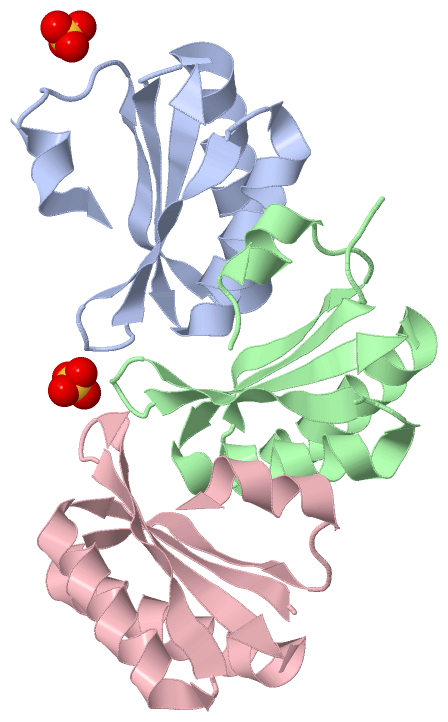
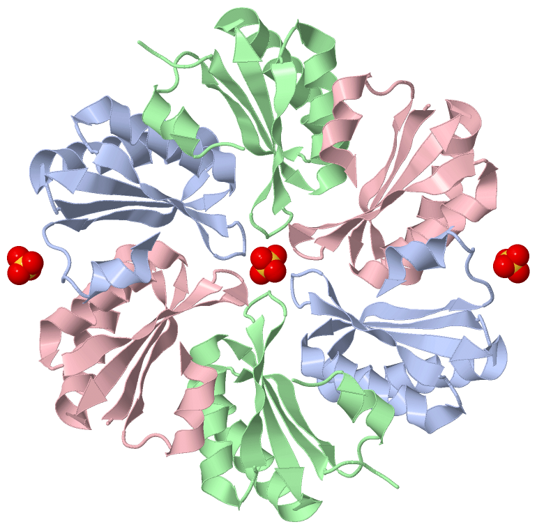
Asymmetric Unit (1, 2)
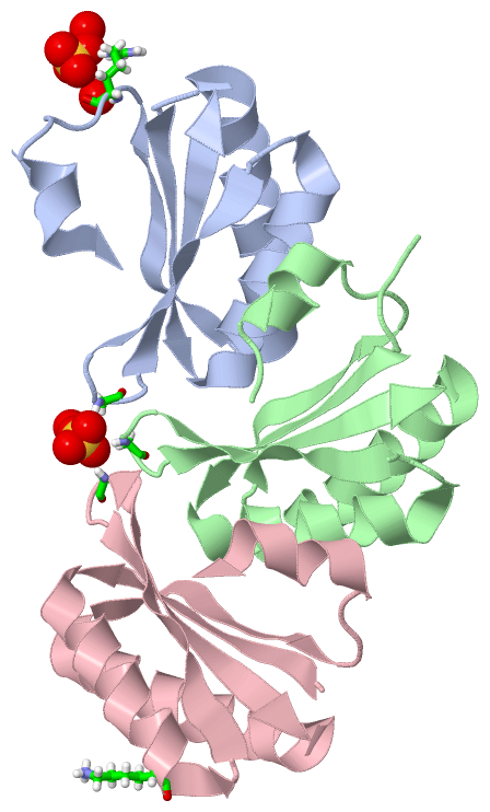
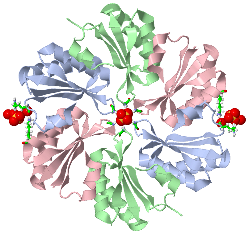
No.NameCountTypeFull Name
1SO42Ligand/IonSULFATE ION
Biological Unit 1 (1, 4)
No.NameCountTypeFull Name
1SO44Ligand/IonSULFATE ION

(-) Sites  (2, 2)

Asymmetric Unit (2, 2)
No.NameEvidenceResiduesDescription
1AC1SOFTWAREGLY A:41 , HOH A:2068 , GLY B:41 , GLY C:41BINDING SITE FOR RESIDUE SO4 A1091
2AC2SOFTWAREARG A:80 , HOH A:2057 , HOH A:2069 , LYS C:27BINDING SITE FOR RESIDUE SO4 A1092

(-) SS Bonds  (0, 0)

(no "SS Bond" information available for 4AXJ)

(-) Cis Peptide Bonds  (0, 0)

(no "Cis Peptide Bond" information available for 4AXJ)

 Sequence-Structure Mapping

(-) SAPs(SNPs)/Variants  (0, 0)

(no "SAP(SNP)/Variant" information available for 4AXJ)

(-) PROSITE Motifs  (0, 0)

(no "PROSITE Motif" information available for 4AXJ)

(-) Exons   (0, 0)

(no "Exon" information available for 4AXJ)

(-) Sequences/Alignments

Asymmetric Unit
   Reformat: Number of residues per line =  ('0' or empty: single-line sequence representation)
  Number of residues per labelling interval =   
  UniProt sequence: complete  aligned part    
   Show mapping: SCOP domains CATH domains Pfam domains Secondary structure (by author)
SAPs(SNPs) PROSITE motifs Exons
(details for a mapped element are shown in a popup box when the mouse pointer rests over it)
Chain A from PDB  Type:PROTEIN  Length:87
 aligned with Q187N0_PEPD6 | Q187N0 from UniProtKB/TrEMBL  Length:95

    Alignment length:87
                                    13        23        33        43        53        63        73        83       
          Q187N0_PEPD6    4 ANALGMIETKGLVGAIEAADAMVKAANVQLVGKEQVGGGLVTVMVRGDVGAVKAATDAGAAAAERVGELISVHVIPRPHFEVDAILP 90
               SCOP domains d4axja_ A: automated matches                                                            SCOP domains
               CATH domains --------------------------------------------------------------------------------------- CATH domains
               Pfam domains --------------------------------------------------------------------------------------- Pfam domains
         Sec.struct. author ..eeeeeeeehhhhhhhhhhhhhhhh..eeeeeee....eeeeeeeehhhhhhhhhhhhhhhhhhhheeeeeeee.....hhhhhh. Sec.struct. author
                 SAPs(SNPs) --------------------------------------------------------------------------------------- SAPs(SNPs)
                    PROSITE --------------------------------------------------------------------------------------- PROSITE
                 Transcript --------------------------------------------------------------------------------------- Transcript
                  4axj A  4 ANALGMIETKGLVGAIEAADAMVKAANVQLVGKEQVGGGLVTVMVRGDVGAVKAATDAGAAAAERVGELISVHVIPRPHFEVDAILP 90
                                    13        23        33        43        53        63        73        83       

Chain B from PDB  Type:PROTEIN  Length:89
 aligned with Q187N0_PEPD6 | Q187N0 from UniProtKB/TrEMBL  Length:95

    Alignment length:89
                                    11        21        31        41        51        61        71        81         
          Q187N0_PEPD6    2 ASANALGMIETKGLVGAIEAADAMVKAANVQLVGKEQVGGGLVTVMVRGDVGAVKAATDAGAAAAERVGELISVHVIPRPHFEVDAILP 90
               SCOP domains d4axjb_ B: automated matches                                                              SCOP domains
               CATH domains ----------------------------------------------------------------------------------------- CATH domains
               Pfam domains ----------------------------------------------------------------------------------------- Pfam domains
         Sec.struct. author ....eeeeeeeehhhhhhhhhhhhhhhh..eeeeeee....eeeeeeeehhhhhhhhhhhhhhhhhhhh.eeeeeee...hhhhhh... Sec.struct. author
                 SAPs(SNPs) ----------------------------------------------------------------------------------------- SAPs(SNPs)
                    PROSITE ----------------------------------------------------------------------------------------- PROSITE
                 Transcript ----------------------------------------------------------------------------------------- Transcript
                  4axj B  2 ASANALGMIETKGLVGAIEAADAMVKAANVQLVGKEQVGGGLVTVMVRGDVGAVKAATDAGAAAAERVGELISVHVIPRPHFEVDAILP 90
                                    11        21        31        41        51        61        71        81         

Chain C from PDB  Type:PROTEIN  Length:86
 aligned with Q187N0_PEPD6 | Q187N0 from UniProtKB/TrEMBL  Length:95

    Alignment length:86
                                    14        24        34        44        54        64        74        84      
          Q187N0_PEPD6    5 NALGMIETKGLVGAIEAADAMVKAANVQLVGKEQVGGGLVTVMVRGDVGAVKAATDAGAAAAERVGELISVHVIPRPHFEVDAILP 90
               SCOP domains d4axjc_ C: automated matches                                                           SCOP domains
               CATH domains -------------------------------------------------------------------------------------- CATH domains
               Pfam domains -------------------------------------------------------------------------------------- Pfam domains
         Sec.struct. author .eeeeeeeehhhhhhhhhhhhhhhh.eeeeeeeeee..eeeeeeeehhhhhhhhhhhhhhhhhhhheeeeeeee...hhhhhhhh. Sec.struct. author
                 SAPs(SNPs) -------------------------------------------------------------------------------------- SAPs(SNPs)
                    PROSITE -------------------------------------------------------------------------------------- PROSITE
                 Transcript -------------------------------------------------------------------------------------- Transcript
                  4axj C  5 NALGMIETKGLVGAIEAADAMVKAANVQLVGKEQVGGGLVTVMVRGDVGAVKAATDAGAAAAERVGELISVHVIPRPHFEVDAILP 90
                                    14        24        34        44        54        64        74        84      

   Legend:   → Mismatch (orange background)
  - → Gap (green background, '-', border residues have a numbering label)
    → Modified Residue (blue background, lower-case, 'x' indicates undefined single-letter code, labelled with number + name)
  x → Chemical Group (purple background, 'x', labelled with number + name, e.g. ACE or NH2)
  extra numbering lines below/above indicate numbering irregularities and modified residue names etc., number ends below/above '|'

 Classification and Annotation

(-) SCOP Domains  (1, 3)

Asymmetric Unit

(-) CATH Domains  (0, 0)

(no "CATH Domain" information available for 4AXJ)

(-) Pfam Domains  (0, 0)

(no "Pfam Domain" information available for 4AXJ)

(-) Gene Ontology  (0, 0)

Asymmetric Unit(hide GO term definitions)
    (no "Gene Ontology" information available for 4AXJ)

 Visualization

(-) Interactive Views

Asymmetric Unit
  Complete Structure
    Jena3D(integrated viewing of ligand, site, SAP, PROSITE, SCOP information)
    WebMol | AstexViewer[tm]@PDBe
(Java Applets, require no local installation except for Java; loading may be slow)
    STRAP
(Java WebStart application, automatic local installation, requires Java; full application with system access!)
    RasMol
(require local installation)
    Molscript (VRML)
(requires installation of a VRML viewer; select preferred view via VRML and generate a mono or stereo PDF format file)
 
  Ligands, Modified Residues, Ions
    SO4  [ RasMol | Jena3D ]  +environment [ RasMol | Jena3D ]
 
  Sites
    AC1  [ RasMol ]  +environment [ RasMol ]
    AC2  [ RasMol ]  +environment [ RasMol ]
 
  Cis Peptide Bonds
(no "Cis Peptide Bonds" information available for 4axj)
 
Biological Unit
  Complete Structure
    Biological Unit 1  [ Jena3D ]

(-) Still Images

Jmol
  protein: cartoon or spacefill or dots and stick; nucleic acid: cartoon and stick; ligands: spacefill; active site: stick
Molscript
  protein, nucleic acid: cartoon; ligands: spacefill; active site: ball and stick

 Databases and Analysis Tools

(-) Databases

Access by PDB/NDB ID
  4axj
    Family and Domain InformationProDom | SYSTERS
    General Structural InformationGlycoscienceDB | MMDB | NDB | OCA | PDB | PDBe | PDBj | PDBsum | PDBWiki | PQS | PROTEOPEDIA
    Orientation in MembranesOPM
    Protein SurfaceSURFACE
    Secondary StructureDSSP (structure derived) | HSSP (homology derived)
    Structural GenomicsGeneCensus
    Structural NeighboursCE | VAST
    Structure ClassificationCATH | Dali | SCOP
    Validation and Original DataBMRB Data View | BMRB Restraints Grid | EDS | PROCHECK | RECOORD | WHAT_CHECK
 
Access by UniProt ID/Accession number
  Q187N0_PEPD6 | Q187N0
    Comparative Protein Structure ModelsModBase
    Genomic InformationEnsembl
    Protein-protein InteractionDIP
    Sequence, Family and Domain InformationInterPro | Pfam | SMART | UniProtKB/TrEMBL
 
Access by Enzyme Classificator   (EC Number)
  (no 'Enzyme Classificator' available)
    General Enzyme InformationBRENDA | EC-PDB | Enzyme | IntEnz
    PathwayKEGG | MetaCyc
 
Access by Disease Identifier   (MIM ID)
  (no 'MIM ID' available)
    Disease InformationOMIM
 
Access by GenAge ID
  (no 'GenAge ID' available)
    Age Related InformationGenAge

(-) Analysis Tools

Access by PDB/NDB ID
    Domain InformationXDom
    Interatomic Contacts of Structural UnitsCSU
    Ligand-protein ContactsLPC
    Protein CavitiescastP
    Sequence and Secondary StructurePDBCartoon
    Structure AlignmentSTRAP(Java WebStart application, automatic local installation, requires Java; full application with system access!)
    Structure and Sequence BrowserSTING
 
Access by UniProt ID/Accession number
  Q187N0_PEPD6 | Q187N0
    Protein Disorder PredictionDisEMBL | FoldIndex | GLOBPLOT (for more information see DisProt)

 Related Entries

(-) Entries Sharing at Least One Protein Chain (UniProt ID)

(no "Entries Sharing at Least One Protein Chain" available for 4AXJ)

(-) Related Entries Specified in the PDB File

(no "Related Entries Specified in the PDB File" available for 4AXJ)