|
|
|
|
 Description
|
|
Compounds
|
||||||||||||||||||||||||||||||||||||||||||||||||||||
Chains, Units
Summary Information (see also Sequences/Alignments below) |
Ligands, Modified Residues, Ions (3, 4)| Asymmetric Unit (3, 4) Biological Unit 1 (0, 0) |
Sites (4, 4)
Asymmetric Unit (4, 4)
|
SS Bonds (1, 1)
Asymmetric Unit
|
||||||||
Cis Peptide Bonds (0, 0)| (no "Cis Peptide Bond" information available for 4AQI) |
SAPs(SNPs)/Variants (2, 2)
Asymmetric Unit (2, 2)
|
||||||||||||||||||||||||||||||||||||||||||||||||||||||||||||||||||||||||||||||||||||||||||||||||||||||||||||||||||||||||||||||||||||
PROSITE Motifs (3, 3)
Asymmetric Unit (3, 3)
|
||||||||||||||||||||||||||||||||||||||||||||||||||||||||||||||||||||||||||||||||
Exons (0, 0)| (no "Exon" information available for 4AQI) |
Sequences/Alignments
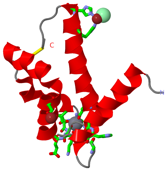
Asymmetric UnitChain A from PDB Type:PROTEIN Length:96 aligned with S1A7A_HUMAN | Q86SG5 from UniProtKB/Swiss-Prot Length:101 Alignment length:96 11 21 31 41 51 61 71 81 91 S1A7A_HUMAN 2 SNTQAERSIIGMIDMFHKYTGRDGKIEKPSLLTMMKENFPNFLSACDKKGIHYLATVFEKKDKNEDKKIDFSEFLSLLGDIAADYHKQSHGAAPCS 97 SCOP domains d4aqia_ A: automated matches SCOP domains CATH domains ------------------------------------------------------------------------------------------------ CATH domains Pfam domains ------------------------------------------------------------------------------------------------ Pfam domains SAPs(SNPs) ---------------------H------------------------------------------------------------T------------- SAPs(SNPs) PROSITE (1) ------------------------------------------------EF_HAND_2 PDB: A:49-84 ------------ PROSITE (1) PROSITE (2) --------------------------------------------------------S100_CABP PDB: A:57-7------------------ PROSITE (2) PROSITE (3) -------------------------------------------------------------EF_HAND_1 ---------------------- PROSITE (3) Transcript ------------------------------------------------------------------------------------------------ Transcript 4aqi A 1 SNTQAERSIIGMIDMFHKYTGRDGKIEKPSLLTMMKENFPNFLSACDKKGIHYLATVFEKKDKNEDKKIDFSEFLSLLGDIAADYHKQSHGAAPCS 96 10 20 30 40 50 60 70 80 90
|
||||||||||||||||||||
SCOP Domains (1, 1)
Asymmetric Unit
 |
CATH Domains (0, 0)| (no "CATH Domain" information available for 4AQI) |
Pfam Domains (0, 0)| (no "Pfam Domain" information available for 4AQI) |
Gene Ontology (4, 4)|
Asymmetric Unit(hide GO term definitions) Chain A (S1A7A_HUMAN | Q86SG5)
|
||||||||||||||||||||||||||||||||||||
Interactive Views
|
|||||||||||||||||||||||||||||||||||||||||||||||||||||||||||||||||||||||||||||||||||||||||||||||||||||||||||||||||||||||||||||||||||||||||||||||||||||||||||||||||||||||||||
Still Images
|
||||||||||||||||
Databases
|
||||||||||||||||||||||||||||||||||||||||||||||||||||||||||||||||||||||||||||||||||||||||||||||||||||||||||||||||||||||||||||||||||||||||||||||||||||||||||||||||
Analysis Tools
|
|||||||||||||||||||||||||||||||||||||||||||||||||||||||||||||
Entries Sharing at Least One Protein Chain (UniProt ID)
Related Entries Specified in the PDB File
|
|