|
|
|
|
 Description
|
|
Compounds
|
||||||||||||||||||||||||||||||||||||||||||||||||
Chains, Units
Summary Information (see also Sequences/Alignments below) |
Ligands, Modified Residues, Ions (2, 3)| Asymmetric Unit (2, 3) Biological Unit 1 (1, 1) Biological Unit 2 (1, 2) |
Sites (3, 3)
Asymmetric Unit (3, 3)
|
SS Bonds (3, 3)
Asymmetric Unit
|
||||||||||||||||
Cis Peptide Bonds (2, 2)
Asymmetric Unit
|
||||||||||||
SAPs(SNPs)/Variants (0, 0)| (no "SAP(SNP)/Variant" information available for 4A3X) |
PROSITE Motifs (0, 0)| (no "PROSITE Motif" information available for 4A3X) |
Exons (0, 0)| (no "Exon" information available for 4A3X) |
Sequences/Alignments
Asymmetric UnitChain A from PDB Type:PROTEIN Length:227 aligned with Q6VBJ0_CANGB | Q6VBJ0 from UniProtKB/TrEMBL Length:1034 Alignment length:227 49 59 69 79 89 99 109 119 129 139 149 159 169 179 189 199 209 219 229 239 249 259 Q6VBJ0_CANGB 40 SKDPTTFPLGCSPDITTPKKGLSMELYSYDFRKKGSYPCWDAAYLDPNYPRTGYKSHRLLAKVDGVTGNINFYYHATKGCTPQLGHLPASYNYPKPLTMTNFTMLLYGYFRPKVTGFHTFTISADDLLFVNFGAGNAFDCCRRDSSADHFGNYQAYAIWGSKTAKDELTVHLDAGVYYPIRLFYNNREYDGALSFTFKTESNENTVSDFSEYFFSLDDTEEGCPGLI 266 SCOP domains ----------------------------------------------------------------------------------------------------------------------------------------------------------------------------------------------------------------------------------- SCOP domains CATH domains ----------------------------------------------------------------------------------------------------------------------------------------------------------------------------------------------------------------------------------- CATH domains Pfam domains ----------------------------------------------------------------------------------------------------------------------------------------------------------------------------------------------------------------------------------- Pfam domains SAPs(SNPs) ----------------------------------------------------------------------------------------------------------------------------------------------------------------------------------------------------------------------------------- SAPs(SNPs) PROSITE ----------------------------------------------------------------------------------------------------------------------------------------------------------------------------------------------------------------------------------- PROSITE Transcript ----------------------------------------------------------------------------------------------------------------------------------------------------------------------------------------------------------------------------------- Transcript 4a3x A 40 SKDPTTFPLGCSPDITTPKKGLSMELYSYDFRKKGSYPCWDAAYLDPNYPRTGYKSHRLLAKVDGVTGNINFYYHATKGCTPQLGHLPASYNYPKPLTMTNFTMLLYGYFRPKVTGFHTFTISADDLLFVNFGAGNAFDCCRRDSSADHFGNYQAYAIWGSKTAKDELTVHLDAGVYYPIRLFYNNREYDGALSFTFKTESNENTVSDFSEYFFSLDDTEEGCPGLI 266 49 59 69 79 89 99 109 119 129 139 149 159 169 179 189 199 209 219 229 239 249 259
|
||||||||||||||||||||
SCOP Domains (0, 0)| (no "SCOP Domain" information available for 4A3X) |
CATH Domains (0, 0)| (no "CATH Domain" information available for 4A3X) |
Pfam Domains (0, 0)| (no "Pfam Domain" information available for 4A3X) |
Gene Ontology (1, 1)|
Asymmetric Unit(hide GO term definitions) Chain A (Q6VBJ0_CANGB | Q6VBJ0)
|
||||||||||||
Interactive Views
|
||||||||||||||||||||||||||||||||||||||||||||||||||||||||||||||||||||||||||||||||||||||||||||||||||||||||||||||||||||||||||||||||||||||||||||||||||||||||||||||||||||||||||
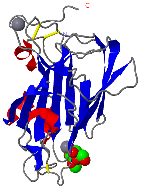
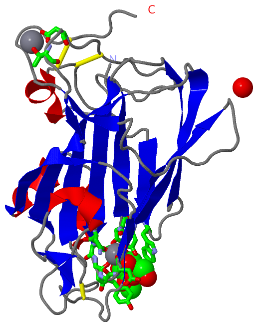
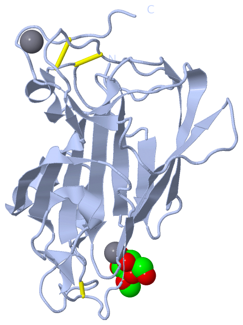
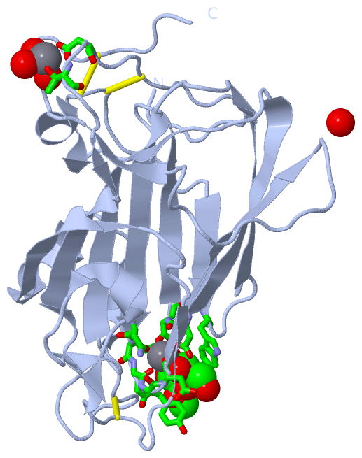
Still Images
|
||||||||||||||||
Databases
|
||||||||||||||||||||||||||||||||||||||||||||||||||||||||||||||||||||||||||||||||||||||||||||||||||||||||||||||||||||||||||||||||||||||||||||||||||||||||||||||||
Analysis Tools
|
|||||||||||||||||||||||||||||||||||||||||||||||||||||||||||||
Entries Sharing at Least One Protein Chain (UniProt ID)
Related Entries Specified in the PDB File
|
|