|
|
|
|
 Description
|
|
Compounds
|
||||||||||||||||||||||||||||||||||||||||||||||||||||
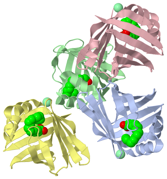
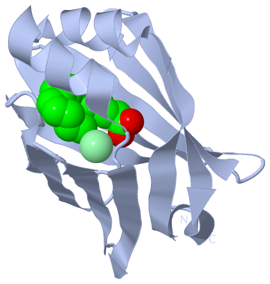
Chains, Units
Summary Information (see also Sequences/Alignments below) |
Ligands, Modified Residues, Ions (2, 7)| Asymmetric Unit (2, 7) Biological Unit 1 (1, 1) Biological Unit 2 (1, 1) Biological Unit 3 (1, 1) Biological Unit 4 (1, 1) |
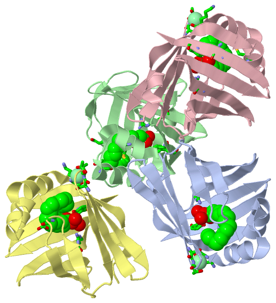
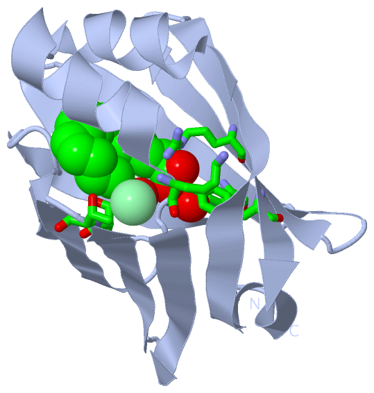
Sites (7, 7)
Asymmetric Unit (7, 7)
|
SS Bonds (0, 0)| (no "SS Bond" information available for 4A1Y) |
Cis Peptide Bonds (0, 0)| (no "Cis Peptide Bond" information available for 4A1Y) |
SAPs(SNPs)/Variants (0, 0)| (no "SAP(SNP)/Variant" information available for 4A1Y) |
PROSITE Motifs (1, 4)
Asymmetric Unit (1, 4)
|
||||||||||||||||||||||||||||||||||||||||||||||||||||||||||||||||||||||||||||||||||||||||||||||||||||||||||||||||||||||||
Exons (0, 0)| (no "Exon" information available for 4A1Y) |
Sequences/Alignments
Asymmetric UnitChain A from PDB Type:PROTEIN Length:133 aligned with MYP2_HUMAN | P02689 from UniProtKB/Swiss-Prot Length:132 Alignment length:133 1 | 9 19 29 39 49 59 69 79 89 99 109 119 129 MYP2_HUMAN - -MSNKFLGTWKLVSSENFDDYMKALGVGLATRKLGNLAKPTVIISKKGDIITIRTESTFKNTEISFKLGQEFEETTADNRKTKSIVTLQRGSLNQVQRWDGKETTIKRKLVNGKMVAECKMKGVVCTRIYEKV 132 SCOP domains d4a1ya_ A: automated matches SCOP domains CATH domains ------------------------------------------------------------------------------------------------------------------------------------- CATH domains Pfam domains ------------------------------------------------------------------------------------------------------------------------------------- Pfam domains SAPs(SNPs) ------------------------------------------------------------------------------------------------------------------------------------- SAPs(SNPs) PROSITE -------FABP PDB: A:6-23 ------------------------------------------------------------------------------------------------------------ PROSITE Transcript ------------------------------------------------------------------------------------------------------------------------------------- Transcript 4a1y A -1 GMSNKFLGTWKLVSSENFDDYMKALGVGLATRKLGNLAKPTVIISKKGDIITIRTESTFKNTEISFQLGQEFEETTADNRKTKSIVTLQRGSLNQVQRWDGKETTIKRKLVNGKMVAECKMKGVVCTRIYEKV 131 8 18 28 38 48 58 68 78 88 98 108 118 128 Chain B from PDB Type:PROTEIN Length:133 aligned with MYP2_HUMAN | P02689 from UniProtKB/Swiss-Prot Length:132 Alignment length:133 1 | 9 19 29 39 49 59 69 79 89 99 109 119 129 MYP2_HUMAN - -MSNKFLGTWKLVSSENFDDYMKALGVGLATRKLGNLAKPTVIISKKGDIITIRTESTFKNTEISFKLGQEFEETTADNRKTKSIVTLQRGSLNQVQRWDGKETTIKRKLVNGKMVAECKMKGVVCTRIYEKV 132 SCOP domains d4a1yb_ B: automated matches SCOP domains CATH domains ------------------------------------------------------------------------------------------------------------------------------------- CATH domains Pfam domains ------------------------------------------------------------------------------------------------------------------------------------- Pfam domains SAPs(SNPs) ------------------------------------------------------------------------------------------------------------------------------------- SAPs(SNPs) PROSITE -------FABP PDB: B:6-23 ------------------------------------------------------------------------------------------------------------ PROSITE Transcript ------------------------------------------------------------------------------------------------------------------------------------- Transcript 4a1y B -1 GMSNKFLGTWKLVSSENFDDYMKALGVGLATRKLGNLAKPTVIISKKGDIITIRTESTFKNTEISFQLGQEFEETTADNRKTKSIVTLQRGSLNQVQRWDGKETTIKRKLVNGKMVAECKMKGVVCTRIYEKV 131 8 18 28 38 48 58 68 78 88 98 108 118 128 Chain C from PDB Type:PROTEIN Length:133 aligned with MYP2_HUMAN | P02689 from UniProtKB/Swiss-Prot Length:132 Alignment length:133 1 | 9 19 29 39 49 59 69 79 89 99 109 119 129 MYP2_HUMAN - -MSNKFLGTWKLVSSENFDDYMKALGVGLATRKLGNLAKPTVIISKKGDIITIRTESTFKNTEISFKLGQEFEETTADNRKTKSIVTLQRGSLNQVQRWDGKETTIKRKLVNGKMVAECKMKGVVCTRIYEKV 132 SCOP domains d4a1yc_ C: automated matches SCOP domains CATH domains ------------------------------------------------------------------------------------------------------------------------------------- CATH domains Pfam domains ------------------------------------------------------------------------------------------------------------------------------------- Pfam domains SAPs(SNPs) ------------------------------------------------------------------------------------------------------------------------------------- SAPs(SNPs) PROSITE -------FABP PDB: C:6-23 ------------------------------------------------------------------------------------------------------------ PROSITE Transcript ------------------------------------------------------------------------------------------------------------------------------------- Transcript 4a1y C -1 GMSNKFLGTWKLVSSENFDDYMKALGVGLATRKLGNLAKPTVIISKKGDIITIRTESTFKNTEISFQLGQEFEETTADNRKTKSIVTLQRGSLNQVQRWDGKETTIKRKLVNGKMVAECKMKGVVCTRIYEKV 131 8 18 28 38 48 58 68 78 88 98 108 118 128 Chain D from PDB Type:PROTEIN Length:133 aligned with MYP2_HUMAN | P02689 from UniProtKB/Swiss-Prot Length:132 Alignment length:133 1 | 9 19 29 39 49 59 69 79 89 99 109 119 129 MYP2_HUMAN - -MSNKFLGTWKLVSSENFDDYMKALGVGLATRKLGNLAKPTVIISKKGDIITIRTESTFKNTEISFKLGQEFEETTADNRKTKSIVTLQRGSLNQVQRWDGKETTIKRKLVNGKMVAECKMKGVVCTRIYEKV 132 SCOP domains d4a1yd_ D: automated matches SCOP domains CATH domains ------------------------------------------------------------------------------------------------------------------------------------- CATH domains Pfam domains ------------------------------------------------------------------------------------------------------------------------------------- Pfam domains SAPs(SNPs) ------------------------------------------------------------------------------------------------------------------------------------- SAPs(SNPs) PROSITE -------FABP PDB: D:6-23 ------------------------------------------------------------------------------------------------------------ PROSITE Transcript ------------------------------------------------------------------------------------------------------------------------------------- Transcript 4a1y D -1 GMSNKFLGTWKLVSSENFDDYMKALGVGLATRKLGNLAKPTVIISKKGDIITIRTESTFKNTEISFQLGQEFEETTADNRKTKSIVTLQRGSLNQVQRWDGKETTIKRKLVNGKMVAECKMKGVVCTRIYEKV 131 8 18 28 38 48 58 68 78 88 98 108 118 128
|
||||||||||||||||||||
SCOP Domains (1, 4)
Asymmetric Unit
 |
CATH Domains (0, 0)| (no "CATH Domain" information available for 4A1Y) |
Pfam Domains (0, 0)| (no "Pfam Domain" information available for 4A1Y) |
Gene Ontology (10, 10)|
Asymmetric Unit(hide GO term definitions) Chain A,B,C,D (MYP2_HUMAN | P02689)
|
||||||||||||||||||||||||||||||||||||||||||||||||||||||||||||||||||||||||||||||
Interactive Views
|
||||||||||||||||||||||||||||||||||||||||||||||||||||||||||||||||||||||||||||||||||||||||||||||||||||||||||||||||||||||||||||||||||||||||||||||||||||||||||||||||||||||||||||||||||||||||||||||||||||||||
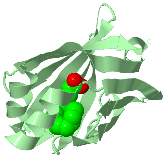
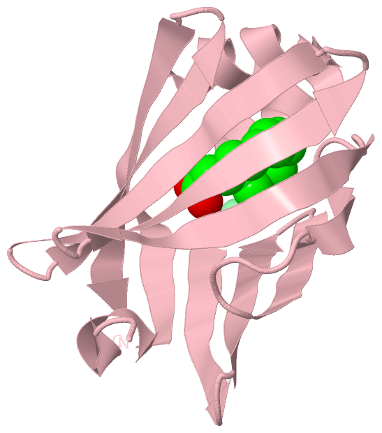
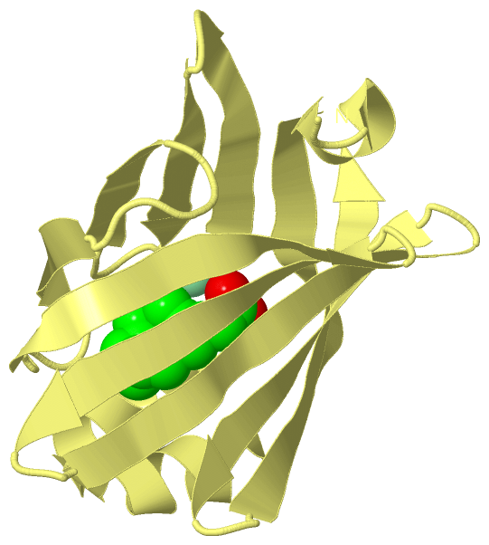
Still Images
|
||||||||||||||||
Databases
|
||||||||||||||||||||||||||||||||||||||||||||||||||||||||||||||||||||||||||||||||||||||||||||||||||||||||||||||||||||||||||||||||||||||||||||||||||||||||||||||||
Analysis Tools
|
|||||||||||||||||||||||||||||||||||||||||||||||||||||||||||||
Entries Sharing at Least One Protein Chain (UniProt ID)
Related Entries Specified in the PDB File
|
|