|
|
|
|
 Description
|
|
Compounds
|
||||||||||||||||||||||||||||||||||||||||
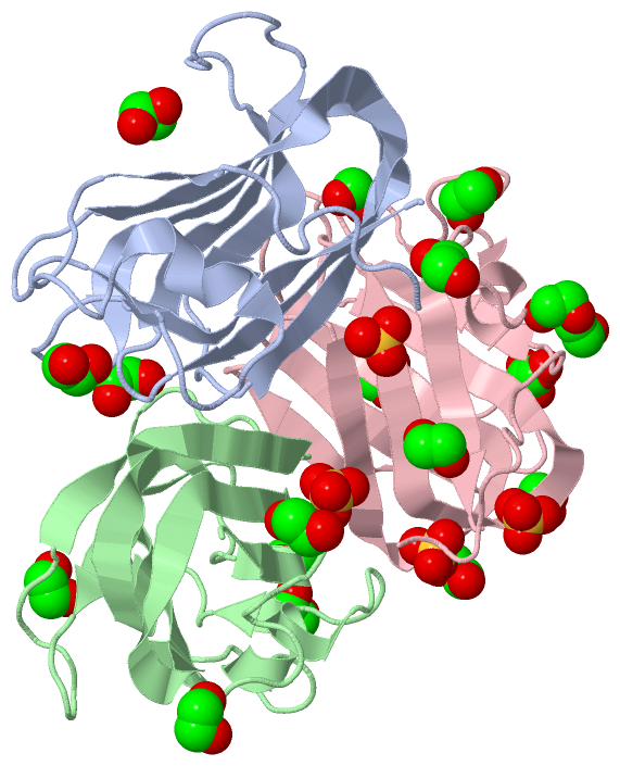
Chains, Units
Summary Information (see also Sequences/Alignments below) |
Ligands, Modified Residues, Ions (2, 22)| Asymmetric Unit (2, 22) Biological Unit 1 (2, 4) Biological Unit 2 (2, 7) Biological Unit 3 (2, 11) |
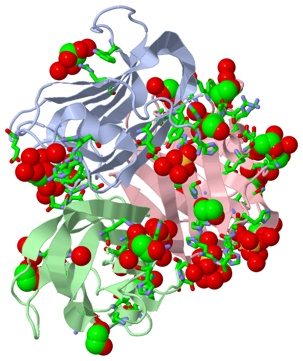
Sites (22, 22)
Asymmetric Unit (22, 22)
|
SS Bonds (0, 0)| (no "SS Bond" information available for 4ZXE) |
Cis Peptide Bonds (3, 3)
Asymmetric Unit
|
||||||||||||||||
SAPs(SNPs)/Variants (0, 0)| (no "SAP(SNP)/Variant" information available for 4ZXE) |
PROSITE Motifs (0, 0)| (no "PROSITE Motif" information available for 4ZXE) |
Exons (0, 0)| (no "Exon" information available for 4ZXE) |
Sequences/Alignments
Asymmetric Unit
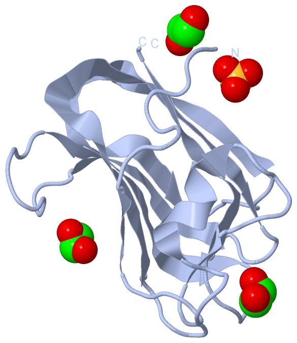
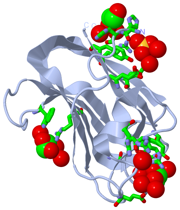
Chain A from PDB Type:PROTEIN Length:132
SCOP domains ------------------------------------------------------------------------------------------------------------------------------------ SCOP domains
CATH domains ------------------------------------------------------------------------------------------------------------------------------------ CATH domains
Pfam domains ------------------------------------------------------------------------------------------------------------------------------------ Pfam domains
SAPs(SNPs) ------------------------------------------------------------------------------------------------------------------------------------ SAPs(SNPs)
PROSITE ------------------------------------------------------------------------------------------------------------------------------------ PROSITE
Transcript ------------------------------------------------------------------------------------------------------------------------------------ Transcript
4zxe A -1 HHNLALNKTATASSIEGAGFEASRAFDGSSTTRWASAEGVDPQWIYVNLGSSQTVNRVKLNWEAAYASSYTIQVSNDSGTPTNWTTVYTTTTGDGGIDDITFTARTAKYVRVHGTVRGTPYGYSLWEFEVYG 130
8 18 28 38 48 58 68 78 88 98 108 118 128
Chain B from PDB Type:PROTEIN Length:132
SCOP domains ------------------------------------------------------------------------------------------------------------------------------------ SCOP domains
CATH domains ------------------------------------------------------------------------------------------------------------------------------------ CATH domains
Pfam domains ------------------------------------------------------------------------------------------------------------------------------------ Pfam domains
SAPs(SNPs) ------------------------------------------------------------------------------------------------------------------------------------ SAPs(SNPs)
PROSITE ------------------------------------------------------------------------------------------------------------------------------------ PROSITE
Transcript ------------------------------------------------------------------------------------------------------------------------------------ Transcript
4zxe B -1 HHNLALNKTATASSIEGAGFEASRAFDGSSTTRWASAEGVDPQWIYVNLGSSQTVNRVKLNWEAAYASSYTIQVSNDSGTPTNWTTVYTTTTGDGGIDDITFTARTAKYVRVHGTVRGTPYGYSLWEFEVYG 130
8 18 28 38 48 58 68 78 88 98 108 118 128
Chain C from PDB Type:PROTEIN Length:136
SCOP domains ---------------------------------------------------------------------------------------------------------------------------------------- SCOP domains
CATH domains ---------------------------------------------------------------------------------------------------------------------------------------- CATH domains
Pfam domains ---------------------------------------------------------------------------------------------------------------------------------------- Pfam domains
SAPs(SNPs) ---------------------------------------------------------------------------------------------------------------------------------------- SAPs(SNPs)
PROSITE ---------------------------------------------------------------------------------------------------------------------------------------- PROSITE
Transcript ---------------------------------------------------------------------------------------------------------------------------------------- Transcript
4zxe C -5 HHHHHHNLALNKTATASSIEGAGFEASRAFDGSSTTRWASAEGVDPQWIYVNLGSSQTVNRVKLNWEAAYASSYTIQVSNDSGTPTNWTTVYTTTTGDGGIDDITFTARTAKYVRVHGTVRGTPYGYSLWEFEVYG 130
4 14 24 34 44 54 64 74 84 94 104 114 124
|
||||||||||||||||||||
SCOP Domains (0, 0)| (no "SCOP Domain" information available for 4ZXE) |
CATH Domains (0, 0)| (no "CATH Domain" information available for 4ZXE) |
Pfam Domains (0, 0)| (no "Pfam Domain" information available for 4ZXE) |
Gene Ontology (7, 7)|
Asymmetric Unit(hide GO term definitions) |
Interactive Views
|
|||||||||||||||||||||||||||||||||||||||||||||||||||||||||||||||||||||||||||||||||||||||||||||||||||||||||||||||||||||||||||||||||||||||||||||||||||||||||||||||||||||||||||||||||||||||||||||||||||||||||||||||||||||||||||||||||||||||||||||||||||||||||||||||||||||||||||||||||||||||||||||||||||||||||||||||||||||||||||
Still Images
|
||||||||||||||||
Databases
|
||||||||||||||||||||||||||||||||||||||||||||||||||||||||||||||||||||||||||||||||||||||||||||||||||||||||||||||||||||||||||||||||||||||||||||||||||||||||||||||||||||||||||||||||
Analysis Tools
|
|||||||||||||||||||||||||||||||||||||||||||||||||||||||||||||
Entries Sharing at Least One Protein Chain (UniProt ID)
Related Entries Specified in the PDB File
|
|