Show PDB file:   
         Plain Text   HTML   (compressed file size)
QuickSearch:   
by PDB,NDB,UniProt,PROSITE Code or Search Term(s)  
(-)Asym./Biol. Unit - manually
(-)Asym./Biol. Unit
(-)Asym./Biol. Unit - sites
collapse expand < >
Image Asym./Biol. Unit - manually
Asym./Biol. Unit - manually  (Jmol Viewer)
Image Asym./Biol. Unit
Asym./Biol. Unit  (Jmol Viewer)
Image Asym./Biol. Unit - sites
Asym./Biol. Unit - sites  (Jmol Viewer)

(-) Description

Title :  RESTRAINED REFINEMENT OF TWO CRYSTALLINE FORMS OF YEAST ASPARTIC ACID AND PHENYLALANINE TRANSFER RNA CRYSTALS
 
Authors :  E. Westhof, P. Dumas, D. Moras
Date :  06 Nov 87  (Deposition) - 06 Nov 87  (Release) - 13 Jul 11  (Revision)
Method :  X-RAY DIFFRACTION
Resolution :  3.00
Chains :  Asym./Biol. Unit :  A
Keywords :  T-Rna, Single Strand, Loops (Keyword Search: [Gene Ontology, PubMed, Web (Google))
 
Reference :  E. Westhof, P. Dumas, D. Moras
Restrained Refinement Of Two Crystalline Forms Of Yeast Aspartic Acid And Phenylalanine Transfer Rna Crystals.
Acta Crystallogr. , Sect. A V. 44 112 1988
PubMed-ID: 3272146  |  Reference-DOI: 10.1107/S010876738700446X
(for further references see the PDB file header)

(-) Compounds

Molecule 1 - TRNAPHE
    ChainsA
    Organism CommonBAKER'S YEAST
    Organism ScientificSACCHAROMYCES CEREVISIAE
    Organism Taxid4932

 Structural Features

(-) Chains, Units

  1
Asymmetric/Biological Unit A

Summary Information (see also Sequences/Alignments below)

(-) Ligands, Modified Residues, Ions  (12, 18)

Asymmetric/Biological Unit (12, 18)
No.NameCountTypeFull Name
11MA1Mod. Nucleotide6-HYDRO-1-METHYLADENOSINE-5'-MONOPHOSPHATE
22MG1Mod. Nucleotide2N-METHYLGUANOSINE-5'-MONOPHOSPHATE
35MC2Mod. Nucleotide5-METHYLCYTIDINE-5'-MONOPHOSPHATE
45MU1Mod. Nucleotide5-METHYLURIDINE 5'-MONOPHOSPHATE
57MG1Mod. Nucleotide7N-METHYL-8-HYDROGUANOSINE-5'-MONOPHOSPHATE
6H2U2Mod. Nucleotide5,6-DIHYDROURIDINE-5'-MONOPHOSPHATE
7M2G1Mod. NucleotideN2-DIMETHYLGUANOSINE-5'-MONOPHOSPHATE
8MG4Ligand/IonMAGNESIUM ION
9OMC1Mod. NucleotideO2'-METHYLYCYTIDINE-5'-MONOPHOSPHATE
10OMG1Mod. NucleotideO2'-METHYLGUANOSINE-5'-MONOPHOSPHATE
11PSU2Mod. NucleotidePSEUDOURIDINE-5'-MONOPHOSPHATE
12YG1Mod. ResidueWYBUTOSINE

(-) Sites  (4, 4)

Asymmetric Unit (4, 4)
No.NameEvidenceResiduesDescription
1AC1SOFTWAREG A:19 , HOH A:185 , HOH A:186 , HOH A:187 , HOH A:188 , HOH A:189BINDING SITE FOR RESIDUE MG A 77
2AC2SOFTWAREG A:20 , A A:21 , HOH A:190 , HOH A:191 , HOH A:192BINDING SITE FOR RESIDUE MG A 78
3AC3SOFTWAREU A:8 , U A:12 , HOH A:193 , HOH A:194 , HOH A:195 , HOH A:196 , HOH A:197 , HOH A:198BINDING SITE FOR RESIDUE MG A 79
4AC4SOFTWAREYG A:37 , A A:38 , HOH A:199 , HOH A:200 , HOH A:201 , HOH A:202 , HOH A:203BINDING SITE FOR RESIDUE MG A 80

(-) SS Bonds  (0, 0)

(no "SS Bond" information available for 4TRA)

(-) Cis Peptide Bonds  (0, 0)

(no "Cis Peptide Bond" information available for 4TRA)

 Sequence-Structure Mapping

(-) SAPs(SNPs)/Variants  (0, 0)

(no "SAP(SNP)/Variant" information available for 4TRA)

(-) PROSITE Motifs  (0, 0)

(no "PROSITE Motif" information available for 4TRA)

(-) Exons   (0, 0)

(no "Exon" information available for 4TRA)

(-) Sequences/Alignments

Asymmetric/Biological Unit
   Reformat: Number of residues per line =  ('0' or empty: single-line sequence representation)
  Number of residues per labelling interval =   
  UniProt sequence: complete  aligned part    
   Show mapping: SCOP domains CATH domains Pfam domains Secondary structure (by author)
SAPs(SNPs) PROSITE motifs Exons
(details for a mapped element are shown in a popup box when the mouse pointer rests over it)
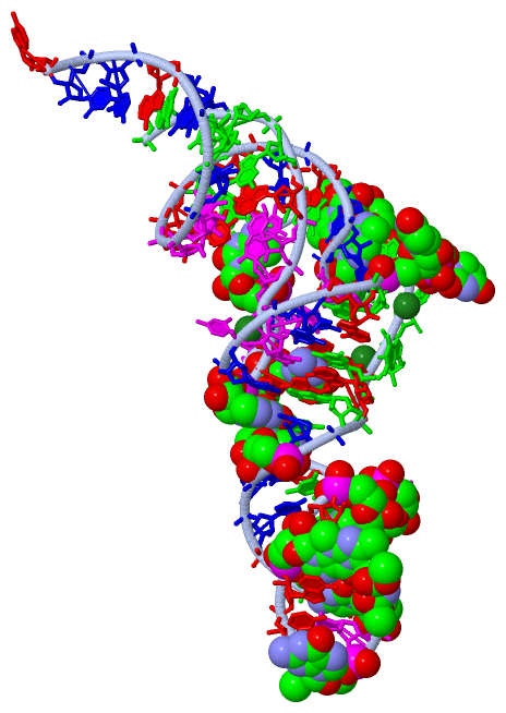
Chain A from PDB  Type:RNA  Length:76
                                                                                                           
                  4tra A  1 GCGGAUUUAgCUCAGuuGGGAGAGCgCCAGAcUgAAgAucUGGAGgUCcUGUGuuCGaUCCACAGAAUUCGCACCA 76
                                    10     || 20     |  30 | |  | 40     |  50   ||  |60        70      
                                    10-2MG ||       26-M2G | |  | ||     |  |    55-PSU                 
                                          16-H2U          32-OMC| ||     |  |    |  58-1MA              
                                           17-H2U           34-OMG||     |  |    |                      
                                                               37-YG     |  |    |                      
                                                                 39-PSU  |  |    |                      
                                                                  40-5MC |  |    |                      
                                                                        46-7MG   |                      
                                                                           49-5MC|                      
                                                                                54-5MU                  

   Legend:   → Mismatch (orange background)
  - → Gap (green background, '-', border residues have a numbering label)
    → Modified Residue (blue background, lower-case, 'x' indicates undefined single-letter code, labelled with number + name)
  x → Chemical Group (purple background, 'x', labelled with number + name, e.g. ACE or NH2)
  extra numbering lines below/above indicate numbering irregularities and modified residue names etc., number ends below/above '|'

 Classification and Annotation

(-) SCOP Domains  (0, 0)

(no "SCOP Domain" information available for 4TRA)

(-) CATH Domains  (0, 0)

(no "CATH Domain" information available for 4TRA)

(-) Pfam Domains  (0, 0)

(no "Pfam Domain" information available for 4TRA)

(-) Gene Ontology  (0, 0)

Asymmetric/Biological Unit(hide GO term definitions)
    (no "Gene Ontology" information available for 4TRA)

 Visualization

(-) Interactive Views

Asymmetric/Biological Unit
  Complete Structure
    Jena3D(integrated viewing of ligand, site, SAP, PROSITE, SCOP information)
    WebMol | AstexViewer[tm]@PDBe
(Java Applets, require no local installation except for Java; loading may be slow)
    STRAP
(Java WebStart application, automatic local installation, requires Java; full application with system access!)
    RasMol
(require local installation)
    Molscript (VRML)
(requires installation of a VRML viewer; select preferred view via VRML and generate a mono or stereo PDF format file)
 
  Ligands, Modified Residues, Ions
    1MA  [ RasMol | Jena3D ]  +environment [ RasMol | Jena3D ]
    2MG  [ RasMol | Jena3D ]  +environment [ RasMol | Jena3D ]
    5MC  [ RasMol | Jena3D ]  +environment [ RasMol | Jena3D ]
    5MU  [ RasMol | Jena3D ]  +environment [ RasMol | Jena3D ]
    7MG  [ RasMol | Jena3D ]  +environment [ RasMol | Jena3D ]
    H2U  [ RasMol | Jena3D ]  +environment [ RasMol | Jena3D ]
    M2G  [ RasMol | Jena3D ]  +environment [ RasMol | Jena3D ]
    MG  [ RasMol | Jena3D ]  +environment [ RasMol | Jena3D ]
    OMC  [ RasMol | Jena3D ]  +environment [ RasMol | Jena3D ]
    OMG  [ RasMol | Jena3D ]  +environment [ RasMol | Jena3D ]
    PSU  [ RasMol | Jena3D ]  +environment [ RasMol | Jena3D ]
    YG  [ RasMol | Jena3D ]  +environment [ RasMol | Jena3D ]
 
  Sites
    AC1  [ RasMol ]  +environment [ RasMol ]
    AC2  [ RasMol ]  +environment [ RasMol ]
    AC3  [ RasMol ]  +environment [ RasMol ]
    AC4  [ RasMol ]  +environment [ RasMol ]
 
  Cis Peptide Bonds
(no "Cis Peptide Bonds" information available for 4tra)
 

(-) Still Images

Jmol
  protein: cartoon or spacefill or dots and stick; nucleic acid: cartoon and stick; ligands: spacefill; active site: stick
Molscript
  protein, nucleic acid: cartoon; ligands: spacefill; active site: ball and stick
Insightii
  ribbon, water, Mg
Setor
  ribbon, sticks
  sticks
  ribbon, Mg
  ribbon, Mg, labeling
  spacefill
Distance Plot
  representative atom O3*

 Databases and Analysis Tools

(-) Databases

Access by PDB/NDB ID
  4tra
    Family and Domain InformationProDom | SYSTERS
    General Structural InformationGlycoscienceDB | MMDB | NDB | OCA | PDB | PDBe | PDBj | PDBsum | PDBWiki | PQS | PROTEOPEDIA
    Orientation in MembranesOPM
    Protein SurfaceSURFACE
    Secondary StructureDSSP | HSSP
    Structural GenomicsGeneCensus
    Structural NeighboursCE | VAST
    Structure ClassificationCATH | Dali | SCOP
    Validation and Original DataBMRB Data View | BMRB Restraints Grid | EDS | PROCHECK | RECOORD | WHAT_CHECK
 
Access by UniProt ID/Accession number
  (no 'UniProt ID/Accession number' available) |
    Comparative Protein Structure ModelsModBase
    Genomic InformationEnsembl
    Protein-protein InteractionDIP
    Sequence, Family and Domain InformationInterPro | Pfam | SMART | UniProt
 
Access by Enzyme Classificator   (EC Number)
  (no 'Enzyme Classificator' available)
    General Enzyme InformationBRENDA | EC-PDB | Enzyme | IntEnz
    PathwayKEGG | MetaCyc
 
Access by Disease Identifier   (MIM ID)
  (no 'MIM ID' available)
    Disease InformationOMIM
 
Access by GenAge ID
  (no 'GenAge ID' available)
    Age Related InformationGenAge

(-) Analysis Tools

Access by PDB/NDB ID
    Domain InformationXDom
    Interatomic Contacts of Structural UnitsCSU
    Ligand-protein ContactsLPC
    Protein CavitiescastP
    Sequence and Secondary StructurePDBCartoon
    Structure AlignmentSTRAP(Java WebStart application, automatic local installation, requires Java; full application with system access!)
    Structure and Sequence BrowserSTING
 
Access by UniProt ID/Accession number
  (no 'UniProt ID/Accession number' available) |
    Protein Disorder PredictionDisEMBL | FoldIndex | GLOBPLOT

 Related Entries

(-) Entries Sharing at Least One Protein Chain (UniProt ID)

(no "Entries Sharing at Least One Protein Chain" available for 4TRA)

(-) Related Entries Specified in the PDB File

(no "Related Entries Specified in the PDB File" available for 4TRA)