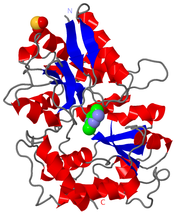
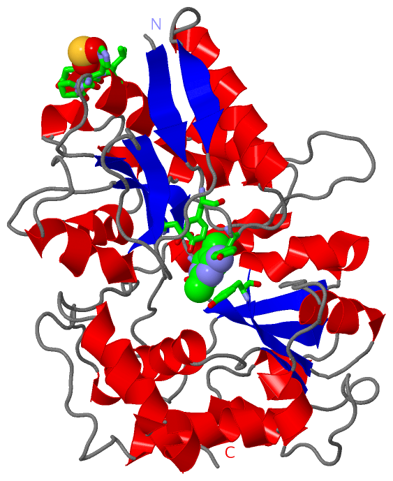
Asymmetric/Biological Unit
Chain A from PDB  Type:PROTEIN  Length:362

               SCOP domains d4thia_ A: Thiaminase I                                                                                                                                                                                                                                                                                                                                                    SCOP domains
               CATH domains 4thiA01 A:9-116,A:264-330 Periplasmic binding protein-like II                                               4thiA02 A:117-263,A:337-370 Periplasmic binding protein-like II                                                                                    4thiA01 A:9-116,A:264-330 Periplasmic binding protein-like II      ------4thiA02 A:117-263,A:337-370        CATH domains
               Pfam domains -------------------------------------------------------------------------------------------------------------------------------------------------------------------------------------------------------------------------------------------------------------------------------------------------------------------------------------------------------------------------- Pfam domains
         Sec.struct. author .eeee........hhhhhhhhhhhhhhh....eeee...............eeeeehhhhhhhhh........hhh...hhh..hhhhhhh........eeee...eeeeee...hhhh....hhhhhhhh...............eee....hhhhhhhhhhhhhhhh................hhhhhhhhhhhhhh.hhhhh.........hhhhhhhh....eeeeehhhhhhhhhhhh.eeee...............eeeee......hhhhhhhhhhhh.hhhhhhhh................hhhhhhhh...hhhhhhhhhh.............hhhhhhhhhh.hhhh.. Sec.struct. author
                 SAPs(SNPs) -------------------------------------------------------------------------------------------------------------------------------------------------------------------------------------------------------------------------------------------------------------------------------------------------------------------------------------------------------------------------- SAPs(SNPs)
                    PROSITE -------------------------------------------------------------------------------------------------------------------------------------------------------------------------------------------------------------------------------------------------------------------------------------------------------------------------------------------------------------------------- PROSITE
                 Transcript -------------------------------------------------------------------------------------------------------------------------------------------------------------------------------------------------------------------------------------------------------------------------------------------------------------------------------------------------------------------------- Transcript
                 4thi A   9 ITLKVAIYPYVPDPARFQAAVLDQWQRQEPGVKLEFTDWDSYSADPPDDLDVFVLDSIFLSHFVDAGYLLPFGSQDIDQAEDVLPFALQGAKRNGEVYGLPQILCTNLLFYRKGDLKIGQVDNIYELYKKIGTSHSEQIPPPQNKGLLINMAGGTTKASMYLEALIDVTGQYTEYDLLPPLDPLNDKVIRGLRLLINMAGEKPSQYVPEDGDAYVRASWFAQGSGRAFIGYSESMMRMGDYAEQVRFKPISSSAGQDIPLFYSDVVSVNSKTAHPELAKKLANVMASADTVEQALRPQADGQYPQYLLPARHQVYEALMQDYPIYSELAQIVNKPSNRVFRLGPEVRTWLKDAKQVLPEALG 370
                                    18        28        38        48        58        68        78        88        98       108       118       128       138       148       158       168       178       188       198       208       218       228       238       248       258       268       278       288       298       308       318       328       338       348       358       368
| Legend: |
|
→ Mismatch |
(orange background) |
| |
- |
→ Gap |
(green background, '-', border residues have a numbering label) |
| |
|
→ Modified Residue |
(blue background, lower-case, 'x' indicates undefined single-letter code, labelled with number + name) |
| |
x |
→ Chemical Group |
(purple background, 'x', labelled with number + name, e.g. ACE or NH2) |
| |
extra numbering lines below/above indicate numbering irregularities and modified residue names etc., number ends below/above '|' |
|
 Description