Show PDB file:   
         Plain Text   HTML   (compressed file size)
QuickSearch:   
by PDB,NDB,UniProt,PROSITE Code or Search Term(s)  
(-)Asym./Biol. Unit
(-)Asym./Biol. Unit - sites
collapse expand < >
Image Asym./Biol. Unit
Asym./Biol. Unit  (Jmol Viewer)
Image Asym./Biol. Unit - sites
Asym./Biol. Unit - sites  (Jmol Viewer)

(-) Description

Title :  2.35 ANGSTROM RESOLUTION CRYSTAL STRUCTURE OF HYPOXANTHINE-GUANINE-XANTHINE PHOSPHORIBOSYLTRANSFERASE FROM LEPTOSPIRA INTERROGANS SEROVAR COPENHAGENI STR. FIOCRUZ L1-130
 
Authors :  A. S. Halavaty, G. Minasov, I. Dubrovska, K. J. Flores, L. Shuvalova, W. F. Anderson, Center For Structural Genomics Of Infectious D (Csgid)
Date :  01 Jul 14  (Deposition) - 16 Jul 14  (Release) - 16 Jul 14  (Revision)
Method :  X-RAY DIFFRACTION
Resolution :  2.35
Chains :  Asym./Biol. Unit :  A,B
Keywords :  Hypoxanthine-Guanine-Xanthine Phosphoribosyltransferase, Leptospira Interrogans Serovar Copenhageni Str. Fiocruz L1-130, Structural Genomics, Center For Structural Genomics Of Infectious Diseases, Glycosyltransferase, Transferase, Csgid, Niaid, National Institute Of Allergy And Infectious Diseases, Purine Salvage Pathway (Keyword Search: [Gene Ontology, PubMed, Web (Google))
 
Reference :  A. S. Halavaty, G. Minasov, I. Dubrovska, K. J. Flores, L. Shuvalova, W. F. Anderson, Center For Structural Genomics Of Infectious Diseases (Csgid)
2. 35 Angstrom Resolution Crystal Structure Of Hypoxanthine-Guanine-Xanthine Phosphoribosyltransferase Fro Leptospira Interrogans Serovar Copenhageni Str. Fiocruz L1-130
To Be Published
PubMed: search

(-) Compounds

Molecule 1 - HYPOXANTHINE-GUANINE-XANTHINE PHOSPHORIBOSYLTRANSFERASE
    ChainsA, B
    EC Number2.4.2.8
    EngineeredYES
    Expression SystemESCHERICHIA COLI
    Expression System PlasmidPMCSG53
    Expression System StrainBL21(DE3)-MAGIC
    Expression System Taxid469008
    Expression System Vector TypePLASMID
    GeneHPT, LA_4295, LIC_13438
    Organism ScientificLEPTOSPIRA INTERROGANS SEROVAR COPENHAGENI
    Organism Taxid267671
    StrainFIOCRUZ L1-130

 Structural Features

(-) Chains, Units

  12
Asymmetric/Biological Unit AB

Summary Information (see also Sequences/Alignments below)

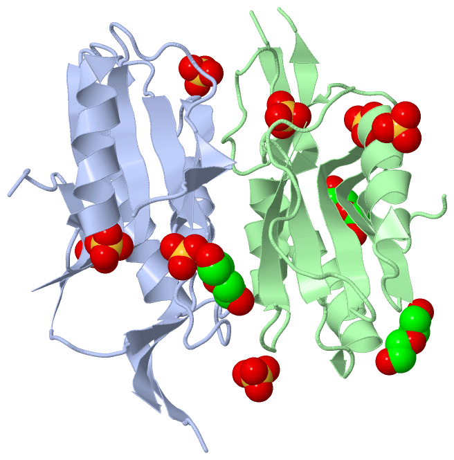
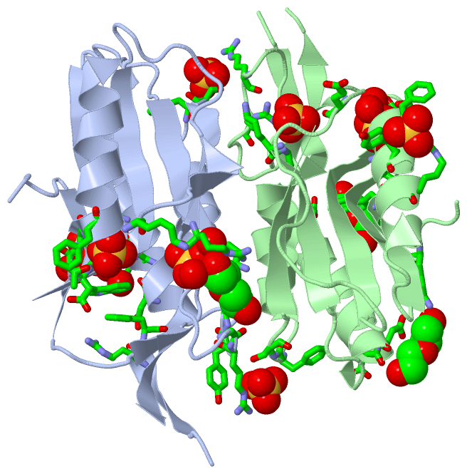
(-) Ligands, Modified Residues, Ions  (2, 11)

Asymmetric/Biological Unit (2, 11)
No.NameCountTypeFull Name
1PEG3Ligand/IonDI(HYDROXYETHYL)ETHER
2SO48Ligand/IonSULFATE ION

(-) Sites  (11, 11)

Asymmetric Unit (11, 11)
No.NameEvidenceResiduesDescription
01AC1SOFTWAREGLN A:20 , LYS A:21 , SER A:24 , HOH A:306 , HOH A:361 , HOH A:362 , PEG B:205BINDING SITE FOR RESIDUE SO4 A 201
02AC2SOFTWARELYS A:45 , GLY A:46 , ARG A:166BINDING SITE FOR RESIDUE SO4 A 202
03AC3SOFTWAREPHE A:60 , SER A:61 , LYS A:80 , ARG B:164BINDING SITE FOR RESIDUE SO4 A 203
04AC4SOFTWARELEU A:103 , ASP A:104 , THR A:105 , GLY A:106 , PHE A:107BINDING SITE FOR RESIDUE SO4 A 204
05AC5SOFTWARELYS A:45 , ARG A:70 , GLY A:163 , ARG A:166 , HOH A:344 , HOH A:360BINDING SITE FOR RESIDUE PEG A 205
06AC6SOFTWARETYR B:111 , ARG B:114BINDING SITE FOR RESIDUE SO4 B 201
07AC7SOFTWAREARG A:164 , TYR A:165 , PHE B:60 , SER B:61BINDING SITE FOR RESIDUE SO4 B 202
08AC8SOFTWARELYS B:45 , GLY B:46 , ARG B:166BINDING SITE FOR RESIDUE SO4 B 203
09AC9SOFTWARELEU B:103 , ASP B:104 , THR B:105 , GLY B:106 , PHE B:107 , HOH B:346 , HOH B:371BINDING SITE FOR RESIDUE SO4 B 204
10BC1SOFTWAREARG A:12 , PHE A:13 , ASP A:17 , TRP A:147 , SO4 A:201 , HOH A:306 , GLU B:31 , ASP B:32 , LYS B:143BINDING SITE FOR RESIDUE PEG B 205
11BC2SOFTWAREASP B:17 , GLU B:82 , TRP B:147BINDING SITE FOR RESIDUE PEG B 206

(-) SS Bonds  (0, 0)

(no "SS Bond" information available for 4QRI)

(-) Cis Peptide Bonds  (2, 2)

Asymmetric/Biological Unit
No.Residues
1Leu A:44 -Lys A:45
2Leu B:44 -Lys B:45

 Sequence-Structure Mapping

(-) SAPs(SNPs)/Variants  (0, 0)

(no "SAP(SNP)/Variant" information available for 4QRI)

(-) PROSITE Motifs  (0, 0)

(no "PROSITE Motif" information available for 4QRI)

(-) Exons   (0, 0)

(no "Exon" information available for 4QRI)

(-) Sequences/Alignments

Asymmetric/Biological Unit
   Reformat: Number of residues per line =  ('0' or empty: single-line sequence representation)
  Number of residues per labelling interval =   
  UniProt sequence: complete  aligned part    
   Show mapping: SCOP domains CATH domains Pfam domains Secondary structure (by author)
SAPs(SNPs) PROSITE motifs Exons
(details for a mapped element are shown in a popup box when the mouse pointer rests over it)
Chain A from PDB  Type:PROTEIN  Length:157
                                                                                                                                                                                             
               SCOP domains ------------------------------------------------------------------------------------------------------------------------------------------------------------- SCOP domains
               CATH domains ------------------------------------------------------------------------------------------------------------------------------------------------------------- CATH domains
               Pfam domains ------------------------------------------------------------------------------------------------------------------------------------------------------------- Pfam domains
         Sec.struct. author ..eeeeeehhhhhhhhhhhhhhhhhhhhh....eeeeee..hhhhhhhhhhh.....eeeeeee....eee.........eeeeeeeee..hhhhhhhhhhhhh....eeeeeeeeee......eeeeee....ee...............eeeee. Sec.struct. author
                 SAPs(SNPs) ------------------------------------------------------------------------------------------------------------------------------------------------------------- SAPs(SNPs)
                    PROSITE ------------------------------------------------------------------------------------------------------------------------------------------------------------- PROSITE
                 Transcript ------------------------------------------------------------------------------------------------------------------------------------------------------------- Transcript
                 4qri A   6 SDILHPRFSREDISQKVKSLALQISEDYKKLNPIFICVLKGGVYFFTDLTREIPFSVEINFVQARKIELLKDIDIDLSDRHVIIVEDILDTGFTLQYLVRHIFTRNPASLEIVTLLLKEEFPVKYIGWRIPDEFLVGYGLDFDGRYRNLPDIHVLEP 176
                                    15        25        35        45        55        65    ||  84        94       104       114       124       139       149       159       169       
                                                                                           70|                                                  133|                                     
                                                                                            80                                                   139                                     

Chain B from PDB  Type:PROTEIN  Length:155
                                                                                                                                                                                           
               SCOP domains ----------------------------------------------------------------------------------------------------------------------------------------------------------- SCOP domains
               CATH domains ----------------------------------------------------------------------------------------------------------------------------------------------------------- CATH domains
               Pfam domains ----------------------------------------------------------------------------------------------------------------------------------------------------------- Pfam domains
         Sec.struct. author ..eeeeeehhhhhhhhhhhhhhhhhhhhh....eeeeee..hhhhhhhhhhh.....eeeeee...............eeeeeeeee..hhhhhhhhhhhhh....eeeeeeeeee.....eeeeee....ee...............eeeee.. Sec.struct. author
                 SAPs(SNPs) ----------------------------------------------------------------------------------------------------------------------------------------------------------- SAPs(SNPs)
                    PROSITE ----------------------------------------------------------------------------------------------------------------------------------------------------------- PROSITE
                 Transcript ----------------------------------------------------------------------------------------------------------------------------------------------------------- Transcript
                 4qri B   6 SDILHPRFSREDISQKVKSLALQISEDYKKLNPIFICVLKGGVYFFTDLTREIPFSVEINFVQAIELLKDIDIDLSDRHVIIVEDILDTGFTLQYLVRHIFTRNPASLEIVTLLLKEFPVKYIGWRIPDEFLVGYGLDFDGRYRNLPDIHVLEPG 177
                                    15        25        35        45        55        65   ||   86        96       106       116       126      |142       152       162       172     
                                                                                          69|                                                 133|                                     
                                                                                           81                                                  140                                     

   Legend:   → Mismatch (orange background)
  - → Gap (green background, '-', border residues have a numbering label)
    → Modified Residue (blue background, lower-case, 'x' indicates undefined single-letter code, labelled with number + name)
  x → Chemical Group (purple background, 'x', labelled with number + name, e.g. ACE or NH2)
  extra numbering lines below/above indicate numbering irregularities and modified residue names etc., number ends below/above '|'

 Classification and Annotation

(-) SCOP Domains  (0, 0)

(no "SCOP Domain" information available for 4QRI)

(-) CATH Domains  (0, 0)

(no "CATH Domain" information available for 4QRI)

(-) Pfam Domains  (0, 0)

(no "Pfam Domain" information available for 4QRI)

(-) Gene Ontology  (6, 6)

Asymmetric/Biological Unit(hide GO term definitions)

 Visualization

(-) Interactive Views

Asymmetric/Biological Unit
  Complete Structure
    Jena3D(integrated viewing of ligand, site, SAP, PROSITE, SCOP information)
    WebMol | AstexViewer[tm]@PDBe
(Java Applets, require no local installation except for Java; loading may be slow)
    STRAP
(Java WebStart application, automatic local installation, requires Java; full application with system access!)
    RasMol
(require local installation)
    Molscript (VRML)
(requires installation of a VRML viewer; select preferred view via VRML and generate a mono or stereo PDF format file)
 
  Ligands, Modified Residues, Ions
    PEG  [ RasMol | Jena3D ]  +environment [ RasMol | Jena3D ]
    SO4  [ RasMol | Jena3D ]  +environment [ RasMol | Jena3D ]
 
  Sites
    AC1  [ RasMol ]  +environment [ RasMol ]
    AC2  [ RasMol ]  +environment [ RasMol ]
    AC3  [ RasMol ]  +environment [ RasMol ]
    AC4  [ RasMol ]  +environment [ RasMol ]
    AC5  [ RasMol ]  +environment [ RasMol ]
    AC6  [ RasMol ]  +environment [ RasMol ]
    AC7  [ RasMol ]  +environment [ RasMol ]
    AC8  [ RasMol ]  +environment [ RasMol ]
    AC9  [ RasMol ]  +environment [ RasMol ]
    BC1  [ RasMol ]  +environment [ RasMol ]
    BC2  [ RasMol ]  +environment [ RasMol ]
 
  Cis Peptide Bonds
    Leu A:44 - Lys A:45   [ RasMol ]  
    Leu B:44 - Lys B:45   [ RasMol ]  
 

(-) Still Images

Jmol
  protein: cartoon or spacefill or dots and stick; nucleic acid: cartoon and stick; ligands: spacefill; active site: stick
Molscript
  protein, nucleic acid: cartoon; ligands: spacefill; active site: ball and stick

 Databases and Analysis Tools

(-) Databases

Access by PDB/NDB ID
  4qri
    Family and Domain InformationProDom | SYSTERS
    General Structural InformationGlycoscienceDB | MMDB | NDB | OCA | PDB | PDBe | PDBj | PDBsum | PDBWiki | PQS | PROTEOPEDIA
    Orientation in MembranesOPM
    Protein SurfaceSURFACE
    Secondary StructureDSSP (structure derived) | HSSP (homology derived)
    Structural GenomicsGeneCensus
    Structural NeighboursCE | VAST
    Structure ClassificationCATH | Dali | SCOP
    Validation and Original DataBMRB Data View | BMRB Restraints Grid | EDS | PROCHECK | RECOORD | WHAT_CHECK
 
Access by UniProt ID/Accession number
  Q72LV5_LEPIC | Q72LV5
    Comparative Protein Structure ModelsModBase
    Genomic InformationEnsembl
    Protein-protein InteractionDIP
    Sequence, Family and Domain InformationInterPro | Pfam | SMART | UniProtKB/TrEMBL
 
Access by Enzyme Classificator   (EC Number)
  2.4.2.8
    General Enzyme InformationBRENDA | EC-PDB | Enzyme | IntEnz
    PathwayKEGG | MetaCyc
 
Access by Disease Identifier   (MIM ID)
  (no 'MIM ID' available)
    Disease InformationOMIM
 
Access by GenAge ID
  (no 'GenAge ID' available)
    Age Related InformationGenAge

(-) Analysis Tools

Access by PDB/NDB ID
    Domain InformationXDom
    Interatomic Contacts of Structural UnitsCSU
    Ligand-protein ContactsLPC
    Protein CavitiescastP
    Sequence and Secondary StructurePDBCartoon
    Structure AlignmentSTRAP(Java WebStart application, automatic local installation, requires Java; full application with system access!)
    Structure and Sequence BrowserSTING
 
Access by UniProt ID/Accession number
  Q72LV5_LEPIC | Q72LV5
    Protein Disorder PredictionDisEMBL | FoldIndex | GLOBPLOT (for more information see DisProt)

 Related Entries

(-) Entries Sharing at Least One Protein Chain (UniProt ID)

(no "Entries Sharing at Least One Protein Chain" available for 4QRI)

(-) Related Entries Specified in the PDB File

(no "Related Entries Specified in the PDB File" available for 4QRI)