|
|
|
|
 Description
|
|
Compounds
|
||||||||||||||||||||||||||||||||||||||||||||||||
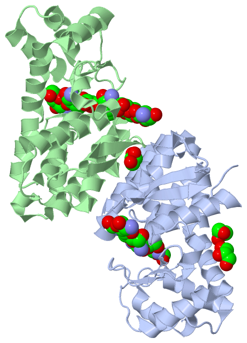
Chains, Units
Summary Information (see also Sequences/Alignments below) |
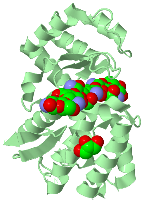
Ligands, Modified Residues, Ions (2, 15)| Asymmetric Unit (2, 15) Biological Unit 1 (2, 8) Biological Unit 2 (2, 7) |
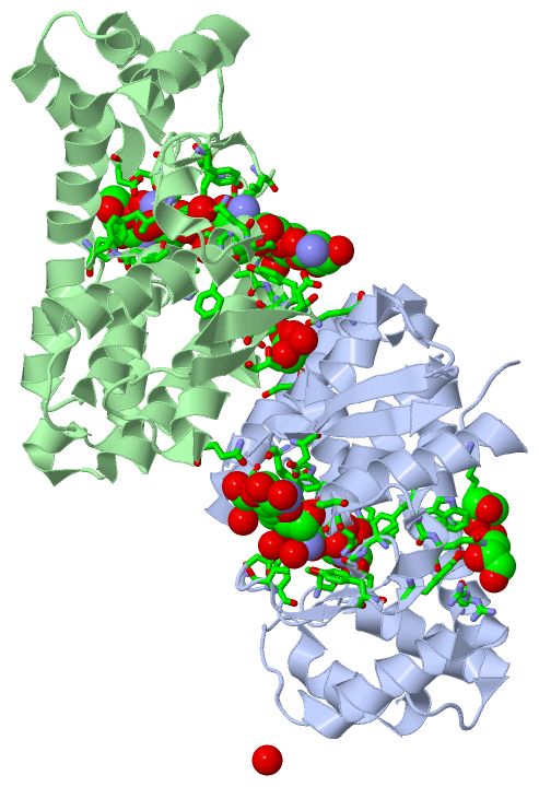
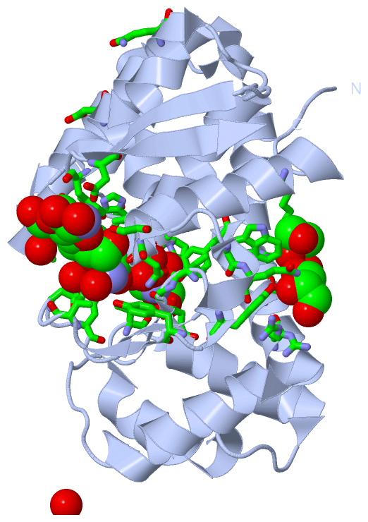
Sites (5, 5)
Asymmetric Unit (5, 5)
|
SS Bonds (0, 0)| (no "SS Bond" information available for 4OLT) |
Cis Peptide Bonds (2, 2)
Asymmetric Unit
|
||||||||||||
SAPs(SNPs)/Variants (0, 0)| (no "SAP(SNP)/Variant" information available for 4OLT) |
PROSITE Motifs (0, 0)| (no "PROSITE Motif" information available for 4OLT) |
Exons (0, 0)| (no "Exon" information available for 4OLT) |
Sequences/Alignments
Asymmetric Unit
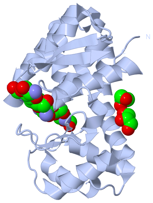
Chain A from PDB Type:PROTEIN Length:236
SCOP domains -------------------------------------------------------------------------------------------------------------------------------------------------------------------------------------------------------------------------------------------- SCOP domains
CATH domains -------------------------------------------------------------------------------------------------------------------------------------------------------------------------------------------------------------------------------------------- CATH domains
Pfam domains -------------------------------------------------------------------------------------------------------------------------------------------------------------------------------------------------------------------------------------------- Pfam domains
SAPs(SNPs) -------------------------------------------------------------------------------------------------------------------------------------------------------------------------------------------------------------------------------------------- SAPs(SNPs)
PROSITE -------------------------------------------------------------------------------------------------------------------------------------------------------------------------------------------------------------------------------------------- PROSITE
Transcript -------------------------------------------------------------------------------------------------------------------------------------------------------------------------------------------------------------------------------------------- Transcript
4olt A 5 TVDLDAPVQKDTAMSLVSSFENSSTDWQAQYGYLEDIADGRGYTGGLIGFTSGTGDMLELVRAYSASSPGNPLEQYIPALEAVNGTDSHAGLGQGFEQAWADAAETSEFRAAQDAERDRVYFDPAVAQGKADGLSALGQFAYYDTLVVHGPGSQRDAFGGIRAEALSAALPPSQGGDETEYLEAFFDARNVIMREEPAHADTSRIDTAQRVFLQNGNFDLERPLTWSVYGDQYSLN 240
14 24 34 44 54 64 74 84 94 104 114 124 134 144 154 164 174 184 194 204 214 224 234
Chain B from PDB Type:PROTEIN Length:237
SCOP domains --------------------------------------------------------------------------------------------------------------------------------------------------------------------------------------------------------------------------------------------- SCOP domains
CATH domains --------------------------------------------------------------------------------------------------------------------------------------------------------------------------------------------------------------------------------------------- CATH domains
Pfam domains --------------------------------------------------------------------------------------------------------------------------------------------------------------------------------------------------------------------------------------------- Pfam domains
SAPs(SNPs) --------------------------------------------------------------------------------------------------------------------------------------------------------------------------------------------------------------------------------------------- SAPs(SNPs)
PROSITE --------------------------------------------------------------------------------------------------------------------------------------------------------------------------------------------------------------------------------------------- PROSITE
Transcript --------------------------------------------------------------------------------------------------------------------------------------------------------------------------------------------------------------------------------------------- Transcript
4olt B 4 GTVDLDAPVQKDTAMSLVSSFENSSTDWQAQYGYLEDIADGRGYTGGLIGFTSGTGDMLELVRAYSASSPGNPLEQYIPALEAVNGTDSHAGLGQGFEQAWADAAETSEFRAAQDAERDRVYFDPAVAQGKADGLSALGQFAYYDTLVVHGPGSQRDAFGGIRAEALSAALPPSQGGDETEYLEAFFDARNVIMREEPAHADTSRIDTAQRVFLQNGNFDLERPLTWSVYGDQYSLN 240
13 23 33 43 53 63 73 83 93 103 113 123 133 143 153 163 173 183 193 203 213 223 233
|
||||||||||||||||||||
SCOP Domains (0, 0)| (no "SCOP Domain" information available for 4OLT) |
CATH Domains (0, 0)| (no "CATH Domain" information available for 4OLT) |
Pfam Domains (0, 0)| (no "Pfam Domain" information available for 4OLT) |
Gene Ontology (6, 6)|
Asymmetric Unit(hide GO term definitions) |
Interactive Views
|
||||||||||||||||||||||||||||||||||||||||||||||||||||||||||||||||||||||||||||||||||||||||||||||||||||||||||||||||||||||||||||||||||||||||||||||||||||||||||||||||||||||||||||||||||||||||
Still Images
|
||||||||||||||||
Databases
|
||||||||||||||||||||||||||||||||||||||||||||||||||||||||||||||||||||||||||||||||||||||||||||||||||||||||||||||||||||||||||||||||||||||||||||||||||||||||||||||||
Analysis Tools
|
|||||||||||||||||||||||||||||||||||||||||||||||||||||||||||||
Entries Sharing at Least One Protein Chain (UniProt ID)
Related Entries Specified in the PDB File
|
|