|
|
|
|
 Description
|
|
Compounds
|
||||||||||||||||||||||||||||||||||||||||||||||||
Chains, Units
Summary Information (see also Sequences/Alignments below) |
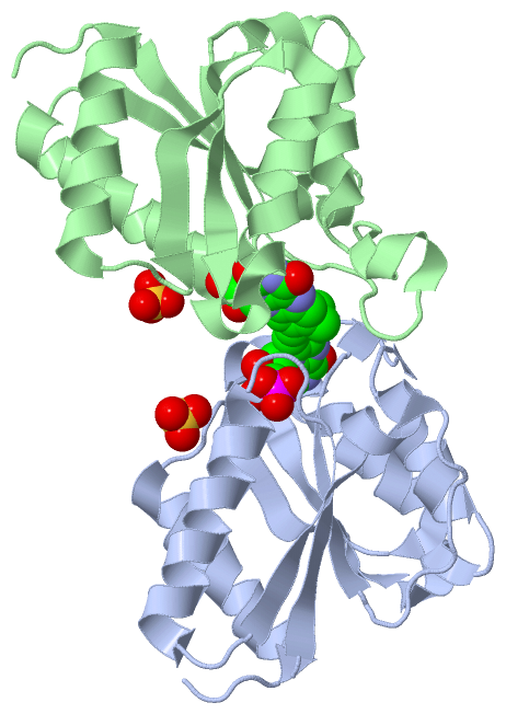
Ligands, Modified Residues, Ions (2, 4)| Asymmetric Unit (2, 4) Biological Unit 1 (2, 2) Biological Unit 2 (2, 2) |
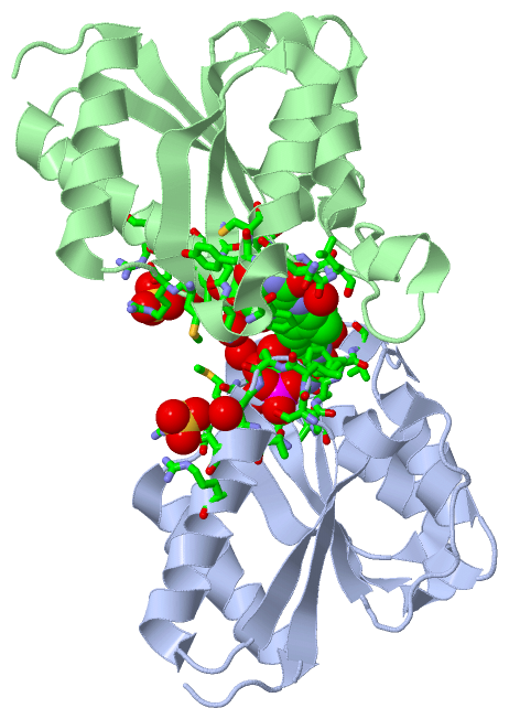
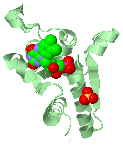
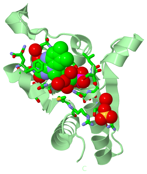
Sites (4, 4)
Asymmetric Unit (4, 4)
|
SS Bonds (0, 0)| (no "SS Bond" information available for 4N82) |
Cis Peptide Bonds (0, 0)| (no "Cis Peptide Bond" information available for 4N82) |
SAPs(SNPs)/Variants (0, 0)| (no "SAP(SNP)/Variant" information available for 4N82) |
PROSITE Motifs (0, 0)| (no "PROSITE Motif" information available for 4N82) |
Exons (0, 0)| (no "Exon" information available for 4N82) |
Sequences/Alignments
Asymmetric Unit
Chain A from PDB Type:PROTEIN Length:148
SCOP domains ---------------------------------------------------------------------------------------------------------------------------------------------------- SCOP domains
CATH domains ---------------------------------------------------------------------------------------------------------------------------------------------------- CATH domains
Pfam domains ---------------------------------------------------------------------------------------------------------------------------------------------------- Pfam domains
SAPs(SNPs) ---------------------------------------------------------------------------------------------------------------------------------------------------- SAPs(SNPs)
PROSITE ---------------------------------------------------------------------------------------------------------------------------------------------------- PROSITE
Transcript ---------------------------------------------------------------------------------------------------------------------------------------------------- Transcript
4n82 A 2 TKVSLVYISLSGNTESFVRRLTDYLLEQHPSLEVEKIHIKDLVKERQPFFEMDNPFIAFLPTYLEDNGDVEILTTDVGDFIAYGQNASKCLGVIGSGNRNFNNQYCLTAKQYSERFGFPVLADFEMRGMLGDIKKVAGIIEELYHIEK 154
11 21 31 41 51 61 || 76 86 96 106 116 126 136 146
66|
72
Chain B from PDB Type:PROTEIN Length:153
SCOP domains --------------------------------------------------------------------------------------------------------------------------------------------------------- SCOP domains
CATH domains --------------------------------------------------------------------------------------------------------------------------------------------------------- CATH domains
Pfam domains --------------------------------------------------------------------------------------------------------------------------------------------------------- Pfam domains
SAPs(SNPs) --------------------------------------------------------------------------------------------------------------------------------------------------------- SAPs(SNPs)
PROSITE --------------------------------------------------------------------------------------------------------------------------------------------------------- PROSITE
Transcript --------------------------------------------------------------------------------------------------------------------------------------------------------- Transcript
4n82 B 2 TKVSLVYISLSGNTESFVRRLTDYLLEQHPSLEVEKIHIKDLVKERQPFFEMDNPFIAFLPTYLEGGNGVDNGDVEILTTDVGDFIAYGQNASKCLGVIGSGNRNFNNQYCLTAKQYSERFGFPVLADFEMRGMLGDIKKVAGIIEELYHIEK 154
11 21 31 41 51 61 71 81 91 101 111 121 131 141 151
|
||||||||||||||||||||
SCOP Domains (0, 0)| (no "SCOP Domain" information available for 4N82) |
CATH Domains (0, 0)| (no "CATH Domain" information available for 4N82) |
Pfam Domains (0, 0)| (no "Pfam Domain" information available for 4N82) |
Gene Ontology (2, 2)|
Asymmetric Unit(hide GO term definitions) |
Interactive Views
|
|||||||||||||||||||||||||||||||||||||||||||||||||||||||||||||||||||||||||||||||||||||||||||||||||||||||||||||||||||||||||||||||||||||||||||||||||||||||||||||||||||||||||
Still Images
|
||||||||||||||||
Databases
|
||||||||||||||||||||||||||||||||||||||||||||||||||||||||||||||||||||||||||||||||||||||||||||||||||||||||||||||||||||||||||||||||||||||||||||||||||||||||||||||||
Analysis Tools
|
|||||||||||||||||||||||||||||||||||||||||||||||||||||||||||||
Entries Sharing at Least One Protein Chain (UniProt ID)
Related Entries Specified in the PDB File
|
|