Show PDB file:   
         Plain Text   HTML   (compressed file size)
QuickSearch:   
by PDB,NDB,UniProt,PROSITE Code or Search Term(s)  
(-)Asymmetric Unit
(-)Asym. Unit - sites
(-)Biological Unit 1
(-)Biol. Unit 1 - sites
(-)Biological Unit 2
collapse expand < >
Image Asymmetric Unit
Asymmetric Unit  (Jmol Viewer)
Image Asym. Unit - sites
Asym. Unit - sites  (Jmol Viewer)
Image Biological Unit 1
Biological Unit 1  (Jmol Viewer)
Image Biol. Unit 1 - sites
Biol. Unit 1 - sites  (Jmol Viewer)
Image Biological Unit 2
Biological Unit 2  (Jmol Viewer)

(-) Description

Title :  CRYSTAL STRUCTURE OF MYOTOXIN II (MJTX-II), A MYOTOXIC LYS49-PHOSPHOLIPASE A2 FROM BOTHROPS MOOJENI.
 
Authors :  G. H. M. Salvador, J. I. Dos Santos, M. R. M. Fontes
Date :  26 Apr 13  (Deposition) - 17 Jul 13  (Release) - 24 Jul 13  (Revision)
Method :  X-RAY DIFFRACTION
Resolution :  1.92
Chains :  Asym. Unit :  A,B
Biol. Unit 1:  B  (1x)
Biol. Unit 2:  A  (1x)
Keywords :  Phospholipase A2-Like Myotoxin, Toxin, Venom Glands (Keyword Search: [Gene Ontology, PubMed, Web (Google))
 
Reference :  G. H. Salvador, W. L. Cavalcante, J. I. Dos Santos, M. Gallacci, A. M. Soares, M. R. Fontes
Structural And Functional Studies With Mytoxin Ii From Bothrops Moojeni Reveal Remarkable Similarities And Differences Compared To Other Catalytically Inactive Phospholipases A2-Like.
Toxicon V. 72C 52 2013
PubMed-ID: 23810946  |  Reference-DOI: 10.1016/J.TOXICON.2013.06.013

(-) Compounds

Molecule 1 - BASIC PHOSPHOLIPASE A2 HOMOLOG 2
    ChainsB, A
    FragmentPHOSPHOLIPASE A2-LIKE MYOTOXIN
    Organism CommonLANCE-HEADED VIPER
    Organism ScientificBOTHROPS MOOJENI
    Organism Taxid98334
    SynonymSVPLA2 HOMOLOG, M-VI, MJTX-II, MYOTOXIN II

 Structural Features

(-) Chains, Units

  12
Asymmetric Unit AB
Biological Unit 1 (1x) B
Biological Unit 2 (1x)A 

Summary Information (see also Sequences/Alignments below)

(-) Ligands, Modified Residues, Ions  (2, 10)

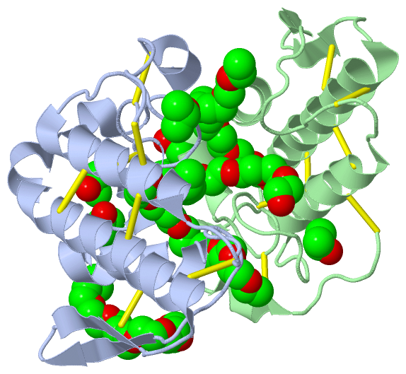
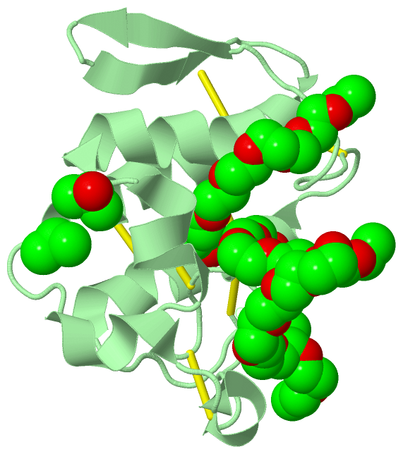
Asymmetric Unit (2, 10)
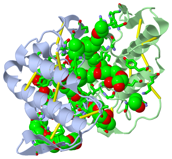
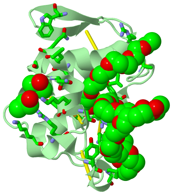
No.NameCountTypeFull Name
1IPA6Ligand/IonISOPROPYL ALCOHOL
2PE44Ligand/Ion2-{2-[2-(2-{2-[2-(2-ETHOXY-ETHOXY)-ETHOXY]-ETHOXY}-ETHOXY)-ETHOXY]-ETHOXY}-ETHANOL
Biological Unit 1 (2, 6)
No.NameCountTypeFull Name
1IPA3Ligand/IonISOPROPYL ALCOHOL
2PE43Ligand/Ion2-{2-[2-(2-{2-[2-(2-ETHOXY-ETHOXY)-ETHOXY]-ETHOXY}-ETHOXY)-ETHOXY]-ETHOXY}-ETHANOL
Biological Unit 2 (2, 4)
No.NameCountTypeFull Name
1IPA3Ligand/IonISOPROPYL ALCOHOL
2PE41Ligand/Ion2-{2-[2-(2-{2-[2-(2-ETHOXY-ETHOXY)-ETHOXY]-ETHOXY}-ETHOXY)-ETHOXY]-ETHOXY}-ETHANOL

(-) Sites  (10, 10)

Asymmetric Unit (10, 10)
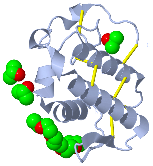
No.NameEvidenceResiduesDescription
01AC1SOFTWARESER A:1 , LEU A:2 , ILE A:9 , TYR A:22 , CYS A:29 , GLY A:30 , LYS A:70 , TYR B:121 , PRO B:125 , PE4 B:203 , HOH B:372 , HOH B:374BINDING SITE FOR RESIDUE PE4 B 201
02AC2SOFTWARELYS A:123 , PRO A:125 , LEU B:2 , ILE B:9 , GLY B:30 , CYS B:45 , LYS B:70 , ARG B:72 , HOH B:385BINDING SITE FOR RESIDUE PE4 B 202
03AC3SOFTWARELEU A:2 , GLY B:23 , GLY B:30 , VAL B:31 , GLY B:32 , LYS B:49 , TYR B:52 , PE4 B:201 , HOH B:338BINDING SITE FOR RESIDUE PE4 B 203
04AC4SOFTWARELYS B:20 , LYS B:115 , ARG B:118 , HOH B:341BINDING SITE FOR RESIDUE IPA B 204
05AC5SOFTWAREARG B:34 , LYS B:53BINDING SITE FOR RESIDUE IPA B 205
06AC6SOFTWAREASN B:17 , LYS B:20 , SER B:21BINDING SITE FOR RESIDUE IPA B 206
07AC7SOFTWAREPHE A:3 , LYS A:7 , LEU A:10 , GLN A:11 , TYR A:75 , TRP A:77 , LEU B:10 , GLN B:11 , GLY B:15 , TRP B:77BINDING SITE FOR RESIDUE PE4 A 201
08AC8SOFTWAREGLY A:15 , LYS A:16 , ASN A:17 , LYS A:20BINDING SITE FOR RESIDUE IPA A 202
09AC9SOFTWAREGLY A:33 , ARG A:34 , LYS A:53BINDING SITE FOR RESIDUE IPA A 203
10BC1SOFTWARELYS A:20 , LYS A:115 , ARG A:118BINDING SITE FOR RESIDUE IPA A 204

(-) SS Bonds  (14, 14)

Asymmetric Unit
No.Residues
1A:27 -A:127
2A:29 -A:45
3A:44 -A:105
4A:50 -A:133
5A:51 -A:98
6A:61 -A:90
7A:84 -A:96
8B:27 -B:127
9B:29 -B:45
10B:44 -B:105
11B:50 -B:133
12B:51 -B:98
13B:61 -B:90
14B:84 -B:96

(-) Cis Peptide Bonds  (0, 0)

(no "Cis Peptide Bond" information available for 4KF3)

 Sequence-Structure Mapping

(-) SAPs(SNPs)/Variants  (0, 0)

(no "SAP(SNP)/Variant" information available for 4KF3)

(-) PROSITE Motifs  (0, 0)

(no "PROSITE Motif" information available for 4KF3)

(-) Exons   (0, 0)

(no "Exon" information available for 4KF3)

(-) Sequences/Alignments

Asymmetric Unit
   Reformat: Number of residues per line =  ('0' or empty: single-line sequence representation)
  Number of residues per labelling interval =   
  UniProt sequence: complete  aligned part    
   Show mapping: SCOP domains CATH domains Pfam domains Secondary structure (by author)
SAPs(SNPs) PROSITE motifs Exons
(details for a mapped element are shown in a popup box when the mouse pointer rests over it)
Chain A from PDB  Type:PROTEIN  Length:122
                                                                                                                                                          
               SCOP domains d4kf3a_ A: automated matches                                                                                               SCOP domains
               CATH domains -------------------------------------------------------------------------------------------------------------------------- CATH domains
               Pfam domains -------------------------------------------------------------------------------------------------------------------------- Pfam domains
         Sec.struct. author .hhhhhhhhhhhhh.hhhhhhh...............hhhhhhhhhhhhhhhh............eeee..eeee...hhhhhhhhhhhhhhhhhhhhhhhhhhhhhh...hhhhh...... Sec.struct. author
                 SAPs(SNPs) -------------------------------------------------------------------------------------------------------------------------- SAPs(SNPs)
                    PROSITE -------------------------------------------------------------------------------------------------------------------------- PROSITE
                 Transcript -------------------------------------------------------------------------------------------------------------------------- Transcript
                 4kf3 A   1 SLFELGKMILQETGKNPAKSYGVYGCNCGVGGRGKPKDATDRCCYVHKCCYKKLTGCDPKKDRYSYSWKDKTIVCGENNSCLKELCECDKAVAICLRENLDTYNKKYRYNYLKPFCKKADPC 133
                                    10  ||    21        31        41        51 ||   ||69        79        89||     100       110       120  ||   131  
                                       13|                                    53|  61|                     90|                            123|        
                                        15                                     57   67                      92                             125        

Chain B from PDB  Type:PROTEIN  Length:122
                                                                                                                                                          
               SCOP domains d4kf3b_ B: automated matches                                                                                               SCOP domains
               CATH domains -------------------------------------------------------------------------------------------------------------------------- CATH domains
               Pfam domains -------------------------------------------------------------------------------------------------------------------------- Pfam domains
         Sec.struct. author hhhhhhhhhhhhhh.hhhhhhh...............hhhhhhhhhhhhhhhh............eeee..eeee...hhhhhhhhhhhhhhhhhhhhh....hhhhhh..hhhhh...... Sec.struct. author
                 SAPs(SNPs) -------------------------------------------------------------------------------------------------------------------------- SAPs(SNPs)
                    PROSITE -------------------------------------------------------------------------------------------------------------------------- PROSITE
                 Transcript -------------------------------------------------------------------------------------------------------------------------- Transcript
                 4kf3 B   1 SLFELGKMILQETGKNPAKSYGVYGCNCGVGGRGKPKDATDRCCYVHKCCYKKLTGCDPKKDRYSYSWKDKTIVCGENNSCLKELCECDKAVAICLRENLDTYNKKYRYNYLKPFCKKADPC 133
                                    10  ||    21        31        41        51 ||   ||69        79        89||     100       110       120  ||   131  
                                       13|                                    53|  61|                     90|                            123|        
                                        15                                     57   67                      92                             125        

   Legend:   → Mismatch (orange background)
  - → Gap (green background, '-', border residues have a numbering label)
    → Modified Residue (blue background, lower-case, 'x' indicates undefined single-letter code, labelled with number + name)
  x → Chemical Group (purple background, 'x', labelled with number + name, e.g. ACE or NH2)
  extra numbering lines below/above indicate numbering irregularities and modified residue names etc., number ends below/above '|'

 Classification and Annotation

(-) SCOP Domains  (1, 2)

Asymmetric Unit

(-) CATH Domains  (0, 0)

(no "CATH Domain" information available for 4KF3)

(-) Pfam Domains  (0, 0)

(no "Pfam Domain" information available for 4KF3)

(-) Gene Ontology  (4, 4)

Asymmetric Unit(hide GO term definitions)

 Visualization

(-) Interactive Views

Asymmetric Unit
  Complete Structure
    Jena3D(integrated viewing of ligand, site, SAP, PROSITE, SCOP information)
    WebMol | AstexViewer[tm]@PDBe
(Java Applets, require no local installation except for Java; loading may be slow)
    STRAP
(Java WebStart application, automatic local installation, requires Java; full application with system access!)
    RasMol
(require local installation)
    Molscript (VRML)
(requires installation of a VRML viewer; select preferred view via VRML and generate a mono or stereo PDF format file)
 
  Ligands, Modified Residues, Ions
    IPA  [ RasMol | Jena3D ]  +environment [ RasMol | Jena3D ]
    PE4  [ RasMol | Jena3D ]  +environment [ RasMol | Jena3D ]
 
  Sites
    AC1  [ RasMol ]  +environment [ RasMol ]
    AC2  [ RasMol ]  +environment [ RasMol ]
    AC3  [ RasMol ]  +environment [ RasMol ]
    AC4  [ RasMol ]  +environment [ RasMol ]
    AC5  [ RasMol ]  +environment [ RasMol ]
    AC6  [ RasMol ]  +environment [ RasMol ]
    AC7  [ RasMol ]  +environment [ RasMol ]
    AC8  [ RasMol ]  +environment [ RasMol ]
    AC9  [ RasMol ]  +environment [ RasMol ]
    BC1  [ RasMol ]  +environment [ RasMol ]
 
  Cis Peptide Bonds
(no "Cis Peptide Bonds" information available for 4kf3)
 
Biological Units
  Complete Structure
    Biological Unit 1  [ Jena3D ]
    Biological Unit 2  [ Jena3D ]

(-) Still Images

Jmol
  protein: cartoon or spacefill or dots and stick; nucleic acid: cartoon and stick; ligands: spacefill; active site: stick
Molscript
  protein, nucleic acid: cartoon; ligands: spacefill; active site: ball and stick

 Databases and Analysis Tools

(-) Databases

Access by PDB/NDB ID
  4kf3
    Family and Domain InformationProDom | SYSTERS
    General Structural InformationGlycoscienceDB | MMDB | NDB | OCA | PDB | PDBe | PDBj | PDBsum | PDBWiki | PQS | PROTEOPEDIA
    Orientation in MembranesOPM
    Protein SurfaceSURFACE
    Secondary StructureDSSP (structure derived) | HSSP (homology derived)
    Structural GenomicsGeneCensus
    Structural NeighboursCE | VAST
    Structure ClassificationCATH | Dali | SCOP
    Validation and Original DataBMRB Data View | BMRB Restraints Grid | EDS | PROCHECK | RECOORD | WHAT_CHECK
 
Access by UniProt ID/Accession number
  PA2H2_BOTMO | Q9I834
    Comparative Protein Structure ModelsModBase
    Genomic InformationEnsembl
    Protein-protein InteractionDIP
    Sequence, Family and Domain InformationInterPro | Pfam | SMART | UniProtKB/SwissProt
 
Access by Enzyme Classificator   (EC Number)
  (no 'Enzyme Classificator' available)
    General Enzyme InformationBRENDA | EC-PDB | Enzyme | IntEnz
    PathwayKEGG | MetaCyc
 
Access by Disease Identifier   (MIM ID)
  (no 'MIM ID' available)
    Disease InformationOMIM
 
Access by GenAge ID
  (no 'GenAge ID' available)
    Age Related InformationGenAge

(-) Analysis Tools

Access by PDB/NDB ID
    Domain InformationXDom
    Interatomic Contacts of Structural UnitsCSU
    Ligand-protein ContactsLPC
    Protein CavitiescastP
    Sequence and Secondary StructurePDBCartoon
    Structure AlignmentSTRAP(Java WebStart application, automatic local installation, requires Java; full application with system access!)
    Structure and Sequence BrowserSTING
 
Access by UniProt ID/Accession number
  PA2H2_BOTMO | Q9I834
    Protein Disorder PredictionDisEMBL | FoldIndex | GLOBPLOT (for more information see DisProt)

 Related Entries

(-) Entries Sharing at Least One Protein Chain (UniProt ID)

UniProtKB/Swiss-Prot
        PA2H2_BOTMO | Q9I8341xxs 4yv5

(-) Related Entries Specified in the PDB File

1xxs MJTX-II COMPLEXED TO FATTY ACID
2q2j PRTX-I
3iq3 BTHTX-I COMPLEXED TO PEG4000
3t0r MJTX-I