Show PDB file:   
         Plain Text   HTML   (compressed file size)
QuickSearch:   
by PDB,NDB,UniProt,PROSITE Code or Search Term(s)  
(-)Asymmetric Unit
(-)Asym. Unit - sites
(-)Biological Unit 1
(-)Biol. Unit 1 - sites
(-)Biological Unit 2
collapse expand < >
Image Asymmetric Unit
Asymmetric Unit  (Jmol Viewer)
Image Asym. Unit - sites
Asym. Unit - sites  (Jmol Viewer)
Image Biological Unit 1
Biological Unit 1  (Jmol Viewer)
Image Biol. Unit 1 - sites
Biol. Unit 1 - sites  (Jmol Viewer)
Image Biological Unit 2
Biological Unit 2  (Jmol Viewer)

(-) Description

Title :  PYRROLOPYRIMIDINE INHIBITORS OF DNA GYRASE B AND TOPOISOMERASE IV, PART I: STRUCTURE GUIDED DISCOVERY AND OPTIMIZATION OF DUAL TARGETING AGENTS WITH POTENT, BROAD-SPECTRUM ENZYMATIC ACTIVITY.
 
Authors :  D. C. Bensen, M Trzoss, L. W. Tari
Date :  14 Nov 12  (Deposition) - 13 Feb 13  (Release) - 27 Feb 13  (Revision)
Method :  X-RAY DIFFRACTION
Resolution :  2.20
Chains :  Asym. Unit :  A,B
Biol. Unit 1:  A  (1x)
Biol. Unit 2:  B  (1x)
Keywords :  Atp-Binding, Nucleotide-Binding, Topoisomerase, Atp-Binding Domain, Isomerase-Isomerase Inhibitor Complex (Keyword Search: [Gene Ontology, PubMed, Web (Google))
 
Reference :  L. W. Tari, M. Trzoss, D. C. Bensen, X. Li, Z. Chen, T. Lam, J. Zhang, C. J. Creighton, M. L. Cunningham, B. Kwan, M. Stidham, K. J. Shaw, F. C. Lightstone, S. E. Wong, T. B. Nguyen, J. Nix, J. Finn
Pyrrolopyrimidine Inhibitors Of Dna Gyrase B (Gyrb) And Topoisomerase Iv (Pare). Part I: Structure Guided Discovery And Optimization Of Dual Targeting Agents With Potent, Broad-Spectrum Enzymatic Activity.
Bioorg. Med. Chem. Lett. V. 23 1529 2013
PubMed-ID: 23352267  |  Reference-DOI: 10.1016/J.BMCL.2012.11.032

(-) Compounds

Molecule 1 - DNA TOPOISOMERASE 4 SUBUNIT B
    ChainsA, B
    EC Number5.99.1.-
    EngineeredYES
    Expression SystemESCHERICHIA COLI
    Expression System StrainBL21(DE3)
    Expression System Taxid469008
    FragmentUNP RESIDUES 12-216
    GeneB3030, DNA TOPOISOMERASE IV SUBUNIT B PARE, JW2998, NFXD, PARE
    Organism ScientificESCHERICHIA COLI
    Organism Taxid83333
    StrainK12
    SynonymTOPOISOMERASE IV SUBUNIT B

 Structural Features

(-) Chains, Units

  12
Asymmetric Unit AB
Biological Unit 1 (1x)A 
Biological Unit 2 (1x) B

Summary Information (see also Sequences/Alignments below)

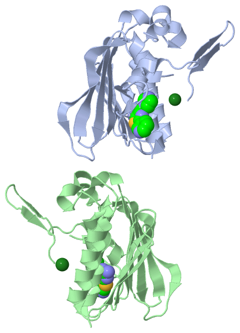
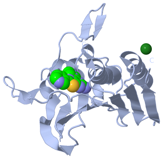
(-) Ligands, Modified Residues, Ions  (2, 4)

Asymmetric Unit (2, 4)
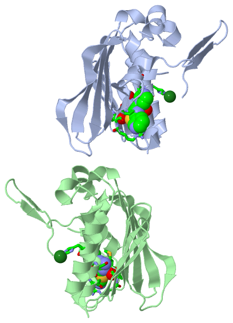
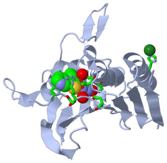
No.NameCountTypeFull Name
11AV2Ligand/Ion7-(1H-IMIDAZOL-1-YL)-2-(PYRIDIN-3-YL)[1,3]THIAZOLO[5,4-D]PYRIMIDIN-5-AMINE
2MG2Ligand/IonMAGNESIUM ION
Biological Unit 1 (1, 1)
No.NameCountTypeFull Name
11AV1Ligand/Ion7-(1H-IMIDAZOL-1-YL)-2-(PYRIDIN-3-YL)[1,3]THIAZOLO[5,4-D]PYRIMIDIN-5-AMINE
2MG-1Ligand/IonMAGNESIUM ION
Biological Unit 2 (1, 1)
No.NameCountTypeFull Name
11AV1Ligand/Ion7-(1H-IMIDAZOL-1-YL)-2-(PYRIDIN-3-YL)[1,3]THIAZOLO[5,4-D]PYRIMIDIN-5-AMINE
2MG-1Ligand/IonMAGNESIUM ION

(-) Sites  (4, 4)

Asymmetric Unit (4, 4)
No.NameEvidenceResiduesDescription
1AC1SOFTWAREASN A:42 , SER A:43 , GLU A:46 , ASP A:69 , ARG A:72 , GLY A:73 , PRO A:75 , ILE A:90 , HOH A:414 , HOH A:430 , HOH A:446BINDING SITE FOR RESIDUE 1AV A 301
2AC2SOFTWAREHIS A:186BINDING SITE FOR RESIDUE MG A 302
3AC3SOFTWAREASN B:42 , SER B:43 , GLU B:46 , ASP B:69 , ARG B:72 , GLY B:73 , MET B:74 , ILE B:90 , HOH B:404 , HOH B:417BINDING SITE FOR RESIDUE 1AV B 301
4AC4SOFTWAREHIS B:186 , HOH B:498BINDING SITE FOR RESIDUE MG B 302

(-) SS Bonds  (0, 0)

(no "SS Bond" information available for 4HZ0)

(-) Cis Peptide Bonds  (2, 2)

Asymmetric Unit
No.Residues
1Ser A:108 -Gly A:109
2Ser B:108 -Gly B:109

 Sequence-Structure Mapping

(-) SAPs(SNPs)/Variants  (0, 0)

(no "SAP(SNP)/Variant" information available for 4HZ0)

(-) PROSITE Motifs  (0, 0)

(no "PROSITE Motif" information available for 4HZ0)

(-) Exons   (0, 0)

(no "Exon" information available for 4HZ0)

(-) Sequences/Alignments

Asymmetric Unit
   Reformat: Number of residues per line =  ('0' or empty: single-line sequence representation)
  Number of residues per labelling interval =   
  UniProt sequence: complete  aligned part    
   Show mapping: SCOP domains CATH domains Pfam domains Secondary structure (by author)
SAPs(SNPs) PROSITE motifs Exons
(details for a mapped element are shown in a popup box when the mouse pointer rests over it)
Chain A from PDB  Type:PROTEIN  Length:201
                                                                                                                                                                                                                                         
               SCOP domains d4hz0a_ A: automated matches                                                                                                                                                                              SCOP domains
               CATH domains --------------------------------------------------------------------------------------------------------------------------------------------------------------------------------------------------------- CATH domains
               Pfam domains --------------------------------------------------------------------------------------------------------------------------------------------------------------------------------------------------------- Pfam domains
         Sec.struct. author ..hhhhhhhhhhh.....hhhhhhhhhhhhhhhhhh....eeeeee.....eeeee........ee....eehhhhhhhh.....eee....eee....hhhhhhhheeeeeeeeee..eeeeeeee..eeeeeeeeeee......eeeeeeee.hhhh.....hhhhhhhhhhhhhhhh...eeeeee....eeeee... Sec.struct. author
                 SAPs(SNPs) --------------------------------------------------------------------------------------------------------------------------------------------------------------------------------------------------------- SAPs(SNPs)
                    PROSITE --------------------------------------------------------------------------------------------------------------------------------------------------------------------------------------------------------- PROSITE
                 Transcript --------------------------------------------------------------------------------------------------------------------------------------------------------------------------------------------------------- Transcript
                 4hz0 A  14 TGLEPVRRRPGMYTDTTRPNHLGQEVIDNSVDEALAGHAKRVDVILHADQSLEVIDDGRGMPVDIHPEEGVPAVELILCRLHAGGKFSNKNYQFSGGLVGISVVNALSKRVEVNVRRDGQVYNIAFENGEKVQDLQVVGTCGKRNTGTSVHFWPDETFFDSPRFSVSRLTHVLKAKAVLCPGVEITFKDEINNTEQRWCYQ 216
                                    23        33        43        53        63        73        83        93       103       115       125       135       145       155       165       175       185       195       205       215 
                                                                                                                           111|                                                                                                      
                                                                                                                            114                                                                                                      

Chain B from PDB  Type:PROTEIN  Length:201
                                                                                                                                                                                                                                         
               SCOP domains d4hz0b_ B: automated matches                                                                                                                                                                              SCOP domains
               CATH domains --------------------------------------------------------------------------------------------------------------------------------------------------------------------------------------------------------- CATH domains
               Pfam domains --------------------------------------------------------------------------------------------------------------------------------------------------------------------------------------------------------- Pfam domains
         Sec.struct. author ..hhhhhhhhhhh.....hhhhhhhhhhhhhhhhhh....eeeeee.....eeeee...............hhhhhhhhh......ee....ee.....hhhhhhhheeeeeeeeee..eeeeeeee..eeeeeeeeeee......eeeeeeee.hhhh.....hhhhhhhhhhhhhhhh...eeeeee....eeeee... Sec.struct. author
                 SAPs(SNPs) --------------------------------------------------------------------------------------------------------------------------------------------------------------------------------------------------------- SAPs(SNPs)
                    PROSITE --------------------------------------------------------------------------------------------------------------------------------------------------------------------------------------------------------- PROSITE
                 Transcript --------------------------------------------------------------------------------------------------------------------------------------------------------------------------------------------------------- Transcript
                 4hz0 B  14 TGLEPVRRRPGMYTDTTRPNHLGQEVIDNSVDEALAGHAKRVDVILHADQSLEVIDDGRGMPVDIHPEEGVPAVELILCRLHAGGKFSNKNYQFSGGLVGISVVNALSKRVEVNVRRDGQVYNIAFENGEKVQDLQVVGTCGKRNTGTSVHFWPDETFFDSPRFSVSRLTHVLKAKAVLCPGVEITFKDEINNTEQRWCYQ 216
                                    23        33        43        53        63        73        83        93       103       115       125       135       145       155       165       175       185       195       205       215 
                                                                                                                           111|                                                                                                      
                                                                                                                            114                                                                                                      

   Legend:   → Mismatch (orange background)
  - → Gap (green background, '-', border residues have a numbering label)
    → Modified Residue (blue background, lower-case, 'x' indicates undefined single-letter code, labelled with number + name)
  x → Chemical Group (purple background, 'x', labelled with number + name, e.g. ACE or NH2)
  extra numbering lines below/above indicate numbering irregularities and modified residue names etc., number ends below/above '|'

 Classification and Annotation

(-) SCOP Domains  (1, 2)

Asymmetric Unit

(-) CATH Domains  (0, 0)

(no "CATH Domain" information available for 4HZ0)

(-) Pfam Domains  (0, 0)

(no "Pfam Domain" information available for 4HZ0)

(-) Gene Ontology  (19, 19)

Asymmetric Unit(hide GO term definitions)

 Visualization

(-) Interactive Views

Asymmetric Unit
  Complete Structure
    Jena3D(integrated viewing of ligand, site, SAP, PROSITE, SCOP information)
    WebMol | AstexViewer[tm]@PDBe
(Java Applets, require no local installation except for Java; loading may be slow)
    STRAP
(Java WebStart application, automatic local installation, requires Java; full application with system access!)
    RasMol
(require local installation)
    Molscript (VRML)
(requires installation of a VRML viewer; select preferred view via VRML and generate a mono or stereo PDF format file)
 
  Ligands, Modified Residues, Ions
    1AV  [ RasMol | Jena3D ]  +environment [ RasMol | Jena3D ]
    MG  [ RasMol | Jena3D ]  +environment [ RasMol | Jena3D ]
 
  Sites
    AC1  [ RasMol ]  +environment [ RasMol ]
    AC2  [ RasMol ]  +environment [ RasMol ]
    AC3  [ RasMol ]  +environment [ RasMol ]
    AC4  [ RasMol ]  +environment [ RasMol ]
 
  Cis Peptide Bonds
    Ser A:108 - Gly A:109   [ RasMol ]  
    Ser B:108 - Gly B:109   [ RasMol ]  
 
Biological Units
  Complete Structure
    Biological Unit 1  [ Jena3D ]
    Biological Unit 2  [ Jena3D ]

(-) Still Images

Jmol
  protein: cartoon or spacefill or dots and stick; nucleic acid: cartoon and stick; ligands: spacefill; active site: stick
Molscript
  protein, nucleic acid: cartoon; ligands: spacefill; active site: ball and stick

 Databases and Analysis Tools

(-) Databases

Access by PDB/NDB ID
  4hz0
    Family and Domain InformationProDom | SYSTERS
    General Structural InformationGlycoscienceDB | MMDB | NDB | OCA | PDB | PDBe | PDBj | PDBsum | PDBWiki | PQS | PROTEOPEDIA
    Orientation in MembranesOPM
    Protein SurfaceSURFACE
    Secondary StructureDSSP (structure derived) | HSSP (homology derived)
    Structural GenomicsGeneCensus
    Structural NeighboursCE | VAST
    Structure ClassificationCATH | Dali | SCOP
    Validation and Original DataBMRB Data View | BMRB Restraints Grid | EDS | PROCHECK | RECOORD | WHAT_CHECK
 
Access by UniProt ID/Accession number
  PARE_ECOLI | P20083
    Comparative Protein Structure ModelsModBase
    Genomic InformationEnsembl
    Protein-protein InteractionDIP
    Sequence, Family and Domain InformationInterPro | Pfam | SMART | UniProtKB/SwissProt
 
Access by Enzyme Classificator   (EC Number)
  5.99.1.-
    General Enzyme InformationBRENDA | EC-PDB | Enzyme | IntEnz
    PathwayKEGG | MetaCyc
 
Access by Disease Identifier   (MIM ID)
  (no 'MIM ID' available)
    Disease InformationOMIM
 
Access by GenAge ID
  (no 'GenAge ID' available)
    Age Related InformationGenAge

(-) Analysis Tools

Access by PDB/NDB ID
    Domain InformationXDom
    Interatomic Contacts of Structural UnitsCSU
    Ligand-protein ContactsLPC
    Protein CavitiescastP
    Sequence and Secondary StructurePDBCartoon
    Structure AlignmentSTRAP(Java WebStart application, automatic local installation, requires Java; full application with system access!)
    Structure and Sequence BrowserSTING
 
Access by UniProt ID/Accession number
  PARE_ECOLI | P20083
    Protein Disorder PredictionDisEMBL | FoldIndex | GLOBPLOT (for more information see DisProt)

 Related Entries

(-) Entries Sharing at Least One Protein Chain (UniProt ID)

UniProtKB/Swiss-Prot
        PARE_ECOLI | P200831s14 1s16 3fv5

(-) Related Entries Specified in the PDB File

4gee 4gfn 4ggl 4hxw 4hxz 4hy1 4hym 4hyp 4hz5