Show PDB file:   
         Plain Text   HTML   (compressed file size)
QuickSearch:   
by PDB,NDB,UniProt,PROSITE Code or Search Term(s)  
(-)Asym./Biol. Unit
(-)Asym./Biol. Unit - sites
collapse expand < >
Image Asym./Biol. Unit
Asym./Biol. Unit  (Jmol Viewer)
Image Asym./Biol. Unit - sites
Asym./Biol. Unit - sites  (Jmol Viewer)

(-) Description

Title :  HEPATITUS C ENVELOPE GLYCOPROTEIN E2 FRAGMENT 412-423 WITH HUMANIZED AND AFFINITY-MATURED ANTIBODY HU5B3.V3
 
Authors :  C. Eigenbrot, M. Ultsch
Date :  29 Oct 12  (Deposition) - 03 Apr 13  (Release) - 29 May 13  (Revision)
Method :  X-RAY DIFFRACTION
Resolution :  2.60
Chains :  Asym./Biol. Unit :  A,H,L
Keywords :  Immune System (Keyword Search: [Gene Ontology, PubMed, Web (Google))
 
Reference :  H. Pantua, J. Diao, M. Ultsch, M. Hazen, M. Mathieu, K. Mccutcheon, K. Takeda, S. Date, T. K. Cheung, Q. Phung, P. Hass, D. Arnott, J. A. Hongo, D. J. Matthews, A. Brown, A. H. Patel, R. F. Kelley, C. Eigenbrot, S. B. Kapadia
Glycan Shifting On Hepatitis C Virus (Hcv) E2 Glycoprotein Is A Mechanism For Escape From Broadly Neutralizing Antibodies.
J. Mol. Biol. V. 425 1899 2013
PubMed-ID: 23458406  |  Reference-DOI: 10.1016/J.JMB.2013.02.025

(-) Compounds

Molecule 1 - E2-PEPTIDE
    ChainsA
    EngineeredYES
    FragmentUNP RESIDUES 50-62
    Organism ScientificHEPATITIS C VIRUS
    Organism Taxid11103
    Other DetailsTHIS SEQUENCE OCCURS NATURALLY IN HEPATITUS C VIRUS
    SyntheticYES
 
Molecule 2 - ANTIBODY HU5B3.V2 FAB HEAVY CHAIN
    ChainsH
    EngineeredYES
    Expression SystemESCHERICHIA COLI
    Expression System Taxid562
    Organism CommonHUMAN
    Organism ScientificHOMO SAPIENS
    Organism Taxid9606
 
Molecule 3 - ANTIBODY HU5B3.V2 FAB LIGHT CHAIN
    ChainsL
    EngineeredYES
    Expression SystemESCHERICHIA COLI
    Expression System Taxid562
    Organism CommonHUMAN
    Organism ScientificHOMO SAPIENS
    Organism Taxid9606

 Structural Features

(-) Chains, Units

  123
Asymmetric/Biological Unit AHL

Summary Information (see also Sequences/Alignments below)
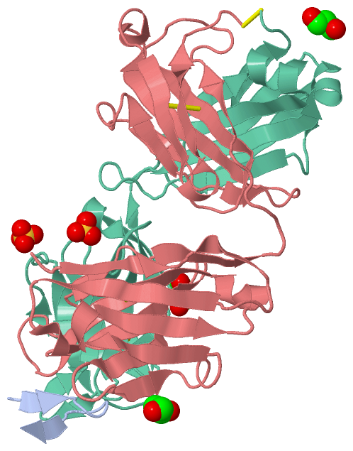
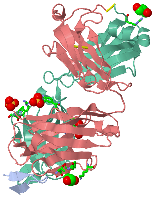
(-) Ligands, Modified Residues, Ions  (2, 5)

Asymmetric/Biological Unit (2, 5)
No.NameCountTypeFull Name
1GOL3Ligand/IonGLYCEROL
2SO42Ligand/IonSULFATE ION

(-) Sites  (5, 5)

Asymmetric Unit (5, 5)
No.NameEvidenceResiduesDescription
1AC1SOFTWARETHR H:57 , ASN H:58 , TYR H:59 , LYS H:64BINDING SITE FOR RESIDUE GOL H 301
2AC2SOFTWAREPRO L:8 , SER L:9 , SER L:10BINDING SITE FOR RESIDUE GOL L 301
3AC3SOFTWARELYS L:39 , LYS L:42BINDING SITE FOR RESIDUE SO4 L 302
4AC4SOFTWAREPRO L:59 , SER L:60 , ARG L:61BINDING SITE FOR RESIDUE SO4 L 303
5AC5SOFTWAREARG L:211BINDING SITE FOR RESIDUE GOL L 304

(-) SS Bonds  (5, 5)

Asymmetric/Biological Unit
No.Residues
1H:22 -H:92
2H:140 -H:196
3H:216 -L:214
4L:23 -L:88
5L:134 -L:194

(-) Cis Peptide Bonds  (5, 5)

Asymmetric/Biological Unit
No.Residues
1Phe H:146 -Pro H:147
2Glu H:148 -Pro H:149
3Ser L:7 -Pro L:8
4Ala L:94 -Pro L:95
5Tyr L:140 -Pro L:141

 Sequence-Structure Mapping

(-) SAPs(SNPs)/Variants  (0, 0)

(no "SAP(SNP)/Variant" information available for 4HS8)

(-) PROSITE Motifs  (0, 0)

(no "PROSITE Motif" information available for 4HS8)

(-) Exons   (0, 0)

(no "Exon" information available for 4HS8)

(-) Sequences/Alignments

Asymmetric/Biological Unit
   Reformat: Number of residues per line =  ('0' or empty: single-line sequence representation)
  Number of residues per labelling interval =   
  UniProt sequence: complete  aligned part    
   Show mapping: SCOP domains CATH domains Pfam domains Secondary structure (by author)
SAPs(SNPs) PROSITE motifs Exons
(details for a mapped element are shown in a popup box when the mouse pointer rests over it)
Chain A from PDB  Type:PROTEIN  Length:12
                                             
               SCOP domains ------------ SCOP domains
               CATH domains ------------ CATH domains
               Pfam domains ------------ Pfam domains
         Sec.struct. author .ee......ee. Sec.struct. author
                 SAPs(SNPs) ------------ SAPs(SNPs)
                    PROSITE ------------ PROSITE
                 Transcript ------------ Transcript
                4hs8 A  412 QLINTNGSWHIN  423
                                   421  

Chain H from PDB  Type:PROTEIN  Length:225
                                                                                                                                                                                                                                                                  
               SCOP domains --------------------------------------------------------------------------------------------------------------------------------------------------------------------------------------------------------------------------------- SCOP domains
               CATH domains --------------------------------------------------------------------------------------------------------------------------------------------------------------------------------------------------------------------------------- CATH domains
               Pfam domains --------------------------------------------------------------------------------------------------------------------------------------------------------------------------------------------------------------------------------- Pfam domains
         Sec.struct. author ..eeeee..eee.....eeeeeeee..hhh.eeeeeeee......eeeeeee....eeee.......eeeeeehhh.eeeeee...hhhhheeeeeeeee..........ee...eeeee........eeeee..........eeeeeeeeeee.....eeee........eee...ee.....eeeeeeeeee.hhh....eeeeeeehhhheeeeeee..... Sec.struct. author
                 SAPs(SNPs) --------------------------------------------------------------------------------------------------------------------------------------------------------------------------------------------------------------------------------- SAPs(SNPs)
                    PROSITE --------------------------------------------------------------------------------------------------------------------------------------------------------------------------------------------------------------------------------- PROSITE
                 Transcript --------------------------------------------------------------------------------------------------------------------------------------------------------------------------------------------------------------------------------- Transcript
                4hs8 H    1 EVQLVESGGGLVQPGGSLRLSCAASGYTFTNYWINWVRQAPGKGLEWVGDIYPSDSFTNYNQNFKDRFTISRDKSKNTAYLQMNSLRAEDTAVYYCARSSIYYGKDYVLDYWGQGTLVTVSSASTKGPSVFPLAPSSKSTSGGTAALGCLVKDYFPEPVTVSWNSGALTSGVHTFPAVLQSSGLYSLSSVVTVPSSSLGTQTYICNVNHKPSNTKVDKKVEPKSC  216
                                    10        20        30        40        50  |     59        69        79   |||  86        96    |||101       111       121       131       141       151       161       171       181       191       201       211     
                                                                              52A                            82A||               100A||||                                                                                                                    
                                                                                                              82B|                100B|||                                                                                                                    
                                                                                                               82C                 100C||                                                                                                                    
                                                                                                                                    100D|                                                                                                                    
                                                                                                                                     100E                                                                                                                    

Chain L from PDB  Type:PROTEIN  Length:218
                                                                                                                                                                                                                                                           
               SCOP domains -------------------------------------------------------------------------------------------------------------------------------------------------------------------------------------------------------------------------- SCOP domains
               CATH domains -------------------------------------------------------------------------------------------------------------------------------------------------------------------------------------------------------------------------- CATH domains
               Pfam domains -------------------------------------------------------------------------------------------------------------------------------------------------------------------------------------------------------------------------- Pfam domains
         Sec.struct. author ...eeee..eeeee....eeeeeee....ee..ee.eeeeee......eeeee...ee.......eeeeee..eeeeee...hhhhheeeeeee......ee...eeeeee......eeeee..hhhhhhh.eeeeeeeeeee.....eeeeee..ee....eeeee.........eeeeeeeeeehhhhh...eeeeeee.......eeeeee.... Sec.struct. author
                 SAPs(SNPs) -------------------------------------------------------------------------------------------------------------------------------------------------------------------------------------------------------------------------- SAPs(SNPs)
                    PROSITE -------------------------------------------------------------------------------------------------------------------------------------------------------------------------------------------------------------------------- PROSITE
                 Transcript -------------------------------------------------------------------------------------------------------------------------------------------------------------------------------------------------------------------------- Transcript
                4hs8 L    1 DIQMTQSPSSLSASVGDRVTITCRASESVDNYGISFMNWFQQKPGKAPKLLIYSASNHASGVPSRFSGSGSGTDFTLTISSLQPEDFATYYCHQSKEAPYAFGQGTKVEIKRTVAAPSVFIFPPSDEQLKSGTASVVCLLNNFYPREAKVQWKVDNALQSGNSQESVTEQDSKDSTYSLSSTLTLSKADYEKHKVYACEVTHQGLSSPVTKSFNRGEC  214
                                    10        20       27C|       36        46        56        66        76        86        96       106       116       126       136       146       156       166       176       186       196       206        
                                                     27A|||                                                                                                                                                                                           
                                                      27B||                                                                                                                                                                                           
                                                       27C|                                                                                                                                                                                           
                                                        27D                                                                                                                                                                                           

   Legend:   → Mismatch (orange background)
  - → Gap (green background, '-', border residues have a numbering label)
    → Modified Residue (blue background, lower-case, 'x' indicates undefined single-letter code, labelled with number + name)
  x → Chemical Group (purple background, 'x', labelled with number + name, e.g. ACE or NH2)
  extra numbering lines below/above indicate numbering irregularities and modified residue names etc., number ends below/above '|'

 Classification and Annotation

(-) SCOP Domains  (0, 0)

(no "SCOP Domain" information available for 4HS8)

(-) CATH Domains  (0, 0)

(no "CATH Domain" information available for 4HS8)

(-) Pfam Domains  (0, 0)

(no "Pfam Domain" information available for 4HS8)

(-) Gene Ontology  (1, 1)

Asymmetric/Biological Unit(hide GO term definitions)

 Visualization

(-) Interactive Views

Asymmetric/Biological Unit
  Complete Structure
    Jena3D(integrated viewing of ligand, site, SAP, PROSITE, SCOP information)
    WebMol | AstexViewer[tm]@PDBe
(Java Applets, require no local installation except for Java; loading may be slow)
    STRAP
(Java WebStart application, automatic local installation, requires Java; full application with system access!)
    RasMol
(require local installation)
    Molscript (VRML)
(requires installation of a VRML viewer; select preferred view via VRML and generate a mono or stereo PDF format file)
 
  Ligands, Modified Residues, Ions
    GOL  [ RasMol | Jena3D ]  +environment [ RasMol | Jena3D ]
    SO4  [ RasMol | Jena3D ]  +environment [ RasMol | Jena3D ]
 
  Sites
    AC1  [ RasMol ]  +environment [ RasMol ]
    AC2  [ RasMol ]  +environment [ RasMol ]
    AC3  [ RasMol ]  +environment [ RasMol ]
    AC4  [ RasMol ]  +environment [ RasMol ]
    AC5  [ RasMol ]  +environment [ RasMol ]
 
  Cis Peptide Bonds
    Ala L:94 - Pro L:95   [ RasMol ]  
    Glu H:148 - Pro H:149   [ RasMol ]  
    Phe H:146 - Pro H:147   [ RasMol ]  
    Ser L:7 - Pro L:8   [ RasMol ]  
    Tyr L:140 - Pro L:141   [ RasMol ]  
 

(-) Still Images

Jmol
  protein: cartoon or spacefill or dots and stick; nucleic acid: cartoon and stick; ligands: spacefill; active site: stick
Molscript
  protein, nucleic acid: cartoon; ligands: spacefill; active site: ball and stick

 Databases and Analysis Tools

(-) Databases

Access by PDB/NDB ID
  4hs8
    Family and Domain InformationProDom | SYSTERS
    General Structural InformationGlycoscienceDB | MMDB | NDB | OCA | PDB | PDBe | PDBj | PDBsum | PDBWiki | PQS | PROTEOPEDIA
    Orientation in MembranesOPM
    Protein SurfaceSURFACE
    Secondary StructureDSSP (structure derived) | HSSP (homology derived)
    Structural GenomicsGeneCensus
    Structural NeighboursCE | VAST
    Structure ClassificationCATH | Dali | SCOP
    Validation and Original DataBMRB Data View | BMRB Restraints Grid | EDS | PROCHECK | RECOORD | WHAT_CHECK
 
Access by UniProt ID/Accession number
  Q9YK84_9HEPC | Q9YK84
    Comparative Protein Structure ModelsModBase
    Genomic InformationEnsembl
    Protein-protein InteractionDIP
    Sequence, Family and Domain InformationInterPro | Pfam | SMART | UniProtKB/TrEMBL
 
Access by Enzyme Classificator   (EC Number)
  (no 'Enzyme Classificator' available)
    General Enzyme InformationBRENDA | EC-PDB | Enzyme | IntEnz
    PathwayKEGG | MetaCyc
 
Access by Disease Identifier   (MIM ID)
  (no 'MIM ID' available)
    Disease InformationOMIM
 
Access by GenAge ID
  (no 'GenAge ID' available)
    Age Related InformationGenAge

(-) Analysis Tools

Access by PDB/NDB ID
    Domain InformationXDom
    Interatomic Contacts of Structural UnitsCSU
    Ligand-protein ContactsLPC
    Protein CavitiescastP
    Sequence and Secondary StructurePDBCartoon
    Structure AlignmentSTRAP(Java WebStart application, automatic local installation, requires Java; full application with system access!)
    Structure and Sequence BrowserSTING
 
Access by UniProt ID/Accession number
  Q9YK84_9HEPC | Q9YK84
    Protein Disorder PredictionDisEMBL | FoldIndex | GLOBPLOT (for more information see DisProt)

 Related Entries

(-) Entries Sharing at Least One Protein Chain (UniProt ID)

UniProtKB/Swiss-Prot
        Q9YK84_9HEPC | Q9YK844dgv 4dgy
UniProtKB/TrEMBL
        Q9YK84_9HEPC | Q9YK844g6a 4hs6

(-) Related Entries Specified in the PDB File

4hs6