| molecular function |
|---|
| | GO:0001664 | | G-protein coupled receptor binding | | Interacting selectively and non-covalently with a G-protein coupled receptor. |
| | GO:0008201 | | heparin binding | | Interacting selectively and non-covalently with heparin, any member of a group of glycosaminoglycans found mainly as an intracellular component of mast cells and which consist predominantly of alternating alpha-(1->4)-linked D-galactose and N-acetyl-D-glucosamine-6-sulfate residues. |
| | GO:0005515 | | protein binding | | Interacting selectively and non-covalently with any protein or protein complex (a complex of two or more proteins that may include other nonprotein molecules). |
| | GO:0005102 | | receptor binding | | Interacting selectively and non-covalently with one or more specific sites on a receptor molecule, a macromolecule that undergoes combination with a hormone, neurotransmitter, drug or intracellular messenger to initiate a change in cell function. |
| biological process |
|---|
| | GO:0016055 | | Wnt signaling pathway | | The series of molecular signals initiated by binding of a Wnt protein to a frizzled family receptor on the surface of the target cell and ending with a change in cell state. |
| | GO:0030177 | | positive regulation of Wnt signaling pathway | | Any process that activates or increases the frequency, rate or extent of Wnt signal transduction. |
| | GO:0090263 | | positive regulation of canonical Wnt signaling pathway | | Any process that increases the rate, frequency, or extent of the Wnt signaling pathway through beta-catenin, the series of molecular signals initiated by binding of a Wnt protein to a frizzled family receptor on the surface of the target cell, followed by propagation of the signal via beta-catenin, and ending with a change in transcription of target genes. |
| | GO:0001934 | | positive regulation of protein phosphorylation | | Any process that activates or increases the frequency, rate or extent of addition of phosphate groups to amino acids within a protein. |
| | GO:0002090 | | regulation of receptor internalization | | Any process that modulates the frequency, rate or extent of receptor internalization. |
| | GO:0050896 | | response to stimulus | | Any process that results in a change in state or activity of a cell or an organism (in terms of movement, secretion, enzyme production, gene expression, etc.) as a result of a stimulus. The process begins with detection of the stimulus and ends with a change in state or activity or the cell or organism. |
| cellular component |
|---|
| | GO:0005576 | | extracellular region | | The space external to the outermost structure of a cell. For cells without external protective or external encapsulating structures this refers to space outside of the plasma membrane. This term covers the host cell environment outside an intracellular parasite. |
| | GO:0005634 | | nucleus | | A membrane-bounded organelle of eukaryotic cells in which chromosomes are housed and replicated. In most cells, the nucleus contains all of the cell's chromosomes except the organellar chromosomes, and is the site of RNA synthesis and processing. In some species, or in specialized cell types, RNA metabolism or DNA replication may be absent. |
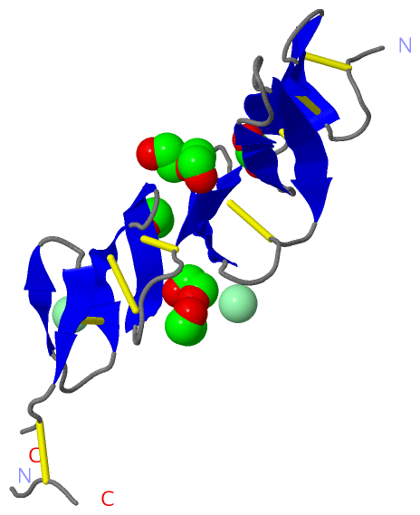
 Description
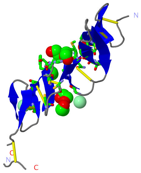