| molecular function |
|---|
| | GO:0005524 | | ATP binding | | Interacting selectively and non-covalently with ATP, adenosine 5'-triphosphate, a universally important coenzyme and enzyme regulator. |
| | GO:0005509 | | calcium ion binding | | Interacting selectively and non-covalently with calcium ions (Ca2+). |
| | GO:0070025 | | carbon monoxide binding | | Interacting selectively and non-covalently with carbon monoxide (CO). |
| | GO:0020037 | | heme binding | | Interacting selectively and non-covalently with heme, any compound of iron complexed in a porphyrin (tetrapyrrole) ring. |
| | GO:0016301 | | kinase activity | | Catalysis of the transfer of a phosphate group, usually from ATP, to a substrate molecule. |
| | GO:0000287 | | magnesium ion binding | | Interacting selectively and non-covalently with magnesium (Mg) ions. |
| | GO:0046872 | | metal ion binding | | Interacting selectively and non-covalently with any metal ion. |
| | GO:0070026 | | nitric oxide binding | | Interacting selectively and non-covalently with nitric oxide (NO). |
| | GO:0019825 | | oxygen binding | | Interacting selectively and non-covalently with oxygen (O2). |
| | GO:0019826 | | oxygen sensor activity | | Interacting selectively and non-covalently with and responding, e.g. by conformational change, to changes in the cellular level of oxygen (O2). |
| | GO:0000155 | | phosphorelay sensor kinase activity | | Catalysis of the phosphorylation of a histidine residue in response to detection of an extracellular signal such as a chemical ligand or change in environment, to initiate a change in cell state or activity. The two-component sensor is a histidine kinase that autophosphorylates a histidine residue in its active site. The phosphate is then transferred to an aspartate residue in a downstream response regulator, to trigger a response. |
| | GO:0046983 | | protein dimerization activity | | The formation of a protein dimer, a macromolecular structure consists of two noncovalently associated identical or nonidentical subunits. |
| | GO:0004673 | | protein histidine kinase activity | | Catalysis of the reaction: ATP + protein L-histidine = ADP + protein phospho-L-histidine. |
| | GO:0004672 | | protein kinase activity | | Catalysis of the phosphorylation of an amino acid residue in a protein, usually according to the reaction: a protein + ATP = a phosphoprotein + ADP. |
| | GO:0016740 | | transferase activity | | Catalysis of the transfer of a group, e.g. a methyl group, glycosyl group, acyl group, phosphorus-containing, or other groups, from one compound (generally regarded as the donor) to another compound (generally regarded as the acceptor). Transferase is the systematic name for any enzyme of EC class 2. |
| biological process |
|---|
| | GO:0070483 | | detection of hypoxia | | The series of events in which a stimulus indicating lowered oxygen tension is received by a cell and converted into a molecular signal. Hypoxia, defined as a decline in O2 levels below normoxic levels of 20.8 - 20.95%, results in metabolic adaptation at both the cellular and organismal level. |
| | GO:0018106 | | peptidyl-histidine phosphorylation | | The phosphorylation of peptidyl-histidine to form peptidyl-1'-phospho-L-histidine (otherwise known as tau-phosphohistidine, tele-phosphohistidine) or peptidyl-3'-phospho-L-histidine (otherwise known as pi-phosphohistidine, pros-phosphohistidine). |
| | GO:0000160 | | phosphorelay signal transduction system | | A conserved series of molecular signals found in prokaryotes and eukaryotes; involves autophosphorylation of a histidine kinase and the transfer of the phosphate group to an aspartate that then acts as a phospho-donor to response regulator proteins. |
| | GO:0016310 | | phosphorylation | | The process of introducing a phosphate group into a molecule, usually with the formation of a phosphoric ester, a phosphoric anhydride or a phosphoric amide. |
| | GO:0046777 | | protein autophosphorylation | | The phosphorylation by a protein of one or more of its own amino acid residues (cis-autophosphorylation), or residues on an identical protein (trans-autophosphorylation). |
| | GO:0023014 | | signal transduction by protein phosphorylation | | A process in which the transfer of one or more phosphate groups to a substrate transmits a signal to the phosphorylated substrate. |
| cellular component |
|---|
| | GO:0005737 | | cytoplasm | | All of the contents of a cell excluding the plasma membrane and nucleus, but including other subcellular structures. |
| | GO:0016021 | | integral component of membrane | | The component of a membrane consisting of the gene products and protein complexes having at least some part of their peptide sequence embedded in the hydrophobic region of the membrane. |
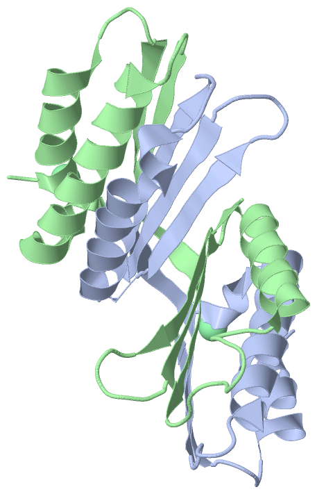
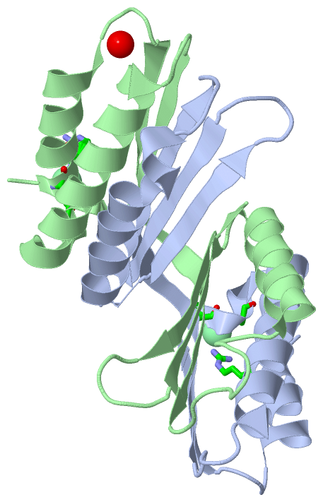
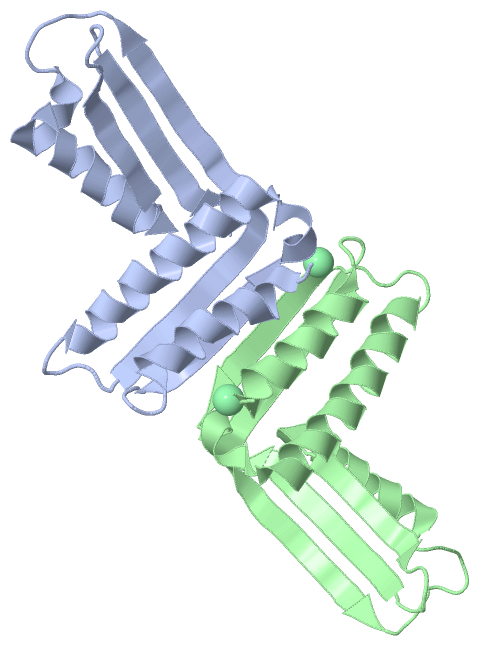
 Description