Show PDB file:   
         Plain Text   HTML   (compressed file size)
QuickSearch:   
by PDB,NDB,UniProt,PROSITE Code or Search Term(s)  
(-)Asym./Biol. Unit
(-)Asym./Biol. Unit - sites
collapse expand < >
Image Asym./Biol. Unit
Asym./Biol. Unit  (Jmol Viewer)
Image Asym./Biol. Unit - sites
Asym./Biol. Unit - sites  (Jmol Viewer)

(-) Description

Title :  STRUCTURE OF BSUDG-P56 COMPLEX
 
Authors :  J. I. Banos-Sanz, L. Mojardin, J. Sanz-Aparicio, B. Gonzalez, M. Salas
Date :  22 Feb 13  (Deposition) - 22 May 13  (Release) - 31 Jul 13  (Revision)
Method :  X-RAY DIFFRACTION
Resolution :  1.45
Chains :  Asym./Biol. Unit :  A,B,C
Keywords :  Hydrolase-Viral Protein Complex (Keyword Search: [Gene Ontology, PubMed, Web (Google))
 
Reference :  J. I. Banos-Sanz, L. Mojardin, J. Sanz-Aparicio, J. M. Lazaro, L. Villar, G. Serrano-Heras, B. Gonzalez, M. Salas
Crystal Structure And Functional Insights Into Uracil-Dna Glycosylase Inhibition By Phage Phi29 Dna Mimic Protein P56
Nucleic Acids Res. V. 41 6761 2013
PubMed-ID: 23671337  |  Reference-DOI: 10.1093/NAR/GKT395

(-) Compounds

Molecule 1 - URACIL-DNA GLYCOSYLASE
    ChainsA
    EC Number3.2.2.27
    EngineeredYES
    Expression SystemESCHERICHIA COLI
    Expression System PlasmidPT7-4-UDG-WT
    Expression System StrainBL21(DE3)
    Expression System Taxid469008
    Expression System VectorPT7-4
    Expression System Vector TypePLASMID
    Organism ScientificBACILLUS SUBTILIS SUBSP. SUBTILIS STR. 168
    Organism Taxid224308
    SynonymUDG
 
Molecule 2 - P56
    ChainsB, C
    EngineeredYES
    Expression SystemESCHERICHIA COLI
    Expression System PlasmidPT7-3-P56-F29
    Expression System StrainBL21(DE3)
    Expression System Taxid469008
    Expression System VectorPT7-3
    Expression System Vector TypePLASMID
    Organism ScientificBACILLUS PHAGE PHI29
    Organism Taxid10756
    SynonymLEFT END OF BACTERIOPHAGE PHI-29 CODING FOR 15 POTENTIAL PROTEINS AMONG THESE ARE THE TERMINAL PROTEIN AND THE PROTEINS ENCODED BY THE GENES 1,2 (SUS), 3, AND (PROBABLY) 4

 Structural Features

(-) Chains, Units

  123
Asymmetric/Biological Unit ABC

Summary Information (see also Sequences/Alignments below)

(-) Ligands, Modified Residues, Ions  (2, 2)

Asymmetric/Biological Unit (2, 2)
No.NameCountTypeFull Name
1CL1Ligand/IonCHLORIDE ION
2GOL1Ligand/IonGLYCEROL

(-) Sites  (2, 2)

Asymmetric Unit (2, 2)
No.NameEvidenceResiduesDescription
1AC1SOFTWAREGLY A:165 , ARG A:166 , HIS A:167 , CL A:1227 , HOH A:2317 , SER B:34 , GLU B:37 , VAL B:38BINDING SITE FOR RESIDUE GOL A1226
2AC2SOFTWARETHR A:186 , HIS A:187 , GOL A:1226 , HOH A:2279 , HOH A:2282BINDING SITE FOR RESIDUE CL A1227

(-) SS Bonds  (1, 1)

Asymmetric/Biological Unit
No.Residues
1A:103 -A:221

(-) Cis Peptide Bonds  (1, 1)

Asymmetric/Biological Unit
No.Residues
1Tyr A:39 -Pro A:40

 Sequence-Structure Mapping

(-) SAPs(SNPs)/Variants  (0, 0)

(no "SAP(SNP)/Variant" information available for 3ZOQ)

(-) PROSITE Motifs  (1, 1)

Asymmetric/Biological Unit (1, 1)
 PROSITEUniProtKBPDB
No.IDACDescriptionIDLocationCountLocation
1U_DNA_GLYCOSYLASEPS00130 Uracil-DNA glycosylase signature.UNG_BACSU58-67  1A:58-67

(-) Exons   (0, 0)

(no "Exon" information available for 3ZOQ)

(-) Sequences/Alignments

Asymmetric/Biological Unit
   Reformat: Number of residues per line =  ('0' or empty: single-line sequence representation)
  Number of residues per labelling interval =   
  UniProt sequence: complete  aligned part    
   Show mapping: SCOP domains CATH domains Pfam domains Secondary structure (by author)
SAPs(SNPs) PROSITE motifs Exons
(details for a mapped element are shown in a popup box when the mouse pointer rests over it)
Chain A from PDB  Type:PROTEIN  Length:223
 aligned with UNG_BACSU | P39615 from UniProtKB/Swiss-Prot  Length:225

    Alignment length:223
                                    12        22        32        42        52        62        72        82        92       102       112       122       132       142       152       162       172       182       192       202       212       222   
            UNG_BACSU     3 QLLQDSWWNQLKEEFEKPYYQELREMLKREYAEQTIYPDSRDIFNALHYTSYDDVKVVILGQDPYHGPGQAQGLSFSVKPGVKQPPSLKNIFLELQQDIGCSIPNHGSLVSWAKQGVLLLNTVLTVRRGQANSHKGKGWERLTDRIIDVLSERERPVIFILWGRHAQMKKERIDTSKHFIIESTHPSPFSARNGFFGSRPFSRANAYLEKMGEAPIDWCIKDL 225
               SCOP domains d3zoqa_ A: automated matches                                                                                                                                                                                                    SCOP domains
               CATH domains ------------------------------------------------------------------------------------------------------------------------------------------------------------------------------------------------------------------------------- CATH domains
               Pfam domains ------------------------------------------------------------------------------------------------------------------------------------------------------------------------------------------------------------------------------- Pfam domains
         Sec.struct. author ...hhhhhhhhhhhhhhhhhhhhhhhhhhhhhhh.ee.hhhhhhhhhhhh......eee.........................hhhhhhhhhhhhhhh.........hhhhhhh.ee.......ee..........hhhhhhhhhhhhhhhh....eeeeehhhhhhhhhhh.....eeeee......hhhhh....hhhhhhhhhhhhh............ Sec.struct. author
                 SAPs(SNPs) ------------------------------------------------------------------------------------------------------------------------------------------------------------------------------------------------------------------------------- SAPs(SNPs)
                    PROSITE -------------------------------------------------------U_DNA_GLYC-------------------------------------------------------------------------------------------------------------------------------------------------------------- PROSITE
                 Transcript ------------------------------------------------------------------------------------------------------------------------------------------------------------------------------------------------------------------------------- Transcript
                 3zoq A   3 QLLQDSWWNQLKEEFEKPYYQELREMLKREYAEQTIYPDSRDIFNALHYTSYDDVKVVILGQDPYHGPGQAQGLSFSVKPGVKQPPSLKNIFLELQQDIGCSIPNHGSLVSWAKQGVLLLNTVLTVRRGQANSHKGKGWERLTDRIIDVLSERERPVIFILWGRHAQMKKERIDTSKHFIIESTHPSPFSARNGFFGSRPFSRANAYLEKMGEAPIDWCIKDL 225
                                    12        22        32        42        52        62        72        82        92       102       112       122       132       142       152       162       172       182       192       202       212       222   

Chain B from PDB  Type:PROTEIN  Length:48
 aligned with P56_BPPH2 | Q38503 from UniProtKB/Swiss-Prot  Length:56

    Alignment length:48
                                    17        27        37        47        
            P56_BPPH2     8 DSYDVTMLLQDDDGKQYYEYHKGLSLSDFEVLYGNTADEIIKLRLDKV  55
               SCOP domains ------------------------------------------------ SCOP domains
               CATH domains ------------------------------------------------ CATH domains
               Pfam domains ------------------------------------------------ Pfam domains
         Sec.struct. author ..eeeeeeee.....eeeeeeeeehhhhhhhhhhh...eeeeeeeee. Sec.struct. author
                 SAPs(SNPs) ------------------------------------------------ SAPs(SNPs)
                    PROSITE ------------------------------------------------ PROSITE
                 Transcript ------------------------------------------------ Transcript
                 3zoq B   8 DSYDVTMLLQDDDGKQYYEYHKGLSLSDFEVLYGNTADEIIKLRLDKV  55
                                    17        27        37        47        

Chain C from PDB  Type:PROTEIN  Length:53
 aligned with P56_BPPH2 | Q38503 from UniProtKB/Swiss-Prot  Length:56

    Alignment length:53
                                    13        23        33        43        53   
            P56_BPPH2     4 NDFVDSYDVTMLLQDDDGKQYYEYHKGLSLSDFEVLYGNTADEIIKLRLDKVL  56
               SCOP domains ----------------------------------------------------- SCOP domains
               CATH domains ----------------------------------------------------- CATH domains
               Pfam domains ----------------------------------------------------- Pfam domains
         Sec.struct. author ......eeeeeeee.....eeeeeeeeehhhhhhhhhhhh..eeeeeeeee.. Sec.struct. author
                 SAPs(SNPs) ----------------------------------------------------- SAPs(SNPs)
                    PROSITE ----------------------------------------------------- PROSITE
                 Transcript ----------------------------------------------------- Transcript
                 3zoq C   4 NDFVDSYDVTMLLQDDDGKQYYEYHKGLSLSDFEVLYGNTADEIIKLRLDKVL  56
                                    13        23        33        43        53   

   Legend:   → Mismatch (orange background)
  - → Gap (green background, '-', border residues have a numbering label)
    → Modified Residue (blue background, lower-case, 'x' indicates undefined single-letter code, labelled with number + name)
  x → Chemical Group (purple background, 'x', labelled with number + name, e.g. ACE or NH2)
  extra numbering lines below/above indicate numbering irregularities and modified residue names etc., number ends below/above '|'

 Classification and Annotation

(-) SCOP Domains  (1, 1)

Asymmetric/Biological Unit

(-) CATH Domains  (0, 0)

(no "CATH Domain" information available for 3ZOQ)

(-) Pfam Domains  (0, 0)

(no "Pfam Domain" information available for 3ZOQ)

(-) Gene Ontology  (11, 11)

Asymmetric/Biological Unit(hide GO term definitions)
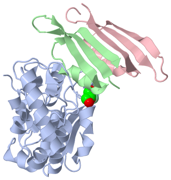
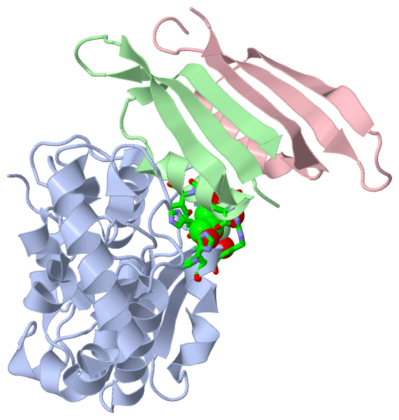
Chain A   (UNG_BACSU | P39615)
molecular function
    GO:0016787    hydrolase activity    Catalysis of the hydrolysis of various bonds, e.g. C-O, C-N, C-C, phosphoric anhydride bonds, etc. Hydrolase is the systematic name for any enzyme of EC class 3.
    GO:0016798    hydrolase activity, acting on glycosyl bonds    Catalysis of the hydrolysis of any glycosyl bond.
    GO:0016799    hydrolase activity, hydrolyzing N-glycosyl compounds    Catalysis of the hydrolysis of any N-glycosyl bond.
    GO:0004844    uracil DNA N-glycosylase activity    Catalysis of the cleavage of the N-C1' glycosidic bond between the damaged DNA base and the deoxyribose sugar, releasing a free base and leaving an apyrimidinic (AP) site. Enzymes with this activity recognize and remove uracil bases in DNA that result from the deamination of cytosine or the misincorporation of dUTP opposite an adenine.
biological process
    GO:0006281    DNA repair    The process of restoring DNA after damage. Genomes are subject to damage by chemical and physical agents in the environment (e.g. UV and ionizing radiations, chemical mutagens, fungal and bacterial toxins, etc.) and by free radicals or alkylating agents endogenously generated in metabolism. DNA is also damaged because of errors during its replication. A variety of different DNA repair pathways have been reported that include direct reversal, base excision repair, nucleotide excision repair, photoreactivation, bypass, double-strand break repair pathway, and mismatch repair pathway.
    GO:0006284    base-excision repair    In base excision repair, an altered base is removed by a DNA glycosylase enzyme, followed by excision of the resulting sugar phosphate. The small gap left in the DNA helix is filled in by the sequential action of DNA polymerase and DNA ligase.
    GO:0006974    cellular response to DNA damage stimulus    Any process that results in a change in state or activity of a cell (in terms of movement, secretion, enzyme production, gene expression, etc.) as a result of a stimulus indicating damage to its DNA from environmental insults or errors during metabolism.
    GO:0008152    metabolic process    The chemical reactions and pathways, including anabolism and catabolism, by which living organisms transform chemical substances. Metabolic processes typically transform small molecules, but also include macromolecular processes such as DNA repair and replication, and protein synthesis and degradation.
cellular component
    GO:0005737    cytoplasm    All of the contents of a cell excluding the plasma membrane and nucleus, but including other subcellular structures.

Chain B,C   (P56_BPPH2 | Q38503)
biological process
    GO:0051701    interaction with host    An interaction between two organisms living together in more or less intimate association. The term host is used for the larger (macro) of the two members of a symbiosis; the various forms of symbiosis include parasitism, commensalism and mutualism.
    GO:0016032    viral process    A multi-organism process in which a virus is a participant. The other participant is the host. Includes infection of a host cell, replication of the viral genome, and assembly of progeny virus particles. In some cases the viral genetic material may integrate into the host genome and only subsequently, under particular circumstances, 'complete' its life cycle.

 Visualization

(-) Interactive Views

Asymmetric/Biological Unit
  Complete Structure
    Jena3D(integrated viewing of ligand, site, SAP, PROSITE, SCOP information)
    WebMol | AstexViewer[tm]@PDBe
(Java Applets, require no local installation except for Java; loading may be slow)
    STRAP
(Java WebStart application, automatic local installation, requires Java; full application with system access!)
    RasMol
(require local installation)
    Molscript (VRML)
(requires installation of a VRML viewer; select preferred view via VRML and generate a mono or stereo PDF format file)
 
  Ligands, Modified Residues, Ions
    CL  [ RasMol | Jena3D ]  +environment [ RasMol | Jena3D ]
    GOL  [ RasMol | Jena3D ]  +environment [ RasMol | Jena3D ]
 
  Sites
    AC1  [ RasMol ]  +environment [ RasMol ]
    AC2  [ RasMol ]  +environment [ RasMol ]
 
  Cis Peptide Bonds
    Tyr A:39 - Pro A:40   [ RasMol ]  
 

(-) Still Images

Jmol
  protein: cartoon or spacefill or dots and stick; nucleic acid: cartoon and stick; ligands: spacefill; active site: stick
Molscript
  protein, nucleic acid: cartoon; ligands: spacefill; active site: ball and stick

 Databases and Analysis Tools

(-) Databases

Access by PDB/NDB ID
  3zoq
    Family and Domain InformationProDom | SYSTERS
    General Structural InformationGlycoscienceDB | MMDB | NDB | OCA | PDB | PDBe | PDBj | PDBsum | PDBWiki | PQS | PROTEOPEDIA
    Orientation in MembranesOPM
    Protein SurfaceSURFACE
    Secondary StructureDSSP (structure derived) | HSSP (homology derived)
    Structural GenomicsGeneCensus
    Structural NeighboursCE | VAST
    Structure ClassificationCATH | Dali | SCOP
    Validation and Original DataBMRB Data View | BMRB Restraints Grid | EDS | PROCHECK | RECOORD | WHAT_CHECK
 
Access by UniProt ID/Accession number
  P56_BPPH2 | Q38503
    Comparative Protein Structure ModelsModBase
    Genomic InformationEnsembl
    Protein-protein InteractionDIP
    Sequence, Family and Domain InformationInterPro | Pfam | SMART | UniProtKB/SwissProt
  UNG_BACSU | P39615
    Comparative Protein Structure ModelsModBase
    Genomic InformationEnsembl
    Protein-protein InteractionDIP
    Sequence, Family and Domain InformationInterPro | Pfam | SMART | UniProtKB/SwissProt
 
Access by Enzyme Classificator   (EC Number)
  3.2.2.27
    General Enzyme InformationBRENDA | EC-PDB | Enzyme | IntEnz
    PathwayKEGG | MetaCyc
 
Access by Disease Identifier   (MIM ID)
  (no 'MIM ID' available)
    Disease InformationOMIM
 
Access by GenAge ID
  (no 'GenAge ID' available)
    Age Related InformationGenAge

(-) Analysis Tools

Access by PDB/NDB ID
    Domain InformationXDom
    Interatomic Contacts of Structural UnitsCSU
    Ligand-protein ContactsLPC
    Protein CavitiescastP
    Sequence and Secondary StructurePDBCartoon
    Structure AlignmentSTRAP(Java WebStart application, automatic local installation, requires Java; full application with system access!)
    Structure and Sequence BrowserSTING
 
Access by UniProt ID/Accession number
  P56_BPPH2 | Q38503
    Protein Disorder PredictionDisEMBL | FoldIndex | GLOBPLOT (for more information see DisProt)
  UNG_BACSU | P39615
    Protein Disorder PredictionDisEMBL | FoldIndex | GLOBPLOT (for more information see DisProt)

 Related Entries

(-) Entries Sharing at Least One Protein Chain (UniProt ID)

UniProtKB/Swiss-Prot
        P56_BPPH2 | Q385032le2
        UNG_BACSU | P396153zor

(-) Related Entries Specified in the PDB File

3zor STRUCTURE OF BSUDG