Show PDB file:   
         Plain Text   HTML   (compressed file size)
QuickSearch:   
by PDB,NDB,UniProt,PROSITE Code or Search Term(s)  
(-)Asymmetric Unit
(-)Asym. Unit - sites
(-)Biological Unit 1
(-)Biol. Unit 1 - sites
(-)Biological Unit 2
collapse expand < >
Image Asymmetric Unit
Asymmetric Unit  (Jmol Viewer)
Image Asym. Unit - sites
Asym. Unit - sites  (Jmol Viewer)
Image Biological Unit 1
Biological Unit 1  (Jmol Viewer)
Image Biol. Unit 1 - sites
Biol. Unit 1 - sites  (Jmol Viewer)
Image Biological Unit 2
Biological Unit 2  (Jmol Viewer)

(-) Description

Title :  CRYSTAL STRUCTURE OF THE ECTODOMAIN OF MOUSE CD81 LARGE EXTRACELLULAR LOOP (MCD81-LEL)
 
Authors :  M. Zhang, S. Cui
Date :  14 Oct 14  (Deposition) - 01 Jul 15  (Release) - 08 Jul 15  (Revision)
Method :  X-RAY DIFFRACTION
Resolution :  1.47
Chains :  Asym. Unit :  A,B
Biol. Unit 1:  A  (1x)
Biol. Unit 2:  B  (1x)
Keywords :  Helical Bundle, Disulfide Bond, Immune Cell Adhesion, Morphology, Activation, Proliferation, Differentiation, Membrane, Cell Adhesion (Keyword Search: [Gene Ontology, PubMed, Web (Google))
 
Reference :  W. Yang, M. Zhang, X. Chi, X. Liu, B. Qin, S. Cui
An Intramolecular Bond At Cluster Of Differentiation 81 Ectodomain Is Important For Hepatitis C Virus Entry
Faseb J. 2015
PubMed-ID: 26116703  |  Reference-DOI: 10.1096/FJ.15-272880

(-) Compounds

Molecule 1 - CD81 ANTIGEN
    ChainsA, B
    EngineeredYES
    Expression SystemESCHERICHIA COLI
    Expression System PlasmidPET-28A
    Expression System StrainROSETTA(DE3)
    Expression System Taxid562
    Expression System Vector TypePLASMID
    FragmentLARGE EXTRACELLULAR LOOP, UNP RESIDUES 113-202
    GeneCD81, TAPA1
    Organism CommonMOUSE
    Organism ScientificMUS MUSCULUS
    Organism Taxid10090
    Synonym26 KDA CELL SURFACE PROTEIN TAPA-1, TARGET OF THE ANTIPROLIFERATIVE ANTIBODY 1

 Structural Features

(-) Chains, Units

  12
Asymmetric Unit AB
Biological Unit 1 (1x)A 
Biological Unit 2 (1x) B

Summary Information (see also Sequences/Alignments below)

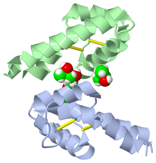
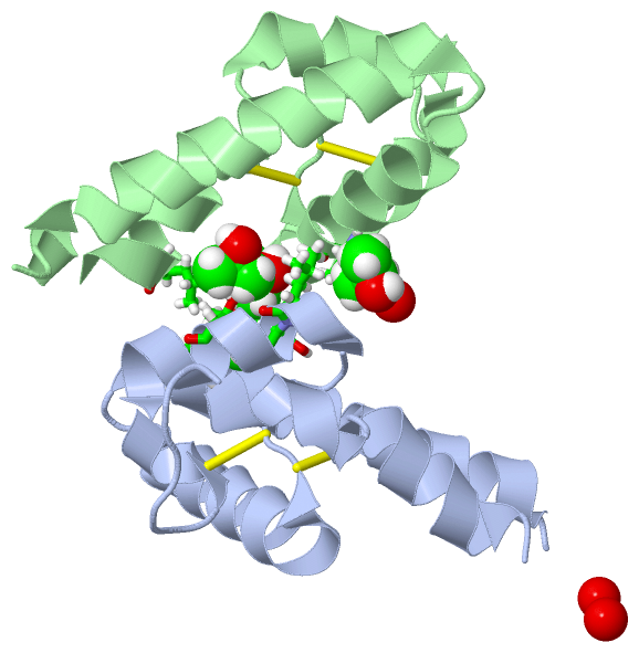
(-) Ligands, Modified Residues, Ions  (1, 4)

Asymmetric Unit (1, 4)
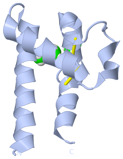
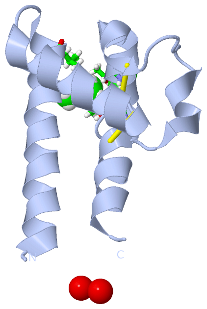
No.NameCountTypeFull Name
1IPA4Ligand/IonISOPROPYL ALCOHOL
Biological Unit 1 (1, 3)
No.NameCountTypeFull Name
1IPA3Ligand/IonISOPROPYL ALCOHOL
Biological Unit 2 (1, 1)
No.NameCountTypeFull Name
1IPA1Ligand/IonISOPROPYL ALCOHOL

(-) Sites  (4, 4)

Asymmetric Unit (4, 4)
No.NameEvidenceResiduesDescription
1AC1SOFTWARELEU A:162 , THR A:166BINDING SITE FOR RESIDUE IPA A 301
2AC2SOFTWARETHR A:163 , THR A:166 , IPA A:303 , LEU B:197BINDING SITE FOR RESIDUE IPA A 302
3AC3SOFTWAREVAL A:135 , IPA A:302BINDING SITE FOR RESIDUE IPA A 303
4AC4SOFTWAREHOH A:418 , HOH A:426 , THR B:149 , HOH B:436BINDING SITE FOR RESIDUE IPA B 301

(-) SS Bonds  (4, 4)

Asymmetric Unit
No.Residues
1A:156 -A:190
2A:157 -A:175
3B:156 -B:190
4B:157 -B:175

(-) Cis Peptide Bonds  (1, 1)

Asymmetric Unit
No.Residues
1Val A:114 -Asn A:115

 Sequence-Structure Mapping

(-) SAPs(SNPs)/Variants  (0, 0)

(no "SAP(SNP)/Variant" information available for 3X0F)

(-) PROSITE Motifs  (0, 0)

(no "PROSITE Motif" information available for 3X0F)

(-) Exons   (0, 0)

(no "Exon" information available for 3X0F)

(-) Sequences/Alignments

Asymmetric Unit
   Reformat: Number of residues per line =  ('0' or empty: single-line sequence representation)
  Number of residues per labelling interval =   
  UniProt sequence: complete  aligned part    
   Show mapping: SCOP domains CATH domains Pfam domains Secondary structure (by author)
SAPs(SNPs) PROSITE motifs Exons
(details for a mapped element are shown in a popup box when the mouse pointer rests over it)
Chain A from PDB  Type:PROTEIN  Length:87
                                                                                                                       
               SCOP domains --------------------------------------------------------------------------------------- SCOP domains
               CATH domains --------------------------------------------------------------------------------------- CATH domains
               Pfam domains --------------------------------------------------------------------------------------- Pfam domains
         Sec.struct. author .hhhhhhhhhhhhhhhhhhhhhhh..hhhhhhhhhhhhhhhh....hhhhhhhhhhhhhhhh......hhhhhh.hhhhhhhhhh.. Sec.struct. author
                 SAPs(SNPs) --------------------------------------------------------------------------------------- SAPs(SNPs)
                    PROSITE --------------------------------------------------------------------------------------- PROSITE
                 Transcript --------------------------------------------------------------------------------------- Transcript
                 3x0f A 114 VNKDQIAKDVKQFYDQALQQAVMDDDANNAKAVVKTFHETLNCCGSNALTTLTTTILRNSLCPSGGNILTPLLQQDCHQKIDELFSG 200
                                   123       133       143       153       163       173       183       193       

Chain B from PDB  Type:PROTEIN  Length:88
                                                                                                                        
               SCOP domains ---------------------------------------------------------------------------------------- SCOP domains
               CATH domains ---------------------------------------------------------------------------------------- CATH domains
               Pfam domains ---------------------------------------------------------------------------------------- Pfam domains
         Sec.struct. author ..hhhhhhhhhhhhhhhhhhhhhhh..hhhhhhhhhhhhhhhh....hhhhhhhhhhhhhhhh.............hhhhhhhhhhh. Sec.struct. author
                 SAPs(SNPs) ---------------------------------------------------------------------------------------- SAPs(SNPs)
                    PROSITE ---------------------------------------------------------------------------------------- PROSITE
                 Transcript ---------------------------------------------------------------------------------------- Transcript
                 3x0f B 113 FVNKDQIAKDVKQFYDQALQQAVMDDDANNAKAVVKTFHETLNCCGSNALTTLTTTILRNSLCPSGGNILTPLLQQDCHQKIDELFSG 200
                                   122       132       142       152       162       172       182       192        

   Legend:   → Mismatch (orange background)
  - → Gap (green background, '-', border residues have a numbering label)
    → Modified Residue (blue background, lower-case, 'x' indicates undefined single-letter code, labelled with number + name)
  x → Chemical Group (purple background, 'x', labelled with number + name, e.g. ACE or NH2)
  extra numbering lines below/above indicate numbering irregularities and modified residue names etc., number ends below/above '|'

 Classification and Annotation

(-) SCOP Domains  (0, 0)

(no "SCOP Domain" information available for 3X0F)

(-) CATH Domains  (0, 0)

(no "CATH Domain" information available for 3X0F)

(-) Pfam Domains  (0, 0)

(no "Pfam Domain" information available for 3X0F)

(-) Gene Ontology  (29, 29)

Asymmetric Unit(hide GO term definitions)

 Visualization

(-) Interactive Views

Asymmetric Unit
  Complete Structure
    Jena3D(integrated viewing of ligand, site, SAP, PROSITE, SCOP information)
    WebMol | AstexViewer[tm]@PDBe
(Java Applets, require no local installation except for Java; loading may be slow)
    STRAP
(Java WebStart application, automatic local installation, requires Java; full application with system access!)
    RasMol
(require local installation)
    Molscript (VRML)
(requires installation of a VRML viewer; select preferred view via VRML and generate a mono or stereo PDF format file)
 
  Ligands, Modified Residues, Ions
    IPA  [ RasMol | Jena3D ]  +environment [ RasMol | Jena3D ]
 
  Sites
    AC1  [ RasMol ]  +environment [ RasMol ]
    AC2  [ RasMol ]  +environment [ RasMol ]
    AC3  [ RasMol ]  +environment [ RasMol ]
    AC4  [ RasMol ]  +environment [ RasMol ]
 
  Cis Peptide Bonds
    Val A:114 - Asn A:115   [ RasMol ]  
 
Biological Units
  Complete Structure
    Biological Unit 1  [ Jena3D ]
    Biological Unit 2  [ Jena3D ]

(-) Still Images

Jmol
  protein: cartoon or spacefill or dots and stick; nucleic acid: cartoon and stick; ligands: spacefill; active site: stick
Molscript
  protein, nucleic acid: cartoon; ligands: spacefill; active site: ball and stick

 Databases and Analysis Tools

(-) Databases

Access by PDB/NDB ID
  3x0f
    Family and Domain InformationProDom | SYSTERS
    General Structural InformationGlycoscienceDB | MMDB | NDB | OCA | PDB | PDBe | PDBj | PDBsum | PDBWiki | PQS | PROTEOPEDIA
    Orientation in MembranesOPM
    Protein SurfaceSURFACE
    Secondary StructureDSSP (structure derived) | HSSP (homology derived)
    Structural GenomicsGeneCensus
    Structural NeighboursCE | VAST
    Structure ClassificationCATH | Dali | SCOP
    Validation and Original DataBMRB Data View | BMRB Restraints Grid | EDS | PROCHECK | RECOORD | WHAT_CHECK
 
Access by UniProt ID/Accession number
  CD81_MOUSE | P35762
    Comparative Protein Structure ModelsModBase
    Genomic InformationEnsembl
    Protein-protein InteractionDIP
    Sequence, Family and Domain InformationInterPro | Pfam | SMART | UniProtKB/SwissProt
 
Access by Enzyme Classificator   (EC Number)
  (no 'Enzyme Classificator' available)
    General Enzyme InformationBRENDA | EC-PDB | Enzyme | IntEnz
    PathwayKEGG | MetaCyc
 
Access by Disease Identifier   (MIM ID)
  (no 'MIM ID' available)
    Disease InformationOMIM
 
Access by GenAge ID
  (no 'GenAge ID' available)
    Age Related InformationGenAge

(-) Analysis Tools

Access by PDB/NDB ID
    Domain InformationXDom
    Interatomic Contacts of Structural UnitsCSU
    Ligand-protein ContactsLPC
    Protein CavitiescastP
    Sequence and Secondary StructurePDBCartoon
    Structure AlignmentSTRAP(Java WebStart application, automatic local installation, requires Java; full application with system access!)
    Structure and Sequence BrowserSTING
 
Access by UniProt ID/Accession number
  CD81_MOUSE | P35762
    Protein Disorder PredictionDisEMBL | FoldIndex | GLOBPLOT (for more information see DisProt)

 Related Entries

(-) Entries Sharing at Least One Protein Chain (UniProt ID)

(no "Entries Sharing at Least One Protein Chain" available for 3X0F)

(-) Related Entries Specified in the PDB File

1g8q CRYSTAL STRUCTURE OF HUMAN CD81 EXTRACELLULAR DOMAIN
1iv5 CRYSTAL STRUCTURE OF HUMAN CD81 EXTRACELLULAR DOMAIN
3x0e
3x0g