|
|
|
|
 Description
|
|
Compounds
|
||||||||||||||||||||||||
Chains, Units
Summary Information (see also Sequences/Alignments below) |
Ligands, Modified Residues, Ions (7, 8)
Asymmetric Unit (7, 8)
|
Sites (8, 8)
Asymmetric Unit (8, 8)
|
SS Bonds (0, 0)| (no "SS Bond" information available for 3WQJ) |
Cis Peptide Bonds (0, 0)| (no "Cis Peptide Bond" information available for 3WQJ) |
SAPs(SNPs)/Variants (0, 0)| (no "SAP(SNP)/Variant" information available for 3WQJ) |
PROSITE Motifs (2, 2)
Asymmetric Unit (2, 2)
|
||||||||||||||||||||||||||||||||||||||||||||||||||||||||||||||||
Exons (0, 0)| (no "Exon" information available for 3WQJ) |
Sequences/Alignments
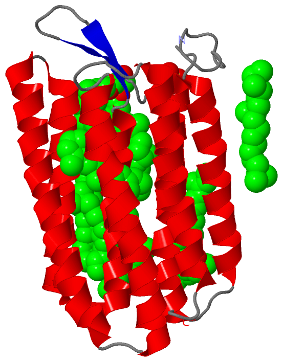
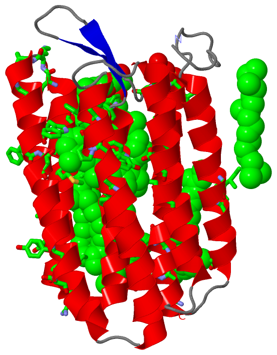
Asymmetric UnitChain A from PDB Type:PROTEIN Length:234 aligned with BACR2_HALS2 | P29563 from UniProtKB/Swiss-Prot Length:259 Alignment length:234 16 26 36 46 56 66 76 86 96 106 116 126 136 146 156 166 176 186 196 206 216 226 236 BACR2_HALS2 7 QAGFDLLNDGRPETLWLGIGTLLMLIGTFYFIARGWGVTDKEAREYYAITILVPGIASAAYLAMFFGIGVTEVELASGTVLDIYYARYADWLFTTPLLLLDLALLAKVDRVTIGTLIGVDALMIVTGLIGALSKTPLARYTWWLFSTIAFLFVLYYLLTSLRSAAAKRSEEVRSTFNTLTALVAVLWTAYPILWIVGTEGAGVVGLGIETLAFMVLDVTAKVGFGFVLLRSRAI 240 SCOP domains ------------------------------------------------------------------------------------------------------------------------------------------------------------------------------------------------------------------------------------------ SCOP domains CATH domains ------------------------------------------------------------------------------------------------------------------------------------------------------------------------------------------------------------------------------------------ CATH domains Pfam domains ------------------------------------------------------------------------------------------------------------------------------------------------------------------------------------------------------------------------------------------ Pfam domains SAPs(SNPs) ------------------------------------------------------------------------------------------------------------------------------------------------------------------------------------------------------------------------------------------ SAPs(SNPs) PROSITE --------------------------------------------------------------------------------------BACTERIAL_OPS-----------------------------------------------------------------------------------------------------------------BACTERIAL_OP---------- PROSITE Transcript ------------------------------------------------------------------------------------------------------------------------------------------------------------------------------------------------------------------------------------------ Transcript 3wqj A 1 QAGFDLLNDGRPETLWLGIGTLLMLIGTFYFIARGWGVTDKEAREYYAITILVPGIASAAYLAMFFGIGVTEVELASGTVLDIYYARYADWLFTTPLLLLDLALLAKVDRVTIGTLIGVDALMIVTGLIGALSKTPLARYTWWLFSTIAFLFVLYYLLTSLRSAAAKRSEEVRSTFNTLTALVAVLWTAYPILWIVGTEGAGVVGLGIETLAFMVLDVTAKVGFGFVLLRSRAI 234 10 20 30 40 50 60 70 80 90 100 110 120 130 140 150 160 170 180 190 200 210 220 230
|
||||||||||||||||||||
SCOP Domains (0, 0)| (no "SCOP Domain" information available for 3WQJ) |
CATH Domains (0, 0)| (no "CATH Domain" information available for 3WQJ) |
Pfam Domains (0, 0)| (no "Pfam Domain" information available for 3WQJ) |
Gene Ontology (12, 12)|
Asymmetric Unit(hide GO term definitions) Chain A (BACR2_HALS2 | P29563)
|
||||||||||||||||||||||||||||||||||||||||||||||||||||||||||||||||||||||||||||||||||||||||||
Interactive Views
|
|||||||||||||||||||||||||||||||||||||||||||||||||||||||||||||||||||||||||||||||||||||||||||||||||||||||||||||||||||||||||||||||||||||||||||||||||||||||||||||||||||||||||||||||||||||||||||||||||||||||||||||||||||||||||||||||||||
Still Images
|
||||||||||||||||
Databases
|
||||||||||||||||||||||||||||||||||||||||||||||||||||||||||||||||||||||||||||||||||||||||||||||||||||||||||||||||||||||||||||||||||||||||||||||||||||||||||||||||
Analysis Tools
|
|||||||||||||||||||||||||||||||||||||||||||||||||||||||||||||
Entries Sharing at Least One Protein Chain (UniProt ID)
Related Entries Specified in the PDB File
|
|