Show PDB file:   
         Plain Text   HTML   (compressed file size)
QuickSearch:   
by PDB,NDB,UniProt,PROSITE Code or Search Term(s)  
(-)Asym./Biol. Unit
(-)Asym./Biol. Unit - sites
collapse expand < >
Image Asym./Biol. Unit
Asym./Biol. Unit  (Jmol Viewer)
Image Asym./Biol. Unit - sites
Asym./Biol. Unit - sites  (Jmol Viewer)

(-) Description

Title :  CRYSTAL STRUCTURE OF PEPTIDYL T-RNA HYDROLASE FROM ACINETOBACTER BAUMANNII IN UNBOUND STATE AT 1.36A RESOLUTION
 
Authors :  S. Kaushik, N. Singh, M. Sinha, P. Kaur, S. Sharma, T. P. Singh
Date :  21 Aug 13  (Deposition) - 02 Oct 13  (Release) - 02 Oct 13  (Revision)
Method :  X-RAY DIFFRACTION
Resolution :  1.36
Chains :  Asym./Biol. Unit :  A
Keywords :  Hydrolase, Translation (Keyword Search: [Gene Ontology, PubMed, Web (Google))
 
Reference :  S. Kaushik, N. Singh, M. Sinha, P. Kaur, S. Sharma, T. P. Singh
Crystal Structure Of Peptidyl T-Rna Hydrolase From Acinetobacter Baumannii In Unbound State At 1. 36A Resolutio
To Be Published
PubMed: search

(-) Compounds

Molecule 1 - PEPTIDYL-TRNA HYDROLASE
    ChainsA
    EC Number3.1.1.29
    EngineeredYES
    Expression SystemESCHERICHIA COLI
    Expression System Taxid562
    GenePTH
    Organism ScientificACINETOBACTER BAUMANNII
    Organism Taxid575584
    StrainATCC 19606
    SynonymPTH

 Structural Features

(-) Chains, Units

  1
Asymmetric/Biological Unit A

Summary Information (see also Sequences/Alignments below)

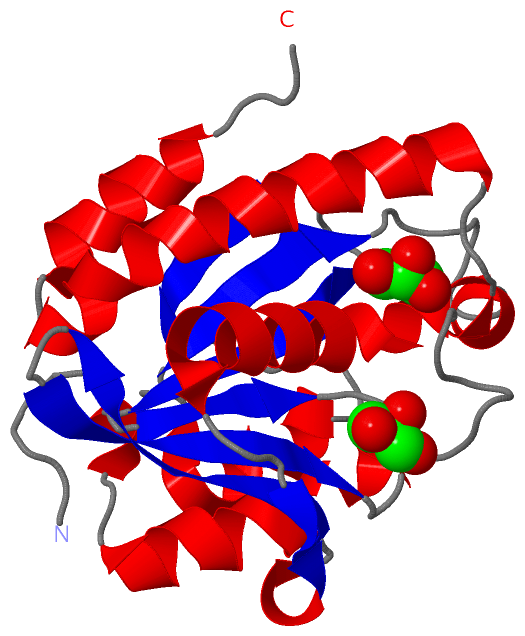
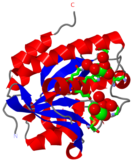
(-) Ligands, Modified Residues, Ions  (1, 2)

Asymmetric/Biological Unit (1, 2)
No.NameCountTypeFull Name
1GOL2Ligand/IonGLYCEROL

(-) Sites  (2, 2)

Asymmetric Unit (2, 2)
No.NameEvidenceResiduesDescription
1AC1SOFTWAREGLY A:14 , GLU A:30 , MET A:66 , HOH A:305 , HOH A:379 , HOH A:448BINDING SITE FOR RESIDUE GOL A 201
2AC2SOFTWAREGLN A:19 , TRP A:27 , GLN A:31 , GLN A:158 , HOH A:477 , HOH A:486BINDING SITE FOR RESIDUE GOL A 202

(-) SS Bonds  (0, 0)

(no "SS Bond" information available for 3WH4)

(-) Cis Peptide Bonds  (0, 0)

(no "Cis Peptide Bond" information available for 3WH4)

 Sequence-Structure Mapping

(-) SAPs(SNPs)/Variants  (0, 0)

(no "SAP(SNP)/Variant" information available for 3WH4)

(-) PROSITE Motifs  (0, 0)

(no "PROSITE Motif" information available for 3WH4)

(-) Exons   (0, 0)

(no "Exon" information available for 3WH4)

(-) Sequences/Alignments

Asymmetric/Biological Unit
   Reformat: Number of residues per line =  ('0' or empty: single-line sequence representation)
  Number of residues per labelling interval =   
  UniProt sequence: complete  aligned part    
   Show mapping: SCOP domains CATH domains Pfam domains Secondary structure (by author)
SAPs(SNPs) PROSITE motifs Exons
(details for a mapped element are shown in a popup box when the mouse pointer rests over it)
Chain A from PDB  Type:PROTEIN  Length:196
 aligned with D0C9L6_ACIB2 | D0C9L6 from UniProtKB/TrEMBL  Length:193

    Alignment length:196
                               1                                                                                                                                                                                                
                               |     7        17        27        37        47        57        67        77        87        97       107       117       127       137       147       157       167       177       187      
         D0C9L6_ACIB2     - ---MSNISLIVGLGNPGSEYAQTRHNAGFWFVEQLADKYGITLKNDPKFHGISGRGNIEGHDVRLLLPMTYMNRSGQSVVPFSKFYQIAPEAILIAHDELDMNPGVIRLKTGGGHGGHNGLRDIVPHIGPNFHRLRIGIGHPGSKERVSGHVLGKAPSNEQSLMDGAIDHALSKVKLLVQGQVPQAMNQINAYKPA 193
               SCOP domains d3wh4a_ A: automated matches                                                                                                                                                                         SCOP domains
               CATH domains ---------------------------------------------------------------------------------------------------------------------------------------------------------------------------------------------------- CATH domains
               Pfam domains ---------------------------------------------------------------------------------------------------------------------------------------------------------------------------------------------------- Pfam domains
         Sec.struct. author ........eeee..........hhhhhhhhhhhhhhhhhh...eeehhh.eeeeeeee..eeeeeeee..hhhhhhhhhhhhhhhh..hhh.eeeeeee.......eeeee........hhhhhhhhhh...eeeeee........hhhhhhh...hhhhhhhhhhhhhhhhhhhhhhhh.hhhhhhhhhhh.... Sec.struct. author
                 SAPs(SNPs) ---------------------------------------------------------------------------------------------------------------------------------------------------------------------------------------------------- SAPs(SNPs)
                    PROSITE ---------------------------------------------------------------------------------------------------------------------------------------------------------------------------------------------------- PROSITE
                 Transcript ---------------------------------------------------------------------------------------------------------------------------------------------------------------------------------------------------- Transcript
                 3wh4 A  -2 GSHMSNISLIVGLGNPGSEYAQTRHNAGFWFVEQLADKYGITLKNDPKFHGISGRGNIEGHDVRLLLPMTYMNRSGQSVVPFSKFYQIAPEAILIAHDELDMNPGVIRLKTGGGHGGHNGLRDIVPHIGPNFHRLRIGIGHPGSKERVSGHVLGKAPSNEQSLMDGAIDHALSKVKLLVQGQVPQAMNQINAYKPA 193
                                     7        17        27        37        47        57        67        77        87        97       107       117       127       137       147       157       167       177       187      

   Legend:   → Mismatch (orange background)
  - → Gap (green background, '-', border residues have a numbering label)
    → Modified Residue (blue background, lower-case, 'x' indicates undefined single-letter code, labelled with number + name)
  x → Chemical Group (purple background, 'x', labelled with number + name, e.g. ACE or NH2)
  extra numbering lines below/above indicate numbering irregularities and modified residue names etc., number ends below/above '|'

 Classification and Annotation

(-) SCOP Domains  (1, 1)

Asymmetric/Biological Unit

(-) CATH Domains  (0, 0)

(no "CATH Domain" information available for 3WH4)

(-) Pfam Domains  (0, 0)

(no "Pfam Domain" information available for 3WH4)

(-) Gene Ontology  (4, 4)

Asymmetric/Biological Unit(hide GO term definitions)
Chain A   (D0C9L6_ACIB2 | D0C9L6)
molecular function
    GO:0004045    aminoacyl-tRNA hydrolase activity    Catalysis of the reaction: N-substituted aminoacyl-tRNA + H2O = N-substituted amino acid + tRNA.
    GO:0016787    hydrolase activity    Catalysis of the hydrolysis of various bonds, e.g. C-O, C-N, C-C, phosphoric anhydride bonds, etc. Hydrolase is the systematic name for any enzyme of EC class 3.
biological process
    GO:0006412    translation    The cellular metabolic process in which a protein is formed, using the sequence of a mature mRNA or circRNA molecule to specify the sequence of amino acids in a polypeptide chain. Translation is mediated by the ribosome, and begins with the formation of a ternary complex between aminoacylated initiator methionine tRNA, GTP, and initiation factor 2, which subsequently associates with the small subunit of the ribosome and an mRNA or circRNA. Translation ends with the release of a polypeptide chain from the ribosome.
cellular component
    GO:0005737    cytoplasm    All of the contents of a cell excluding the plasma membrane and nucleus, but including other subcellular structures.

 Visualization

(-) Interactive Views

Asymmetric/Biological Unit
  Complete Structure
    Jena3D(integrated viewing of ligand, site, SAP, PROSITE, SCOP information)
    WebMol | AstexViewer[tm]@PDBe
(Java Applets, require no local installation except for Java; loading may be slow)
    STRAP
(Java WebStart application, automatic local installation, requires Java; full application with system access!)
    RasMol
(require local installation)
    Molscript (VRML)
(requires installation of a VRML viewer; select preferred view via VRML and generate a mono or stereo PDF format file)
 
  Ligands, Modified Residues, Ions
    GOL  [ RasMol | Jena3D ]  +environment [ RasMol | Jena3D ]
 
  Sites
    AC1  [ RasMol ]  +environment [ RasMol ]
    AC2  [ RasMol ]  +environment [ RasMol ]
 
  Cis Peptide Bonds
(no "Cis Peptide Bonds" information available for 3wh4)
 

(-) Still Images

Jmol
  protein: cartoon or spacefill or dots and stick; nucleic acid: cartoon and stick; ligands: spacefill; active site: stick
Molscript
  protein, nucleic acid: cartoon; ligands: spacefill; active site: ball and stick

 Databases and Analysis Tools

(-) Databases

Access by PDB/NDB ID
  3wh4
    Family and Domain InformationProDom | SYSTERS
    General Structural InformationGlycoscienceDB | MMDB | NDB | OCA | PDB | PDBe | PDBj | PDBsum | PDBWiki | PQS | PROTEOPEDIA
    Orientation in MembranesOPM
    Protein SurfaceSURFACE
    Secondary StructureDSSP (structure derived) | HSSP (homology derived)
    Structural GenomicsGeneCensus
    Structural NeighboursCE | VAST
    Structure ClassificationCATH | Dali | SCOP
    Validation and Original DataBMRB Data View | BMRB Restraints Grid | EDS | PROCHECK | RECOORD | WHAT_CHECK
 
Access by UniProt ID/Accession number
  D0C9L6_ACIB2 | D0C9L6
    Comparative Protein Structure ModelsModBase
    Genomic InformationEnsembl
    Protein-protein InteractionDIP
    Sequence, Family and Domain InformationInterPro | Pfam | SMART | UniProtKB/TrEMBL
 
Access by Enzyme Classificator   (EC Number)
  3.1.1.29
    General Enzyme InformationBRENDA | EC-PDB | Enzyme | IntEnz
    PathwayKEGG | MetaCyc
 
Access by Disease Identifier   (MIM ID)
  (no 'MIM ID' available)
    Disease InformationOMIM
 
Access by GenAge ID
  (no 'GenAge ID' available)
    Age Related InformationGenAge

(-) Analysis Tools

Access by PDB/NDB ID
    Domain InformationXDom
    Interatomic Contacts of Structural UnitsCSU
    Ligand-protein ContactsLPC
    Protein CavitiescastP
    Sequence and Secondary StructurePDBCartoon
    Structure AlignmentSTRAP(Java WebStart application, automatic local installation, requires Java; full application with system access!)
    Structure and Sequence BrowserSTING
 
Access by UniProt ID/Accession number
  D0C9L6_ACIB2 | D0C9L6
    Protein Disorder PredictionDisEMBL | FoldIndex | GLOBPLOT (for more information see DisProt)

 Related Entries

(-) Entries Sharing at Least One Protein Chain (UniProt ID)

UniProtKB/TrEMBL
        D0C9L6_ACIB2 | D0C9L64fop 4fot 4hoy 4iko 4jwk 4jx9 4jy7 4lwq 4lwr 4olj

(-) Related Entries Specified in the PDB File

4lwq THE SAME PROTEIN AT 1.38A RESOLUTION