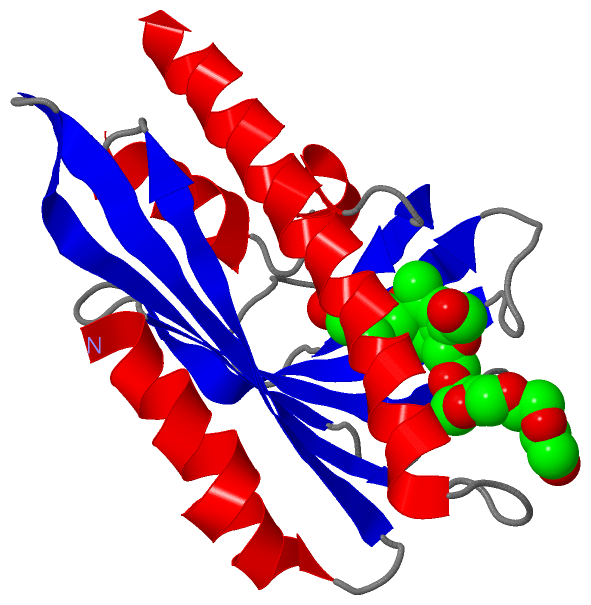
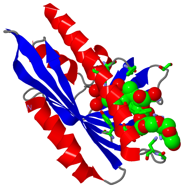
Show PDB file:   
         Plain Text   HTML   (compressed file size)
QuickSearch:   
by PDB,NDB,UniProt,PROSITE Code or Search Term(s)  
(-)Asym./Biol. Unit
(-)Asym./Biol. Unit - sites
collapse expand < >
Image Asym./Biol. Unit
Asym./Biol. Unit  (Jmol Viewer)
Image Asym./Biol. Unit - sites
Asym./Biol. Unit - sites  (Jmol Viewer)

(-) Description

Title :  CRYSTAL STRUCTURE OF THE HIGH-AFFINITY ABSCISIC ACID RECEPTOR PYL9/RCAR9 BOUND TO ABA
 
Authors :  M. Nakagawa, Y. Hirano, M. Kagiyama, N. Shibata, T. Hakoshima
Date :  13 Apr 13  (Deposition) - 09 Apr 14  (Release) - 09 Apr 14  (Revision)
Method :  X-RAY DIFFRACTION
Resolution :  1.90
Chains :  Asym./Biol. Unit :  A
Keywords :  Abscisic Acid Receptor, Drought Tolerance, Protein Phosphatase Inhibitor, Start/Bet V1 Family, Hormone Receptor (Keyword Search: [Gene Ontology, PubMed, Web (Google))
 
Reference :  M. Nakagawa, M. Kagiyama, N. Shibata, Y. Hirano, T. Hakoshima
Mechanism Of High-Affinity Abscisic Acid Binding To Pyl9/Rcar1
Genes Cells 2014
PubMed-ID: 24645846  |  Reference-DOI: 10.1111/GTC.12140

(-) Compounds

Molecule 1 - ABSCISIC ACID RECEPTOR PYL9
    ChainsA
    EngineeredYES
    Expression SystemESCHERICHIA COLI
    Expression System PlasmidPET49B
    Expression System StrainROSETTA2 (DE3)
    Expression System Taxid562
    Expression System Vector TypePLASMID
    GeneAT1G01360, F6F3.16, PYL9, RCAR1
    Organism CommonMOUSE-EAR CRESS, THALE-CRESS
    Organism ScientificARABIDOPSIS THALIANA
    Organism Taxid3702
    SynonymABI1-BINDING PROTEIN 4, PYR1-LIKE PROTEIN 9, REGULATORY COMPONENTS OF ABA RECEPTOR 1

 Structural Features

(-) Chains, Units

  1
Asymmetric/Biological Unit A

Summary Information (see also Sequences/Alignments below)

(-) Ligands, Modified Residues, Ions  (2, 2)

Asymmetric/Biological Unit (2, 2)
No.NameCountTypeFull Name
1A8S1Ligand/Ion(2Z,4E)-5-[(1S)-1-HYDROXY-2,6,6-TRIMETHYL-4-OXOCYCLOHEX-2-EN-1-YL]-3-METHYLPENTA-2,4-DIENOIC ACID
2P6G1Ligand/IonHEXAETHYLENE GLYCOL

(-) Sites  (2, 2)

Asymmetric Unit (2, 2)
No.NameEvidenceResiduesDescription
1AC1SOFTWARELYS A:63 , ALA A:91 , SER A:94 , TYR A:122 , ASN A:169 , HOH A:301 , HOH A:302 , HOH A:304 , HOH A:307 , HOH A:312BINDING SITE FOR RESIDUE A8S A 201
2AC2SOFTWAREPHE A:65 , SER A:87 , ARG A:118 , GLY A:152 , ASN A:153 , THR A:158 , PHE A:161 , HOH A:354BINDING SITE FOR RESIDUE P6G A 202

(-) SS Bonds  (0, 0)

(no "SS Bond" information available for 3W9R)

(-) Cis Peptide Bonds  (0, 0)

(no "Cis Peptide Bond" information available for 3W9R)

 Sequence-Structure Mapping

(-) SAPs(SNPs)/Variants  (0, 0)

(no "SAP(SNP)/Variant" information available for 3W9R)

(-) PROSITE Motifs  (0, 0)

(no "PROSITE Motif" information available for 3W9R)

(-) Exons   (0, 0)

(no "Exon" information available for 3W9R)

(-) Sequences/Alignments

Asymmetric/Biological Unit
   Reformat: Number of residues per line =  ('0' or empty: single-line sequence representation)
  Number of residues per labelling interval =   
  UniProt sequence: complete  aligned part    
   Show mapping: SCOP domains CATH domains Pfam domains Secondary structure (by author)
SAPs(SNPs) PROSITE motifs Exons
(details for a mapped element are shown in a popup box when the mouse pointer rests over it)
Chain A from PDB  Type:PROTEIN  Length:171
 aligned with PYL9_ARATH | Q84MC7 from UniProtKB/Swiss-Prot  Length:187

    Alignment length:171
                                    22        32        42        52        62        72        82        92       102       112       122       132       142       152       162       172       182 
           PYL9_ARATH    13 GGLETVQYVRTHHQHLCRENQCTSALVKHIKAPLHLVWSLVRRFDQPQKYKPFVSRCTVIGDPEIGSLREVNVKSGLPATTSTERLELLDDEEHILGIKIIGGDHRLKNYSSILTVHPEIIEGRAGTMVIESFVVDVPQGNTKDETCYFVEALIRCNLKSLADVSERLASQ 183
               SCOP domains --------------------------------------------------------------------------------------------------------------------------------------------------------------------------- SCOP domains
               CATH domains --------------------------------------------------------------------------------------------------------------------------------------------------------------------------- CATH domains
               Pfam domains --------------------------------------------------------------------------------------------------------------------------------------------------------------------------- Pfam domains
         Sec.struct. author hhhhhhhhhhhhhhhhh...eeeeeeeeee..hhhhhhhhhh...hhhhh...eeeee.........eeeeee.......eeeeeeeeee....eeeeeeeee.......eeeeeeeeeee..eeeeeeeeeeeee.....hhhhhhhhhhhhhhhhhhhhhhhhhhhhh. Sec.struct. author
                 SAPs(SNPs) --------------------------------------------------------------------------------------------------------------------------------------------------------------------------- SAPs(SNPs)
                    PROSITE --------------------------------------------------------------------------------------------------------------------------------------------------------------------------- PROSITE
                 Transcript --------------------------------------------------------------------------------------------------------------------------------------------------------------------------- Transcript
                 3w9r A  13 GGLETVQYVRTHHQHLCRENQCTSALVKHIKAPLHLVWSLVRRFDQPQKYKPFVSRCTVIGDPEIGSLREVNVKSGLPATTSTERLELLDDEEHILGIKIIGGDHRLKNYSSILTVHPEIIEGRAGTMVIESFVVDVPQGNTKDETCYFVEALIRCNLKSLADVSERLASQ 183
                                    22        32        42        52        62        72        82        92       102       112       122       132       142       152       162       172       182 

   Legend:   → Mismatch (orange background)
  - → Gap (green background, '-', border residues have a numbering label)
    → Modified Residue (blue background, lower-case, 'x' indicates undefined single-letter code, labelled with number + name)
  x → Chemical Group (purple background, 'x', labelled with number + name, e.g. ACE or NH2)
  extra numbering lines below/above indicate numbering irregularities and modified residue names etc., number ends below/above '|'

 Classification and Annotation

(-) SCOP Domains  (0, 0)

(no "SCOP Domain" information available for 3W9R)

(-) CATH Domains  (0, 0)

(no "CATH Domain" information available for 3W9R)

(-) Pfam Domains  (0, 0)

(no "Pfam Domain" information available for 3W9R)

(-) Gene Ontology  (12, 12)

Asymmetric/Biological Unit(hide GO term definitions)
Chain A   (PYL9_ARATH | Q84MC7)
molecular function
    GO:0010427    abscisic acid binding    Interacting selectively and non-covalently with abscisic acid, plant hormones that regulate aspects of plant growth.
    GO:0005515    protein binding    Interacting selectively and non-covalently with any protein or protein complex (a complex of two or more proteins that may include other nonprotein molecules).
    GO:0042803    protein homodimerization activity    Interacting selectively and non-covalently with an identical protein to form a homodimer.
    GO:0004864    protein phosphatase inhibitor activity    Stops, prevents or reduces the activity of a protein phosphatase, an enzyme that hydrolyzes phosphate groups from phosphorylated proteins.
    GO:0004872    receptor activity    Combining with an extracellular or intracellular messenger to initiate a change in cell activity.
biological process
    GO:0009738    abscisic acid-activated signaling pathway    A series of molecular signals generated by the binding of the plant hormone abscisic acid (ABA) to a receptor, and ending with modulation of a cellular process, e.g. transcription.
    GO:0043086    negative regulation of catalytic activity    Any process that stops or reduces the activity of an enzyme.
    GO:0080163    regulation of protein serine/threonine phosphatase activity    Any process that modulates the frequency, rate or extent of protein serine/threonine phosphatase activity: catalysis of the reaction: protein serine/threonine phosphate + H2O = protein serine/threonine + phosphate.
cellular component
    GO:0005737    cytoplasm    All of the contents of a cell excluding the plasma membrane and nucleus, but including other subcellular structures.
    GO:0016020    membrane    A lipid bilayer along with all the proteins and protein complexes embedded in it an attached to it.
    GO:0005634    nucleus    A membrane-bounded organelle of eukaryotic cells in which chromosomes are housed and replicated. In most cells, the nucleus contains all of the cell's chromosomes except the organellar chromosomes, and is the site of RNA synthesis and processing. In some species, or in specialized cell types, RNA metabolism or DNA replication may be absent.
    GO:0005886    plasma membrane    The membrane surrounding a cell that separates the cell from its external environment. It consists of a phospholipid bilayer and associated proteins.

 Visualization

(-) Interactive Views

Asymmetric/Biological Unit
  Complete Structure
    Jena3D(integrated viewing of ligand, site, SAP, PROSITE, SCOP information)
    WebMol | AstexViewer[tm]@PDBe
(Java Applets, require no local installation except for Java; loading may be slow)
    STRAP
(Java WebStart application, automatic local installation, requires Java; full application with system access!)
    RasMol
(require local installation)
    Molscript (VRML)
(requires installation of a VRML viewer; select preferred view via VRML and generate a mono or stereo PDF format file)
 
  Ligands, Modified Residues, Ions
    A8S  [ RasMol | Jena3D ]  +environment [ RasMol | Jena3D ]
    P6G  [ RasMol | Jena3D ]  +environment [ RasMol | Jena3D ]
 
  Sites
    AC1  [ RasMol ]  +environment [ RasMol ]
    AC2  [ RasMol ]  +environment [ RasMol ]
 
  Cis Peptide Bonds
(no "Cis Peptide Bonds" information available for 3w9r)
 

(-) Still Images

Jmol
  protein: cartoon or spacefill or dots and stick; nucleic acid: cartoon and stick; ligands: spacefill; active site: stick
Molscript
  protein, nucleic acid: cartoon; ligands: spacefill; active site: ball and stick

 Databases and Analysis Tools

(-) Databases

Access by PDB/NDB ID
  3w9r
    Family and Domain InformationProDom | SYSTERS
    General Structural InformationGlycoscienceDB | MMDB | NDB | OCA | PDB | PDBe | PDBj | PDBsum | PDBWiki | PQS | PROTEOPEDIA
    Orientation in MembranesOPM
    Protein SurfaceSURFACE
    Secondary StructureDSSP (structure derived) | HSSP (homology derived)
    Structural GenomicsGeneCensus
    Structural NeighboursCE | VAST
    Structure ClassificationCATH | Dali | SCOP
    Validation and Original DataBMRB Data View | BMRB Restraints Grid | EDS | PROCHECK | RECOORD | WHAT_CHECK
 
Access by UniProt ID/Accession number
  PYL9_ARATH | Q84MC7
    Comparative Protein Structure ModelsModBase
    Genomic InformationEnsembl
    Protein-protein InteractionDIP
    Sequence, Family and Domain InformationInterPro | Pfam | SMART | UniProtKB/SwissProt
 
Access by Enzyme Classificator   (EC Number)
  (no 'Enzyme Classificator' available)
    General Enzyme InformationBRENDA | EC-PDB | Enzyme | IntEnz
    PathwayKEGG | MetaCyc
 
Access by Disease Identifier   (MIM ID)
  (no 'MIM ID' available)
    Disease InformationOMIM
 
Access by GenAge ID
  (no 'GenAge ID' available)
    Age Related InformationGenAge

(-) Analysis Tools

Access by PDB/NDB ID
    Domain InformationXDom
    Interatomic Contacts of Structural UnitsCSU
    Ligand-protein ContactsLPC
    Protein CavitiescastP
    Sequence and Secondary StructurePDBCartoon
    Structure AlignmentSTRAP(Java WebStart application, automatic local installation, requires Java; full application with system access!)
    Structure and Sequence BrowserSTING
 
Access by UniProt ID/Accession number
  PYL9_ARATH | Q84MC7
    Protein Disorder PredictionDisEMBL | FoldIndex | GLOBPLOT (for more information see DisProt)

 Related Entries

(-) Entries Sharing at Least One Protein Chain (UniProt ID)

UniProtKB/Swiss-Prot
        PYL9_ARATH | Q84MC73oqu

(-) Related Entries Specified in the PDB File

(no "Related Entries Specified in the PDB File" available for 3W9R)