|
|
|
|
 Description
|
|
Compounds
|
||||||||||||||||||||||||||||||||||||||||||||||||
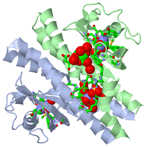
Chains, Units
Summary Information (see also Sequences/Alignments below) |
Ligands, Modified Residues, Ions (3, 12)| Asymmetric/Biological Unit (3, 12) |
Sites (12, 12)
Asymmetric Unit (12, 12)
|
SS Bonds (0, 0)| (no "SS Bond" information available for 3UB8) |
Cis Peptide Bonds (0, 0)| (no "Cis Peptide Bond" information available for 3UB8) |
SAPs(SNPs)/Variants (0, 0)| (no "SAP(SNP)/Variant" information available for 3UB8) |
PROSITE Motifs (0, 0)| (no "PROSITE Motif" information available for 3UB8) |
Exons (0, 0)| (no "Exon" information available for 3UB8) |
Sequences/Alignments
Asymmetric/Biological UnitChain A from PDB Type:PROTEIN Length:157 aligned with B6JPK4_HELP2 | B6JPK4 from UniProtKB/TrEMBL Length:561 Alignment length:157 50 60 70 80 90 100 110 120 130 140 150 160 170 180 190 B6JPK4_HELP2 41 MEHLETGQYKKREKTLAYMTKILEQGIHEYYKNFDNATARKMALDYFKRINDDKGMIYMVVVDKNGVVLFDPVNPKTVGQSGLDAQSVDGVYYVRGYLEAAKKGGGYTYYKMPKYDGGVPEKKFAYSHYDEVSQMVIAATSYYTDINTENQAIKEGV 197 SCOP domains ------------------------------------------------------------------------------------------------------------------------------------------------------------- SCOP domains CATH domains ------------------------------------------------------------------------------------------------------------------------------------------------------------- CATH domains Pfam domains ------------------------------------------------------------------------------------------------------------------------------------------------------------- Pfam domains SAPs(SNPs) ------------------------------------------------------------------------------------------------------------------------------------------------------------- SAPs(SNPs) PROSITE ------------------------------------------------------------------------------------------------------------------------------------------------------------- PROSITE Transcript ------------------------------------------------------------------------------------------------------------------------------------------------------------- Transcript 3ub8 A 44 MEHLETGQYKKREKTLAYMTKILEQGIHEYYKSFDNDTARKMALDYFKRINDDKGMIYMVVVDKNGVVLFDPVNPKTVGQSGLDAQSVDGVYYVRGYLEAAKKGGGYTYYKMPKYDGGVPEKKFAYSHYDEVSQMVIAATSYYTDINTENKAIKEGV 200 53 63 73 83 93 103 113 123 133 143 153 163 173 183 193 Chain B from PDB Type:PROTEIN Length:158 aligned with B6JPK4_HELP2 | B6JPK4 from UniProtKB/TrEMBL Length:561 Alignment length:158 50 60 70 80 90 100 110 120 130 140 150 160 170 180 190 B6JPK4_HELP2 41 MEHLETGQYKKREKTLAYMTKILEQGIHEYYKNFDNATARKMALDYFKRINDDKGMIYMVVVDKNGVVLFDPVNPKTVGQSGLDAQSVDGVYYVRGYLEAAKKGGGYTYYKMPKYDGGVPEKKFAYSHYDEVSQMVIAATSYYTDINTENQAIKEGVN 198 SCOP domains -------------------------------------------------------------------------------------------------------------------------------------------------------------- SCOP domains CATH domains -------------------------------------------------------------------------------------------------------------------------------------------------------------- CATH domains Pfam domains -------------------------------------------------------------------------------------------------------------------------------------------------------------- Pfam domains SAPs(SNPs) -------------------------------------------------------------------------------------------------------------------------------------------------------------- SAPs(SNPs) PROSITE -------------------------------------------------------------------------------------------------------------------------------------------------------------- PROSITE Transcript -------------------------------------------------------------------------------------------------------------------------------------------------------------- Transcript 3ub8 B 44 MEHLETGQYKKREKTLAYMTKILEQGIHEYYKSFDNDTARKMALDYFKRINDDKGMIYMVVVDKNGVVLFDPVNPKTVGQSGLDAQSVDGVYYVRGYLEAAKKGGGYTYYKMPKYDGGVPEKKFAYSHYDEVSQMVIAATSYYTDINTENKAIKEGVN 201 53 63 73 83 93 103 113 123 133 143 153 163 173 183 193
|
||||||||||||||||||||
SCOP Domains (0, 0)| (no "SCOP Domain" information available for 3UB8) |
CATH Domains (0, 0)| (no "CATH Domain" information available for 3UB8) |
Pfam Domains (0, 0)| (no "Pfam Domain" information available for 3UB8) |
Gene Ontology (4, 4)|
Asymmetric/Biological Unit(hide GO term definitions) Chain A,B (B6JPK4_HELP2 | B6JPK4)
|
||||||||||||||||||||||||||||||||||||||||||
Interactive Views
|
|||||||||||||||||||||||||||||||||||||||||||||||||||||||||||||||||||||||||||||||||||||||||||||||||||||||||||||||||||||||||||||||||||||||||||||||||||||||||||||||||||||||||||||||||||||||||||||||||||||||||||||||||
Still Images
|
||||||||||||||||
Databases
|
||||||||||||||||||||||||||||||||||||||||||||||||||||||||||||||||||||||||||||||||||||||||||||||||||||||||||||||||||||||||||||||||||||||||||||||||||||||||||||||||
Analysis Tools
|
|||||||||||||||||||||||||||||||||||||||||||||||||||||||||||||
Entries Sharing at Least One Protein Chain (UniProt ID)
Related Entries Specified in the PDB File
|
|