|
|
|
|
 Description
|
|
Compounds
|
||||||||||||||||||||||||||||||||||||||||||||||||||||||||
Chains, Units
Summary Information (see also Sequences/Alignments below) |
Ligands, Modified Residues, Ions (4, 6)| Asymmetric/Biological Unit (4, 6) |
Sites (6, 6)
Asymmetric Unit (6, 6)
|
SS Bonds (0, 0)| (no "SS Bond" information available for 3TT4) |
Cis Peptide Bonds (1, 1)
Asymmetric/Biological Unit
|
||||||||
SAPs(SNPs)/Variants (3, 3)
Asymmetric/Biological Unit (3, 3)
|
|||||||||||||||||||||||||||||||||||||||||||||||||||||||||||||||||||||||||||||||
PROSITE Motifs (1, 1)
Asymmetric/Biological Unit (1, 1)
|
||||||||||||||||||||||||
Exons (5, 5)
Sequences/Alignments
Asymmetric/Biological UnitChain A from PDB Type:PROTEIN Length:159 aligned with MMP8_HUMAN | P22894 from UniProtKB/Swiss-Prot Length:467 Alignment length:159 113 123 133 143 153 163 173 183 193 203 213 223 233 243 253 MMP8_HUMAN 104 GNPKWERTNLTYRIRNYTPQLSEAEVERAIKDAFELWSVASPLIFTRISQGEADINIAFYQRDHGDNSPFDGPNGILAHAFQPGQGIGGDAHFDAEETWTNTSANYNLFLVAAHEFGHSLGLAHSSDPGALMYPNYAFRETSNYSLPQDDIDGIQAIYG 262 SCOP domains d3tt4a_ A: automated matches SCOP domains CATH domains --------------------------------------------------------------------------------------------------------------------------------------------------------------- CATH domains Pfam domains --------------------------------------------------------------------------------------------------------------------------------------------------------------- Pfam domains SAPs(SNPs) --------------------------------------------------E--------------------------------------V----------------------------------------------------Y---------------- SAPs(SNPs) PROSITE --------------------------------------------------------------------------------------------------------------ZINC_PROTE--------------------------------------- PROSITE Transcript 1 (1) Exon 1.6c -------------------------------------------------Exon 1.8b PDB: A:146-188 UniProt: 166-208 -----------------------------------------------------1 Transcript 1 (1) Transcript 1 (2) ------------Exon 1.7 PDB: A:96-146 UniProt: 116-166 -----------------------------------------Exon 1.9 PDB: A:188-242 UniProt: 208-262 Transcript 1 (2) 3tt4 A 84 GNPKWERTNLTYRIRNYTPQLSEAEVERAIKDAFELWSVASPLIFTRISQGEADINIAFYQRDHGDNSPFDGPNGILAHAFQPGQGIGGDAHFDAEETWTNTSANYNLFLVAAHEFGHSLGLAHSSDPGALMYPNYAFRETSNYSLPQDDIDGIQAIYG 242 93 103 113 123 133 143 153 163 173 183 193 203 213 223 233
|
||||||||||||||||||||
SCOP Domains (1, 1)
Asymmetric/Biological Unit
 |
CATH Domains (0, 0)| (no "CATH Domain" information available for 3TT4) |
Pfam Domains (0, 0)| (no "Pfam Domain" information available for 3TT4) |
Gene Ontology (16, 16)|
Asymmetric/Biological Unit(hide GO term definitions) Chain A (MMP8_HUMAN | P22894)
|
||||||||||||||||||||||||||||||||||||||||||||||||||||||||||||||||||||||||||||||||||||||||||||||||||||||||||||||||||
Interactive Views
|
|||||||||||||||||||||||||||||||||||||||||||||||||||||||||||||||||||||||||||||||||||||||||||||||||||||||||||||||||||||||||||||||||||||||||||||||||||||||||||||||||||||||||||||||
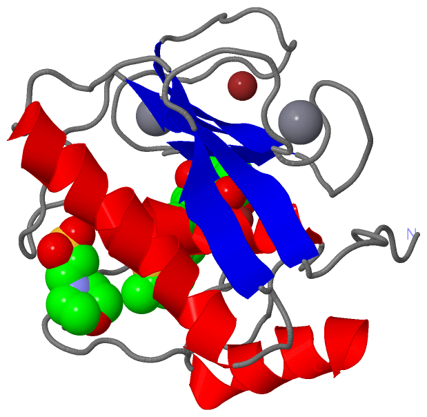
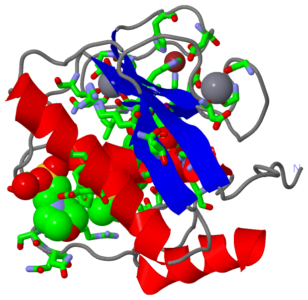
Still Images
|
||||||||||||||||
Databases
|
||||||||||||||||||||||||||||||||||||||||||||||||||||||||||||||||||||||||||||||||||||||||||||||||||||||||||||||||||||||||||||||||||||||||||||||||||||||||||||||||
Analysis Tools
|
|||||||||||||||||||||||||||||||||||||||||||||||||||||||||||||
Entries Sharing at Least One Protein Chain (UniProt ID)
Related Entries Specified in the PDB File
|
|